Automated image analysis of nuclear shape: what can we learn from a prematurely aged cell?
- PMID: 22354768
- PMCID: PMC3314174
- DOI: 10.18632/aging.100434
Automated image analysis of nuclear shape: what can we learn from a prematurely aged cell?
Abstract
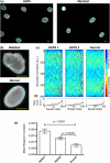
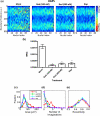
The premature aging disorder, Hutchinson-Gilford progeria syndrome (HGPS), is caused by mutant lamin A, which affects the nuclear scaffolding. The phenotypic hallmark of HGPS is nuclear blebbing. Interestingly, similar nuclear blebbing has also been observed in aged cells from healthy individuals. Recent work has shown that treatment with rapamycin, an inhibitor of the mTOR pathway, reduced nuclear blebbing in HGPS fibroblasts. However, the extent of blebbing varies considerably within each cell population, which makes manual blind counting challenging and subjective. Here, we show a novel, automated and high throughput nuclear shape analysis that quantitatively measures curvature, area, perimeter, eccentricity and additional metrics of nuclear morphology for large populations of cells. We examined HGPS fibroblast cells treated with rapamycin and RAD001 (an analog to rapamycin). Our analysis shows that treatment with RAD001 and rapamycin reduces nuclear blebbing, consistent with blind counting controls. In addition, we find that rapamycin treatment reduces the area of the nucleus, but leaves the eccentricity unchanged. Our nuclear shape analysis provides an unbiased, multidimensional "fingerprint" for a population of cells, which can be used to quantify treatment efficacy and analyze cellular aging.
Conflict of interest statement
The authors of this manuscript have no conflict of interest to declare.
Figures


Similar articles
-
Depressing time: Waiting, melancholia, and the psychoanalytic practice of care.In: Kirtsoglou E, Simpson B, editors. The Time of Anthropology: Studies of Contemporary Chronopolitics. Abingdon: Routledge; 2020. Chapter 5. In: Kirtsoglou E, Simpson B, editors. The Time of Anthropology: Studies of Contemporary Chronopolitics. Abingdon: Routledge; 2020. Chapter 5. PMID: 36137063 Free Books & Documents. Review.
-
Dynamic Field Theory of Executive Function: Identifying Early Neurocognitive Markers.Monogr Soc Res Child Dev. 2024 Dec;89(3):7-109. doi: 10.1111/mono.12478. Monogr Soc Res Child Dev. 2024. PMID: 39628288 Free PMC article.
-
DNA damage responses in progeroid syndromes arise from defective maturation of prelamin A.J Cell Sci. 2006 Nov 15;119(Pt 22):4644-9. doi: 10.1242/jcs.03263. Epub 2006 Oct 24. J Cell Sci. 2006. PMID: 17062639 Free PMC article.
-
Defining the optimum strategy for identifying adults and children with coeliac disease: systematic review and economic modelling.Health Technol Assess. 2022 Oct;26(44):1-310. doi: 10.3310/ZUCE8371. Health Technol Assess. 2022. PMID: 36321689 Free PMC article.
-
Trends in Surgical and Nonsurgical Aesthetic Procedures: A 14-Year Analysis of the International Society of Aesthetic Plastic Surgery-ISAPS.Aesthetic Plast Surg. 2024 Oct;48(20):4217-4227. doi: 10.1007/s00266-024-04260-2. Epub 2024 Aug 5. Aesthetic Plast Surg. 2024. PMID: 39103642 Review.
Cited by
-
An inhibitory role of progerin in the gene induction network of adipocyte differentiation from iPS cells.Aging (Albany NY). 2013 Apr;5(4):288-303. doi: 10.18632/aging.100550. Aging (Albany NY). 2013. PMID: 23596277 Free PMC article.
-
Accurate Detection of Dysmorphic Nuclei Using Dynamic Programming and Supervised Classification.PLoS One. 2017 Jan 26;12(1):e0170688. doi: 10.1371/journal.pone.0170688. eCollection 2017. PLoS One. 2017. PMID: 28125723 Free PMC article.
-
Tumor promoter-induced cellular senescence: cell cycle arrest followed by geroconversion.Oncotarget. 2014 Dec 30;5(24):12715-27. doi: 10.18632/oncotarget.3011. Oncotarget. 2014. PMID: 25587030 Free PMC article.
-
A comprehensive review of computational and image analysis techniques for quantitative evaluation of striated muscle tissue architecture.Biophys Rev (Melville). 2022 Dec;3(4):041302. doi: 10.1063/5.0057434. Epub 2022 Nov 4. Biophys Rev (Melville). 2022. PMID: 36407035 Free PMC article. Review.
-
Deep Learning in Label-free Cell Classification.Sci Rep. 2016 Mar 15;6:21471. doi: 10.1038/srep21471. Sci Rep. 2016. PMID: 26975219 Free PMC article.
References
-
- Capell BC, Collins FS. Human laminopathies: nuclei gone genetically awry. Nat Rev Genet. 2006;7:940–952. - PubMed
-
- Gordon LB, Harling-Berg CJ, Rothman FG. Highlights of the 2007 Progeria Research Foundation scientific workshop: progress in translational science. J Gerontol A Biol Sci Med Sci. 2008;63:777–787. - PubMed
-
- Gordon LB, McCarten KM, Giobbie-Hurder A, Machan JT, Campbell SE, Berns SD, Kieran MW. Disease progression in Hutchinson-Gilford progeria syndrome: impact on growth and development. Pediatrics. 2007;120:824–833. - PubMed
Publication types
MeSH terms
Substances
Grants and funding
LinkOut - more resources
Full Text Sources
Miscellaneous

