Neutralizing antibodies against the preactive form of respiratory syncytial virus fusion protein offer unique possibilities for clinical intervention
- PMID: 22323598
- PMCID: PMC3286924
- DOI: 10.1073/pnas.1115941109
Neutralizing antibodies against the preactive form of respiratory syncytial virus fusion protein offer unique possibilities for clinical intervention
Abstract
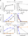
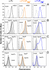
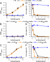
Human respiratory syncytial virus (hRSV) is the most important viral agent of pediatric respiratory infections worldwide. The only specific treatment available today is a humanized monoclonal antibody (Palivizumab) directed against the F glycoprotein, administered prophylactically to children at very high risk of severe hRSV infections. Palivizumab, as most anti-F antibodies so far described, recognizes an epitope that is shared by the two conformations in which hRSV_F can fold, the metastable prefusion form and the highly stable postfusion conformation. We now describe a unique class of antibodies specific for the prefusion form of this protein that account for most of the neutralizing activity of either a rabbit serum raised against a vaccinia virus recombinant expressing hRSV_F or a human Ig preparation (Respigam), which was used for prophylaxis before Palivizumab. These antibodies therefore offer unique possibilities for immune intervention against hRSV, and their production should be assessed in trials of hRSV vaccines.
Conflict of interest statement
The authors declare no conflict of interest.
Figures


Similar articles
-
Influence of Respiratory Syncytial Virus F Glycoprotein Conformation on Induction of Protective Immune Responses.J Virol. 2016 May 12;90(11):5485-5498. doi: 10.1128/JVI.00338-16. Print 2016 Jun 1. J Virol. 2016. PMID: 27009962 Free PMC article.
-
Potent Human Single-Domain Antibodies Specific for a Novel Prefusion Epitope of Respiratory Syncytial Virus F Glycoprotein.J Virol. 2021 Aug 25;95(18):e0048521. doi: 10.1128/JVI.00485-21. Epub 2021 Aug 25. J Virol. 2021. PMID: 34160257 Free PMC article.
-
Enhanced Neutralizing Antibody Response Induced by Respiratory Syncytial Virus Prefusion F Protein Expressed by a Vaccine Candidate.J Virol. 2015 Sep;89(18):9499-510. doi: 10.1128/JVI.01373-15. Epub 2015 Jul 8. J Virol. 2015. PMID: 26157122 Free PMC article.
-
Clinical Potential of Prefusion RSV F-specific Antibodies.Trends Microbiol. 2018 Mar;26(3):209-219. doi: 10.1016/j.tim.2017.09.009. Epub 2017 Oct 17. Trends Microbiol. 2018. PMID: 29054341 Review.
-
Human respiratory syncytial virus: pathogenesis, immune responses, and current vaccine approaches.Eur J Clin Microbiol Infect Dis. 2018 Oct;37(10):1817-1827. doi: 10.1007/s10096-018-3289-4. Epub 2018 Jun 6. Eur J Clin Microbiol Infect Dis. 2018. PMID: 29876771 Review.
Cited by
-
Biology of Infection and Disease Pathogenesis to Guide RSV Vaccine Development.Front Immunol. 2019 Jul 25;10:1675. doi: 10.3389/fimmu.2019.01675. eCollection 2019. Front Immunol. 2019. PMID: 31402910 Free PMC article. Review.
-
Structure-based design of prefusion-stabilized human metapneumovirus fusion proteins.Nat Commun. 2022 Mar 14;13(1):1299. doi: 10.1038/s41467-022-28931-3. Nat Commun. 2022. PMID: 35288548 Free PMC article.
-
A recombinant anchorless respiratory syncytial virus (RSV) fusion (F) protein/monophosphoryl lipid A (MPL) vaccine protects against RSV-induced replication and lung pathology.Vaccine. 2014 Mar 14;32(13):1495-500. doi: 10.1016/j.vaccine.2013.11.032. Epub 2013 Nov 16. Vaccine. 2014. PMID: 24252693 Free PMC article.
-
Development of a Virosomal RSV Vaccine Containing 3D-PHAD® Adjuvant: Formulation, Composition, and Long-Term Stability.Pharm Res. 2018 Jul 3;35(9):172. doi: 10.1007/s11095-018-2453-y. Pharm Res. 2018. PMID: 29971500 Free PMC article.
-
Reverse vaccinology 2.0: Human immunology instructs vaccine antigen design.J Exp Med. 2016 Apr 4;213(4):469-81. doi: 10.1084/jem.20151960. Epub 2016 Mar 28. J Exp Med. 2016. PMID: 27022144 Free PMC article. Review.
References
-
- Falsey AR, Hennessey PA, Formica MA, Cox C, Walsh EE. Respiratory syncytial virus infection in elderly and high-risk adults. N Engl J Med. 2005;352:1749–1759. - PubMed
-
- Kim HW, et al. Respiratory syncytial virus disease in infants despite prior administration of antigenic inactivated vaccine. Am J Epidemiol. 1969;89:422–434. - PubMed
-
- Collins PL, Crowe JE. 2007 Respiratory syncytial virus and metapneumovirus. Fields Virology (Wolters Kluwer/Lippincott Williams & Wilkins, Philadelphia), 5th Ed, pp 1601–1646.
Publication types
MeSH terms
Substances
LinkOut - more resources
Full Text Sources
Other Literature Sources
Medical

