. 2012 Feb 7;3(1):e00301-11.
doi: 10.1128/mBio.00301-11. Print 2012.

Host cell nucleolin is required to maintain the architecture of human cytomegalovirus replication compartments

Affiliations

Host cell nucleolin is required to maintain the architecture of human cytomegalovirus replication compartments

Blair L Strang et al. mBio. .

Abstract

Drastic reorganization of the nucleus is a hallmark of herpesvirus replication. This reorganization includes the formation of viral replication compartments, the subnuclear structures in which the viral DNA genome is replicated. The architecture of replication compartments is poorly understood. However, recent work with human cytomegalovirus (HCMV) showed that the viral DNA polymerase subunit UL44 concentrates and viral DNA synthesis occurs at the periphery of these compartments. Any cellular factors involved in replication compartment architecture are largely unknown. Previously, we found that nucleolin, a major protein component of nucleoli, associates with HCMV UL44 in infected cells and is required for efficient viral DNA synthesis. Here, we show that nucleolin binds to purified UL44. Confocal immunofluorescence analysis demonstrated colocalization of nucleolin with UL44 at the periphery of replication compartments. Pharmacological inhibition of viral DNA synthesis prevented the formation of replication compartments but did not abrogate association of UL44 and nucleolin. Thus, association of UL44 and nucleolin is unlikely to be a nonspecific effect related to development of replication compartments. No detectable colocalization of 5-ethynyl-2'-deoxyuridine (EdU)-labeled viral DNA with nucleolin was observed, suggesting that nucleolin is not directly involved in viral DNA synthesis. Small interfering RNA (siRNA)-mediated knockdown of nucleolin caused improper localization of UL44 and a defect in EdU incorporation into viral DNA. We propose a model in which nucleolin anchors UL44 at the periphery of replication compartments to maintain their architecture and promote viral DNA synthesis.

Importance: Human cytomegalovirus (HCMV) is an important human pathogen. HCMV infection causes considerable rearrangement of the structure of the nucleus, largely due to the formation of viral replication compartments within the nucleus. Within these compartments, the virus replicates its DNA genome. We previously demonstrated that nucleolin is required for efficient viral DNA synthesis and now find that the nucleolar protein nucleolin interacts with a subunit of the viral DNA polymerase, UL44, specifically at the periphery of replication compartments. Moreover, we find that nucleolin is required to properly localize UL44 at this region. Nucleolin is, therefore, involved in the organization of proteins within replication compartments. This, to our knowledge, is the first report identifying a cellular protein required for maintaining replication compartment architecture.


Figures
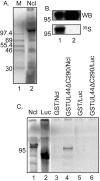
FIG 1
FIG 1
Binding of UL44 and nucleolin in vitro. (A) Radiolabeled nucleolin (Ncl) was produced by in vitro transcription-translation (lane 2), and its mobility in SDS-PAGE was compared to those of radiolabeled proteins of known molecular masses (lane 1). The molecular masses (kDa) of the proteins in lane 1 are noted to the left of the panel. (B) The mobility of radiolabeled nucleolin (lane 1) was compared to the mobility of nucleolin in infected cell lysate (HFF cells infected for 72 h with AD169 at an MOI of 3) and analyzed by Western blotting (WB) using polyclonal rabbit antisera recognizing nucleolin (top panel). Signal from radiolabeled protein (35S) detected on a phosphorimager screen is shown in the bottom panel. The position of a molecular mass marker (kDa) is noted to the left of each panel. (C) GST or GSTUL44ΔC290 was incubated with radiolabeled nucleolin (Ncl) or radiolabeled luciferase (Luc), passed over a glutathione column, and analyzed by SDS-PAGE and autoradiography. The GST and radiolabeled proteins used in each reaction are noted above the figure. The nucleolin and luciferase proteins used in each reaction are shown in lanes 1 and 2. The radiolabeled proteins eluted from each reaction are shown in lanes 3 to 6. The position of a molecular mass marker (kDa) is noted to the left of the panel.
FIG 2
FIG 2
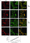
Localization of UL44 and nucleolin in HCMV-infected cells. HFF cells were infected at an MOI of 3 with HCMV strain AD169. Cells were prepared for IF analysis by spinning disk confocal microscopy at the indicated time points. Cells were stained with a MAb recognizing UL44 (αICP36) or nucleolin (Ncl). UL44 and nucleolin were detected using secondary antibodies conjugated to red and green fluorophores, respectively. Panels i to vi show the images obtained by acquiring sequential optical planes in the z axis. Each panel shows the xz and yz axis of the cells and a single focal plane from the z axis (xy). The proteins detected by IF in each experiment are noted above the panels in the left and middle columns. Panels in the right column show the images in the left and middle columns merged. The time points assayed are noted to the left of the figure. The arrow in panels ii and v indicates nucleoli in uninfected and infected cells. The white box in panel vi represents a magnified area.
FIG 3
FIG 3
Localization of UL44 and nucleolin in the presence of an inhibitor of viral DNA synthesis. HFF cells were infected with AD169 at an MOI of 3 in the presence or absence of phosphonoformic acid (PFA) (520 µM). Forty-eight hours postinfection, infected cells were analyzed by IF staining with antibodies recognizing UL44 (αICP36 MAb) and nucleolin (Ncl). UL44 and nucleolin were detected using secondary antibodies conjugated to red and green fluorophores, respectively. The images in each panel were obtained by acquiring sequential optical planes in the z axis. Each panel shows the xz and yz axis of the cells and a single focal plane from the z axis (xy). Panels in the left column show UL44 staining, and the middle column shows nucleolin staining. Panels in the right column show the images in the left and middle columns merged. (A to C) Uninfected (0 h.p.i.) cells. (D to F) Infected cells 48 h.p.i. (G to I) Infected cells 48 h.p.i. in the presence of PFA.
FIG 4
FIG 4
Localization of UL44, nucleolin, and EdU-labeled viral DNA in HCMV-infected cells. HFF cells were infected at an MOI of 3 with HCMV strain AD169. At 96 h.p.i., infected cells were incubated in the presence or absence of EdU for 60 minutes. Cells were prepared for IF analysis by spinning disk confocal microscopy. Cells were treated using a green fluorescent azide to detect EdU incorporated into DNA and stained with a MAb recognizing UL44 (αICP36) (A) or nucleolin (Ncl) (B). UL44 and nucleolin were detected using secondary antibodies conjugated to red fluorophores. Panels i to vi of panels A and B show the images obtained by acquiring sequential optical planes in the z axis. Each panel shows the xz and yz axis of the cells and a single focal plane from the z axis (xy). EdU or the protein detected by IF in each experiment is noted above the panels in the left and middle columns. Panels in the right column show the images in the left and middle columns merged. The time points assayed are noted to the left of the figure. The arrows in panels iii and vi of panel B indicate nucleoli in uninfected and infected cells. The white boxes in panel vi of panels A and B represent magnified areas. The arrow within the box in panel A indicates colocalization of UL44 with punctate EdU staining at the periphery of the replication compartment.
FIG 5
FIG 5
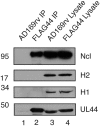
Analysis of viral and cellular proteins in infected cells by immunoprecipitation. Lysates from AD169rv (lane 3)- and HCMV-FLAG44 (lane 4)-infected cells and proteins immunoprecipitated using an anti-FLAG antibody from those lysates (lanes 1 and 2, respectively) were separated on a 10% polyacrylamide gel. Proteins in each lane were examined by Western blotting for the presence of nucleolin (Ncl), histone H1 (H1), histone H2 (H2), or UL44 using antibodies recognizing these proteins, as indicated to the right of the figure. The positions of molecular mass markers (kDa) are indicated to the left of the figure.
FIG 6
FIG 6
Localization of UL44 in Ncl siRNA-treated cells. (A and B) HFF cells transfected with control or nucleolin siRNA were infected at an MOI of 3 with HCMV strain AD169. Ninety-six hours postinfection, infected cells were analyzed by IF staining with UL44 MAb αICP36 or antibody recognizing nucleolin (Ncl). UL44 and nucleolin were detected using secondary antibodies conjugated to red and green fluorophores, respectively. The images in each panel were obtained by acquiring sequential optical planes in the z axis. Each panel shows a single focal plane from the z axis. Panels in the right column show the images in the left and middle columns merged. Cells shown in panels i to ix of panel B are from the same experiment. Arrows show “holes” in staining. (C) The mean pixel intensity of UL44 and nucleolin (Ncl) staining in the nucleus of each cell in panel B was measured and plotted with respect to those of the other cells. Each data point represents a single cell. The r2 value and P value (t test for r) of the trend line shown in the figure are noted.
FIG 7
FIG 7
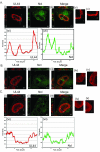
Localization of UL44 in Ncl siRNA-treated cells. HFF cells transfected with nucleolin siRNA (A to C) were infected at an MOI of 3 with HCMV strain AD169. Ninety-six hours postinfection, infected cells were analyzed by IF staining with UL44 MAb αICP36 or antibody recognizing nucleolin (Ncl). UL44 and nucleolin were detected using secondary antibodies conjugated to red and green fluorophores, respectively. The images in each panel were obtained by acquiring sequential optical planes in the z axis. Each panel shows the xz and yz axis of the cells and a single focal plane from the z axis (xy). Panels A, B, and C show three different cells from the same experiment that contain different levels of nucleolin. (i to iii) Panels in the left column show UL44 staining, and those in the middle column show nucleolin staining. Panels in the right column show the images in the left and middle columns merged. (iv) Enlarged area from box 1 in panel i. (v) Enlarged area from box 2 in panel i. (vi and vii) Pixel intensities of UL44 (red) and nucleolin (green) staining, respectively, from along the white lines in panels iii of panels A and C. Pixel intensity from the bottom to the top of each line is shown.
FIG 8
FIG 8
EdU incorporation into viral DNA in siRNA-treated cells. (i) HFF cells transfected with nucleolin siRNA were infected at an MOI of 3 with HCMV virus AD169. Ninety-six hours postinfection, infected cells were incubated with EdU (60 min) and then analyzed by IF. Cells were stained with a MAb recognizing UL44 (αICP36 MAb) and treated with a green fluorescent azide to detect EdU incorporated into DNA. UL44 was detected using a secondary antibody conjugated to a red fluorophore. Each panel shows the xz and yz axis of the cells and a single focal plane from the z axis (xy). Each panel shows the merged signals from the red and green fluorophores. Cells shown are from the same experiment. (ii) Enlarged areas from the boxes indicated in panel i. (iii) Pixel intensities of EdU (green) staining from along the white lines in panels A and B. Pixel intensity from the bottom to the top of each line is shown.

Similar articles

Cited by

References

    1. Quinlan MP, Chen LB, Knipe DM. 1984. The intranuclear location of a herpes simplex virus DNA-binding protein is determined by the status of viral DNA replication. Cell 36:857–868 - PubMed
    1. Zhong L, Hayward GS. 1997. Assembly of complete, functionally active herpes simplex virus DNA replication compartments and recruitment of associated viral and cellular proteins in transient cotransfection assays. J. Virol. 71:3146–3160 - PMC - PubMed
    1. Lukonis CJ, Burkham J, Weller SK. 1997. Herpes simplex virus type 1 prereplicative sites are a heterogeneous population: only a subset are likely to be precursors to replication compartments. J. Virol. 71:4771–4781 - PMC - PubMed
    1. Lukonis CJ, Weller SK. 1996. Characterization of nuclear structures in cells infected with herpes simplex virus type 1 in the absence of viral DNA replication. J. Virol. 70:1751–1758 - PMC - PubMed
    1. Lukonis CJ, Weller SK. 1997. Formation of herpes simplex virus type 1 replication compartments by transfection: requirements and localization to nuclear domain 10. J. Virol. 71:2390–2399 - PMC - PubMed

Publication types