. 2012 Sep;61(9):1451-61.
doi: 10.1007/s00262-012-1212-x. Epub 2012 Feb 5.

Expression of IL-15 in NK cells results in rapid enrichment and selective cytotoxicity of gene-modified effectors that carry a tumor-specific antigen receptor

Affiliations

Expression of IL-15 in NK cells results in rapid enrichment and selective cytotoxicity of gene-modified effectors that carry a tumor-specific antigen receptor

Christiane Sahm et al. Cancer Immunol Immunother. 2012 Sep.

Abstract

Natural killer (NK) cells hold promise for adoptive cancer immunotherapy but are dependent on cytokines such as interleukin (IL)-2 for growth and cytotoxicity. Here, we investigated the consequences of ectopic expression of IL-15 in human NK cells. IL-2 and IL-15 belong to the common γ chain family of cytokines and have overlapping activities. Transduction of clinically applicable NK-92 cells with lentiviral vectors encoding human IL-15 resulted in predominantly intracellular expression of the cytokine, and STAT5 activation, proliferation and cytotoxicity of the producer cells in the absence of IL-2. Growth of non-transduced bystander cells was not supported, allowing rapid enrichment of gene-modified cells solely by IL-2 withdrawal. This was also the case upon transduction of NK-92 and NKL cells with a bicistronic lentiviral vector encoding IL-15 and a chimeric antigen receptor (CAR) targeting the pancarcinoma antigen EpCAM. Effector cells co-expressing CAR and IL-15 continued to proliferate in the absence of exogenous cytokines and displayed high and selective cell-killing activity against EpCAM-expressing breast carcinoma cells that were resistant to the natural cytotoxicity of unmodified NK cells. This strategy facilitates rapid isolation and continuous expansion of retargeted NK cells and may extend their potential clinical utility.


Conflict of interest statement

The authors declare that they have no conflict of interest.

Figures

Fig. 1
Fig. 1
Expression of IL-15 in NK-92 cells. a Schematic representation of the lentiviral transfer vector pS-IL15-IEW that encodes under the control of the Spleen Focus Forming Virus promoter (SFFV) human IL-15, followed by an internal ribosome entry site (IRES) and cDNA encoding enhanced green fluorescent protein (EGFP) as a marker. After transduction with S-IL15-IEW vector particles, EGFP-expressing NK-92/IL-15-EGFP cells were enriched by flow cytometric cell sorting and tested for IL-15 expression. b IL-15 mRNA expression in NK-92/IL-15-EGFP cells grown in the presence and absence of exogenous IL-2 was verified by semi-quantitative RT-PCR. c The presence of IL-15 protein in NK-92/IL-15-EGFP cells was investigated by intracellular cytokine staining with anti-IL-15 antibody and flow cytometry (open area). Parental NK-92 cells served as a control (filled area). d The amount of IL-15 in cell lysate and conditioned culture supernatant was quantified by IL-15-specific ELISA. Parental NK-92 cells served as a control. Mean values ± SEM are shown (n = 3)
Fig. 2
Fig. 2
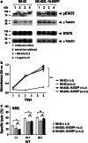
IL-15-expressing NK-92 cells proliferate in the absence of exogenous IL-2 and retain their cytotoxicity. a Cytokine-induced activation of STAT5. Cell lysates of parental NK-92 cells from continuous culture in IL-2-containing medium and of NK-92/IL-15-EGFP cells from continuous culture in medium lacking IL-2 (lanes 1) after starvation overnight in medium without serum or cytokines (lanes 2) or after activation of starved cells for 15 min with either 500 IU/ml IL-2 (lanes 3) or 5 ng/ml IL-15 (lanes 4) were analyzed by immunoblotting with phospho-STAT5-specific antibody as indicated. For comparison, also the levels of total STAT5 were determined. γ-Tubulin was analyzed as a loading control. b Growth of NK cells in the absence of exogenous cytokines. NK-92/IL-15-EGFP and parental NK-92 were placed in growth medium containing or lacking IL-2, and proliferation was followed for six consecutive days by MTT metabolization assays and determination of the absorbance at 595 nm as a measure for the relative number of viable cells. Mean values ± SEM are shown (n = 3); ***p < 0.0001. c Natural cytotoxicity of NK-92/IL-15-EGFP cells after culture in the presence or absence of IL-2 toward NK-sensitive K562 cells was determined in FACS-based cytotoxicity assays at different effector to target ratios (E/T). Parental NK-92 cells grown in the presence of IL-2 were included for comparison. Mean values ± SD are shown (n = 2); *p < 0.05; ns, p > 0.05
Fig. 3
Fig. 3
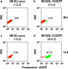
IL-15 serves as a selectable marker for rapid enrichment of gene-modified NK cells. NK-92 cells were freshly transduced with S-IL15-IEW lentiviral vector, kept in IL-2-containing medium for 48 h, and then grown in medium without IL-2 for another 14 days. Non-transduced parental NK-92 control cells (a, c), unsorted NK-92 cells in IL-2-containing medium 48 h after transduction (b), and 14 days after IL-2 withdrawal (d) were analyzed for EGFP expression by flow cytometry. EGFP-negative (red) and EGFP-positive cell populations (green) are indicated
Fig. 4
Fig. 4
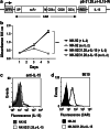
IL-2 withdrawal results in enrichment of NK-92 cells co-expressing IL-15 and a chimeric antigen receptor. a Schematic representation of the lentiviral transfer vector pS-31.28.z-I-IL15-W that encodes under the control of the Spleen Focus Forming Virus promoter (SFFV) the chimeric antigen receptor 31.28.z (CAR) that consists of an immunoglobulin heavy chain signal peptide (SP), the EpCAM-specific scFv(MOC31) antibody fragment (scFv), a Myc-tag (M), the CD8 α hinge region (CD8α), transmembrane and intracellular domains of CD28 (CD28), and the intracellular domain of CD3 ζ chain (CD3ζ), followed by an internal ribosome entry site (IRES) and cDNA encoding human IL-15. b After transduction with S-31.28.z-I-IL15-W vector particles, IL-15-expressing cells were selected by IL-2 withdrawal for 14 days. Then, proliferation of the selected cells in growth medium containing or lacking IL-2 was analyzed in MTT metabolization assays by determination of the absorbance at 595 nm as a measure for the relative number of viable cells. Parental NK-92 cells were included for comparison. Mean values ± SEM are shown (n = 3); ***p < 0.0001. NK-92/31.28.z-IL-15 cells with high CAR surface expression were isolated from the selected pool by flow cytometric cell sorting with Myc-tag-specific antibody 9E10. c The presence of IL-15 protein in sorted NK-92/31.28.z-IL-15 cells was investigated by intracellular cytokine staining with anti-IL-15 antibody and flow cytometry (open area). Parental NK-92 cells served as a control (gray area). d Homogeneous CAR surface expression in NK-92/31.28.z-IL-15 cells was confirmed by flow cytometry with 9E10 antibody (open area). Parental NK-92 cells were included for comparison (filled area)
Fig. 5
Fig. 5
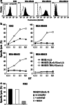
NK-92/31.28.z-IL-15 cells display targeted cytotoxicity in the absence of exogenous cytokines. a Expression of EpCAM on the surface of erythroleukemic K562 cells, MDA-MB468 and MDA-MB453 breast carcinoma cells, and MDA-MB435 melanoma cells was determined by flow cytometry with EpCAM-specific MOC31 antibody (filled areas). Cells only treated with APC-coupled secondary antibody served as controls (open areas). b Cytotoxicity of NK-92/31.28.z-IL-15 cells grown in the absence of exogenous IL-2 toward K562, MDA-MB468, MDA-MB453, and MDA-MB435 cells was determined in FACS-based cytotoxicity assays at different effector to target ratios (E/T) as indicated (open squares). Parental NK-92 cells grown in the presence of IL-2 were included for comparison (filled circles). Cytotoxicity of NK-92/31.TM-IL-15 cells that express IL-15 and an EpCAM-specific CAR that lack signaling domains was analyzed using EpCAM-expressing MDA-MB468 and MDA-MB453 cells (gray squares). c Specificity of target cell recognition and cell killing by NK-92/31.28.z-IL-15 cells was analyzed in FACS-based cytotoxicity assays with K562 cells at an effector to target ratio of 5:1. Cells were co-incubated in the absence of competitor (filled bar), or in the presence of EpCAM-specific antibody MOC31 (gray bar) or isotype-matched control antibody (open bar)
Fig. 6
Fig. 6
Selective enrichment and targeted cytotoxicity of NKL cells co-expressing IL-15 and EpCAM-specific CAR 31.28.z. a Human NKL cells were transduced with S-31.28.z-I-IL15-W vector particles and selected by IL-2 withdrawal for 14 days. Then, proliferation of the selected cells in growth medium containing or lacking IL-2 was analyzed in MTT metabolization assays by determination of the absorbance at 595 nm as a measure for the relative number of viable cells. Parental NKL cells were included for comparison. Mean values ± SEM are shown (n = 3). b NKL/31.28.z-IL-15 cells with high CAR surface expression were isolated from the selected pool by flow cytometric cell sorting with Myc-tag-specific antibody 9E10 (open area). Parental NKL cells were included for comparison (filled area). c Cytotoxicity of NKL/31.28.z-IL-15 cells grown in the absence of IL-2 toward EpCAM-positive MDA-MB453 and EpCAM-negative MDA-MB435 cells was determined in FACS-based cytotoxicity assays at different effector to target ratios (E/T) as indicated (open squares). Parental NKL cells grown in the presence of IL-2 were included for comparison (filled circles)

Similar articles

Cited by

References

    1. Gross G, Waks T, Eshhar Z. Expression of immunoglobulin-T-cell receptor chimeric molecules as functional receptors with antibody-type specificity. Proc Natl Acad Sci USA. 1989;86:10024–10028. doi: 10.1073/pnas.86.24.10024. - DOI - PMC - PubMed
    1. Uherek C, Groner B, Wels W. Chimeric antigen receptors for the retargeting of cytotoxic effector cells. J Hematother Stem Cell Res. 2001;10:523–534. doi: 10.1089/15258160152509136. - DOI - PubMed
    1. Hombach A, Abken H. Costimulation tunes tumor-specific activation of redirected T cells in adoptive immunotherapy. Cancer Immunol Immunother. 2007;56:731–737. doi: 10.1007/s00262-006-0249-0. - DOI - PMC - PubMed
    1. Ngo MC, Rooney CM, Howard JM, Heslop HE. Ex vivo gene transfer for improved adoptive immunotherapy of cancer. Hum Mol Genet. 2011;20:R93–R99. doi: 10.1093/hmg/ddr102. - DOI - PMC - PubMed
    1. Porter DL, Levine BL, Kalos M, Bagg A, June CH. Chimeric antigen receptor-modified T cells in chronic lymphoid leukemia. N Engl J Med. 2011;365:725–733. doi: 10.1056/NEJMoa1103849. - DOI - PMC - PubMed

Publication types

MeSH terms

LinkOut - more resources