. 2013 Jun;34(24):1834-45.
doi: 10.1093/eurheartj/ehs011. Epub 2012 Feb 3.

Cardiomyocyte-expressed farnesoid-X-receptor is a novel apoptosis mediator and contributes to myocardial ischaemia/reperfusion injury

Affiliations

Cardiomyocyte-expressed farnesoid-X-receptor is a novel apoptosis mediator and contributes to myocardial ischaemia/reperfusion injury

Jun Pu et al. Eur Heart J. 2013 Jun.

Abstract

Aims: Emerging evidence indicates that nuclear receptors play a critical regulatory role in cardiovascular physiology/pathology. Recently, farnesoid-X-receptor (FXR), a member of the metabolic nuclear receptor superfamily, has been demonstrated to be expressed in vascular cells, with important roles in vascular physiology/pathology. However, the potential cardiac function of FXR remains unclear. We investigated the cardiac expression and biological function of FXR.

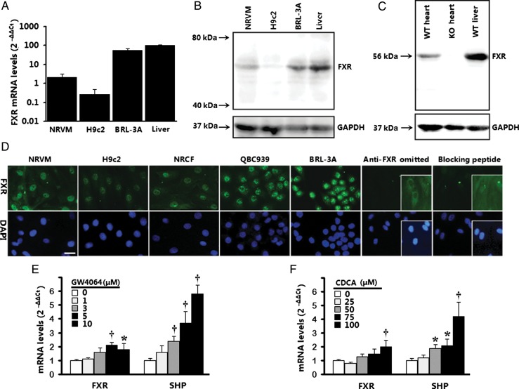
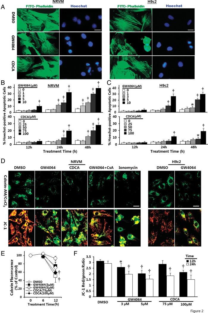
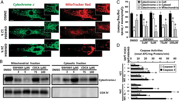
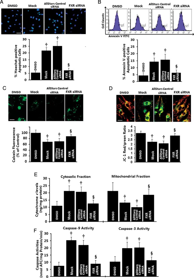
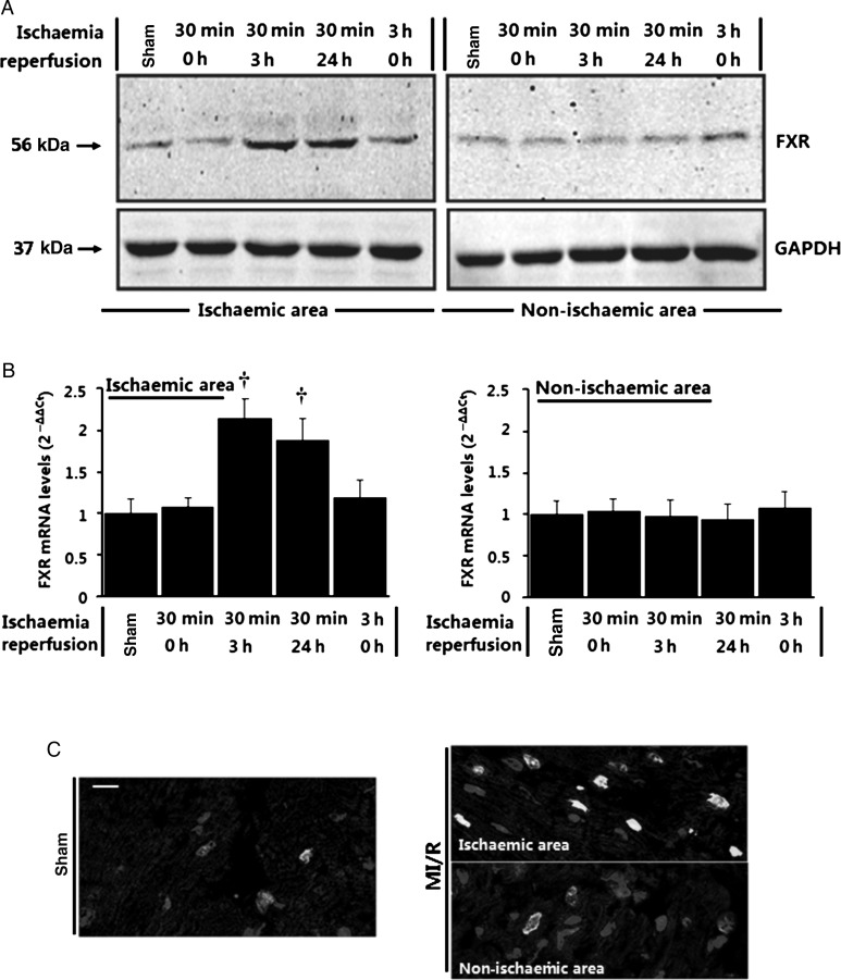
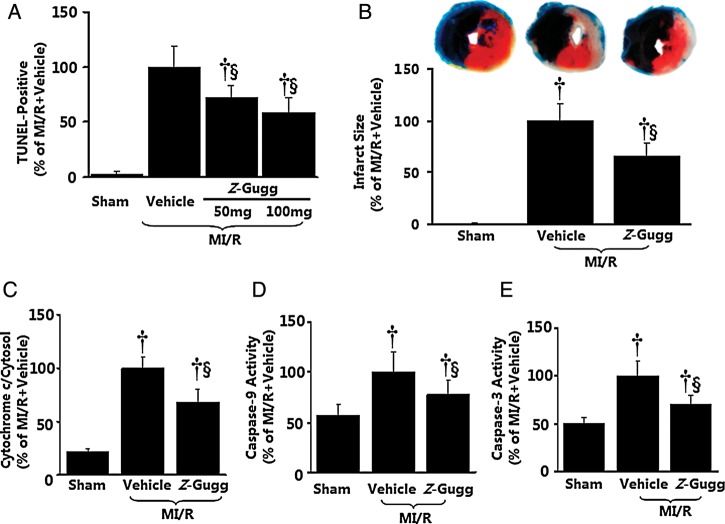
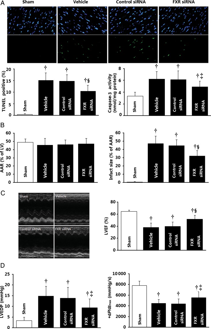
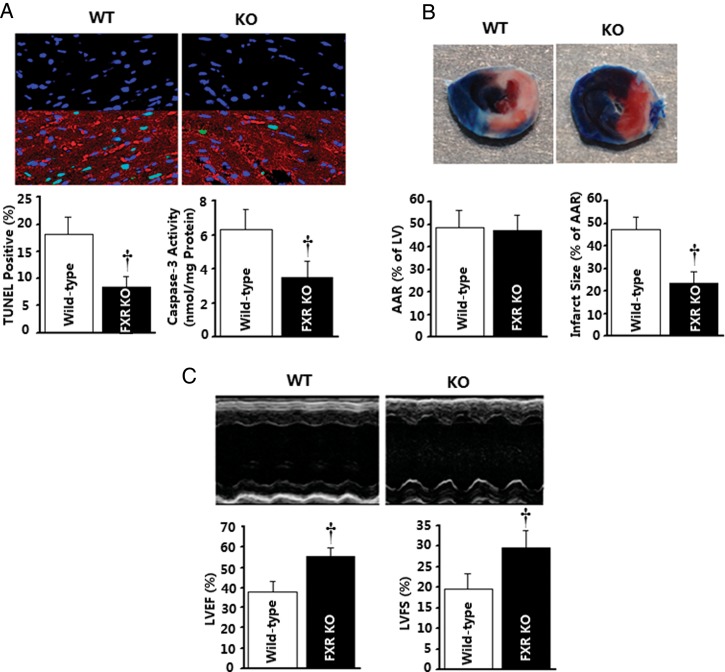
Methods and results: Farnesoid-X-receptor was detected in both isolated neonatal rat cardiac myocytes and fibroblasts. Natural and synthetic FXR agonists upregulated cardiac FXR expression, stimulated myocyte apoptosis, and reduced myocyte viability dose- and time-dependently. Mechanistic studies demonstrated that FXR agonists disrupted mitochondria, characterized by mitochondrial permeability transition pores activation, mitochondrial potential dissipation, cytochrome c release, and both caspase-9 and -3 activation. Such mitochondrial apoptotic responses were abolished by siRNA-mediated silencing of endogenous FXR or pharmacological inhibition of mitochondrial death signalling. Furthermore, low levels of FXR were detected in the adult mouse heart, with significant (∼2.0-fold) upregulation after myocardial ischaemia/reperfusion (MI/R). Pharmacological inhibition or genetic ablation of FXR significantly reduced myocardial apoptosis by 29.0-53.4%, decreased infarct size by 23.4-49.7%, and improved cardiac function in ischaemic/reperfused myocardium.

Conclusion: These results demonstrate that nuclear receptor FXR acts as a novel functional receptor in cardiac tissue, regulates apoptosis in cardiomyocytes, and contributes to MI/R injury.

Keywords: Apoptosis; Myocytes; Nuclear receptors.


Figures

Figure 1
Figure 1
Expression of farnesoid-X-receptor (FXR) in cardiac cells and heart tissues. (A) Farnesoid-X-receptor gene expression by real-time PCR in neonatal rat ventricular myocytes (NRVMs) and H9c2 ventricular cells. Rat liver tissues and BRL-3A rat liver cells served as positive control. Results are representative of three separate cultures. (B) Farnesoid-X-receptor protein expression by western blot in neonatal rat ventricular myocytes and H9c2 ventricular cells. Bands (∼56 kDa) are representative of three separate experiments. (C) Farnesoid-X-receptor protein expression by western blot in heart tissue from wild-type (WT) and farnesoid-X-receptor-knockout (KO) mice. Rat liver lysates from wild-type mice served as positive controls. (D) Immunolocalization of farnesoid-X-receptor in neonatal rat ventricular myocytes and H9c2 cells. Neonatal rat cardiac fibroblasts (NRCFs) are also shown. Primary antibody omission or pre-absorption with blocking peptide served as negative control. Bar = 20 μm. (E and F) Induction of farnesoid-X-receptor and its target gene SHP (small heterodimer partner) by farnesoid-X-receptor agonists (24 h; n= 3–4 per concentration) in neonatal rat ventricular myocytes using real-time PCR. Results were normalized against GAPDH and converted to percentage induction relative to vehicle controls. *P< 0.05, P< 0.01 vs. vehicle-treated control.
Figure 2
Figure 2
Induction of nuclear condensation and mitochondrial permeability transition pore (MPTP) activation by farnesoid-X-receptor (FXR) activation. (A) Representative confocal images of neonatal rat ventricular myocytes (NRVMs) or H9c2 ventricular cells double-labelled with Hoechst 33258 for nuclei (blue) and phalloidin for cytoskeletal F-actin (green). Typical images shown are from cardiomyocytes after 24 h incubation with GW4064 (5 μmol/L) or chenodeoxycholic acid (CDCA) (100 μmol/L). Bar = 20 μm. (B and C) Time-course of induction of late apoptosis after GW4064 or chenodeoxycholic acid treatment in neonatal rat ventricular myocytes (B) or H9c2 (C) cardiomyocytes. n = 4–6 independent cultures. *P< 0.05, P< 0.01 vs. vehicle-treated control. (D) Representative confocal images of mitochondrial permeability transition pore activation by calcein AM/CoCl2 labelling (top panel) and mitochondrial depolarization by JC-1 staining (bottom panel). Typical images shown are from cells after incubation with GW4064 (5 μmol/L) or chenodeoxycholic acid (100 μmol/L) for 12 h (neonatal rat ventricular myocytes) or 20 h (H9c2). Bar = 20 μm. (E) Time-courses of changes in mitochondrial calcein signal by flow cytometry after farnesoid-X-receptor agonist treatment (n = 3–4 independent experiments). P< 0.01 compared with time-matched vehicle-treated control. (F) Quantitative data of mitochondrial ΔΨm by flow cytometry in neonatal rat ventricular myocytes (n = 3–4 independent experiments). *P< 0.05, P< 0.01 compared with corresponding vehicle-treated control.
Figure 3
Figure 3
Induction of cytochrome c release and caspases activation by farnesoid-X-receptor (FXR) activation. (A) Confocal images of neonatal rat ventricular myocytes (NRVMs) stained with 2CYTC-199 antibody for cytochrome c (green) and MitoTracker for mitochondria (red). Typical images shown are from neonatal rat ventricular myocytes after incubation with GW4064 (5 μmol/L) for 12 and 24 h (n = 3–5 independent experiments, the same for all other assays). Amplified images show the alteration in mitochondrial organization after treatment with farnesoid-X-receptor agonists. Bar = 10 μm. (B) Western blot of cytochrome c distribution was done on mitochondrial and cytosolic fractions from neonatal rat ventricular myocytes exposed to farnesoid-X-receptor agonists for 12 h. Cytochrome oxidase IV (COX IV) was utilized to verify the quality of the extraction procedure. (C) Quantitative analysis of cytochrome c in mitochondrial and cytosolic fractions by sandwich ELISA. *P< 0.05, P< 0.01 vs. vehicle-treated control. (D) Caspase-9-like and caspase-3-like activities in neonatal rat ventricular myocytes were assessed by fluorometric assay. *P< 0.05, P< 0.01 vs. corresponding vehicle-treated control.
Figure 4
Figure 4
Effects of in vitro farnesoid-X-receptor (FXR) silencing on apoptotic mitochondrial changes in cardiomyocytes. Neonatal rat ventricular myocyte (NRVMs) were transfected with 20 nmol/L of farnesoid-X-receptor siRNA, AllStars Negative siRNA, or mock-treated for 24 h, followed by GW4064 (5 μmol/L) treatment for 12 (BF) or 24 h (A). DMSO-treated cells were utilized as control. (A and B) Late apoptosis assessed by Hoechst staining (A, n= 4 independent experiments; bar= 40 μm) and early apoptotic cells as assessed by annexin V staining (B, n= 3 independent experiments). (C) Representative confocal images of mitochondrial permeability transition pore opening measured by calcein-AM/CoCl2 quenching (upper) and quantitative data by flow cytometry (bottom, n= 3–4 independent experiments). (D) Representative confocal images of mitochondrial depolarization by JC-1 staining (upper) and quantitative data by flow cytometry (bottom, n= 3–4 independent experiments). (E) Mitochondrial and cytosolic cytochrome c levels measured by sandwich ELISA (n= 4 independent experiments). (F) Caspase-9-like and caspase-3-like activities were measured using the colorimetric substrate LEHD-AFC or DEVD-AFC (n= 3–4 independent experiments). *P< 0.05, P< 0.01 compared with vehicle-treated cells, P< 0.05, §P< 0.01 compared with GW4064-treated cells.
Figure 5
Figure 5
Upregulation of farnesoid-X-receptor (FXR) by ischaemia/reperfusion stimuli in the heart. (A and B) Time course of farnesoid-X-receptor activation detected by western blots (A) and real-time quantitative PCR (B) in the heart (n= 5–6 per time point, ischaemic and non-ischaemic areas) subjected to in vivo ischaemia/reperfusion (I/R) for the indicated times. Sham-operated animals were utilized as control. Results were normalized against GAPDH and converted to fold induction relative to sham-operated controls. *P< 0.05, P< 0.01 compared with sham-operated controls. (C) Confocal immunofluorescence analysis on heart sections from sham-operated and ischaemia/reperfusion mice 24 h after surgery. Sections were subjected to immunofluorescence for farnesoid-X-receptor (green) and α-actinin (red) and to staining for nuclei with DAPI (blue), and overlays are shown (Bar= 20 μm).
Figure 6
Figure 6
Effect of pharmacological inhibition of farnesoid-X-receptor (FXR) on myocardial ischaemia/reperfusion injury. (AE) Farnesoid-X-receptor antagonist Z-guggulsterone (Z-Gugg) reduced post-ischaemic myocardial apoptotic responses determined by TUNEL labelling (A, n= 8–10 hearts per group), cytochrome c release (C, n= 7–8 hearts per group), caspase-9 and -3 activation (D and E, n= 8–12 hearts per group), and myocardial infarct size determined by Evans blue/TTC double-staining (B, n= 10–12 hearts per group). Mean values obtained from vehicle-treated myocardial ischaemia/reperfusion animals were treated as 100%, and individual values from each animal were normalized against the mean values. P< 0.01 vs. sham; §P< 0.01 vs. myocardial ischaemia/reperfusion + vehicle.
Figure 7
Figure 7
Effect of genetic inhibition of farnesoid-X-receptor (FXR) by in vivo siRNA transfection upon ischaemia/reperfusion injury. (A) Apoptosis as determined by TUNEL labelling (n= 6–8 hearts per group) and caspase-3 activation (n= 8–11 hearts per group). (B) Myocardial infarction was determined by Evans blue/TTC double-staining (n= 8–10 hearts per group). (C and D) Cardiac function was determined by echocardiography (n= 8–10 animals per group) and catheter-based haemodynamic measurements (n= 8–9 animals per group). Myocardial ischaemia/reperfusion procedures were performed 48 h after intramyocardial siRNA delivery. *P< 0.05, P< 0.01 vs. the sham group; P< 0.05, §P< 0.01 vs. the control siRNA group. AAR, area at risk; LVEF, left ventricular ejection fraction; LVEDP, left ventricular end-diastolic pressure.
Figure 8
Figure 8
Myocardial injury caused by ischaemia/reperfusion is reduced in farnesoid-X-receptor (FXR)-deficient mice. Farnesoid-X-receptor-knockout (KO) mice and wild-type (WT) mice were subjected to 30 min of ischaemia and 3 (A) or 24 h (B and C) of reperfusion. (A) Myocardial apoptosis as determined by TUNEL labelling (n= 6 hearts per group) and caspase-3 activation (n= 6 hearts per group). (B) Myocardial infarction was determined by Evans blue/TTC double-staining (n= 5–6 hearts per group). (C) Cardiac function was determined by echocardiography (n= 6 animals per group). P< 0.01 vs. the wild-type group. AAR, area at risk; LVEF, left ventricular ejection fraction.

Similar articles

Cited by

References

    1. Movassagh M, Foo RS. Simplified apoptotic cascades. Heart Fail Rev. 2008;13:111–119. - PubMed
    1. Son NH, Yu S, Tuinei J, Arai K, Hamai H, Homma S, Shulman GI, Abel ED, Goldberg IJ. PPARγ-induced cardiolipotoxicity in mice is ameliorated by PPARα deficiency despite increases in fatty acid oxidation. J Clin Invest. 2010;120:3443–3454. - PMC - PubMed
    1. Choudhary R, Baker KM, Pan J. All-trans retinoic acid prevents angiotensin II- and mechanical stretch-induced reactive oxygen species generation and cardiomyocyte apoptosis. J Cell Physiol. 2008;215:172–181. - PubMed
    1. Morrissy S, Xu B, Aguilar D, Zhang J, Chen QM. Inhibition of apoptosis by progesterone in cardiomyocytes. Aging Cell. 2010;9:799–809. - PMC - PubMed
    1. Shan P, Pu J, Yuan A, Shen L, Shen L, Chai D, He B. RXR agonists inhibit oxidative stress-induced apoptosis in H9c2 rat ventricular cells. Biochem Biophys Res Commun. 2008;375:628–633. - PubMed

Publication types

MeSH terms