. 2012 Mar 30;287(14):10977-89.
doi: 10.1074/jbc.M111.324616. Epub 2012 Feb 2.

Sphingolipid-modulated exosome secretion promotes clearance of amyloid-β by microglia

Affiliations

Sphingolipid-modulated exosome secretion promotes clearance of amyloid-β by microglia

Kohei Yuyama et al. J Biol Chem. .

Abstract

Amyloid β-peptide (Aβ), the pathogenic agent of Alzheimer disease, is a physiological metabolite whose levels are constantly controlled in normal brain. Recent studies have demonstrated that a fraction of extracellular Aβ is associated with exosomes, small membrane vesicles of endosomal origin, although the fate of Aβ in association with exosome is largely unknown. In this study, we identified novel roles for neuron-derived exosomes acting on extracellular Aβ, i.e. exosomes drive conformational changes in Aβ to form nontoxic amyloid fibrils and promote uptake of Aβ by microglia. The Aβ internalized together with exosomes was further transported to lysosomes and degraded. We also found that blockade of phosphatidylserine on the surface of exosomes by annexin V not only prevented exosome uptake but also suppressed Aβ incorporation into microglia. In addition, we demonstrated that secretion of neuron-derived exosomes was modulated by the activities of sphingolipid-metabolizing enzymes, including neutral sphingomyelinase 2 (nSMase2) and sphingomyelin synthase 2 (SMS2). In transwell experiments, up-regulation of exosome secretion from neuronal cells by treatment with SMS2 siRNA enhanced Aβ uptake into microglial cells and significantly decreased extracellular levels of Aβ. Our findings indicate a novel mechanism responsible for clearance of Aβ through its association with exosomes. The modulation of the vesicle release and/or elimination may alter the risk of AD.


Figures
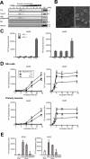
FIGURE 1.
FIGURE 1.
Aβ amyloidogenesis by neuronal exosomes. A, exosomes were collected from the culture supernatant of Neuroblastoma N2a cells, by sequential centrifugation as indicated under “Experimental Procedures.” The 100,000 × g pellets were further subjected to sucrose gradient centrifugation, and the resulting fractions were analyzed for the exosomal proteins Alix and Tsg101 and for the GM1 ganglioside. B, purified exosomes (100,000 × g pellet) underwent negative staining with phosphotungstic acid and were examined by electron microscopy. Scale bars, right panel, 500 nm; left panel, 100 nm. C, culture medium from N2a cells was subjected to sequential centrifugation. The resulting pellets, 3,000 × g (P3), 4,000 × g (P4), 10,000 × g (P10), and 100,000 × g (P100), were mixed with soluble 25 μm seed-free soluble Aβ40 or Aβ42 and incubated for 24 h at 37 °C. Amyloid fibrils formed in the incubation mixtures were measured with a ThT assay. The indicated values for relative fluorescence (AU) are means ± S.E. D, after the indicated time of incubation, ThT fluorescence intensities were measured in mixtures containing 25 μm Aβ and the indicated amount of exosomes derived from the culture supernatant of N2a cells or cortical neurons. Values provided as the means ± S.E. are as follows: *, p < 0.05; **, p < 0.01; ***, p < 0.001; t test. E, after CTB or EGCase treatment, the exosomes (Exo) were mixed with 25 μm Aβ and incubated at 37 °C for 5 h. Ctrl, control. Values are the means ± S.E. *, p < 0.05; **, p < 0.01; t test.
FIGURE 2.
FIGURE 2.
Effects of exosomes on oligomerization and toxicity of Aβ. A, purified N2a-derived exosomes were mixed with soluble 25 μm Aβ42 and incubated at 37 °C for the indicated times. The incubation mixtures were subjected to dot blot analysis using anti-oligomer (A11) and anti-Aβ (6E10) antibodies. B, 25 μm Aβ42 was incubated at 37 °C for 5 h to form oligomeric Aβ. N2a-derived exosomes were added to the solution, which contained the oligomeric Aβ, and further incubated for the indicated times at 37 °C. The incubation mixtures were subjected to dot blotting. C–E, Aβ42 (25 μm) was incubated with or without N2a-derived exosomes at 37 °C for 5 h. The incubation mixtures were subsequently added to cortical neurons, and after 24 h, the cell viabilities were determined using a WST-1 assay (C) or LIVE/DEAD viability kit (D and E). Neurons were stained with SYTO10/RED DEADTM, showing green staining for all cells and red staining for dead cells. Scale bar, 20 μm. Data are represented as the means ± S.E. **, p < 0.01; ***, p < 0.001; t test.
FIGURE 3.
FIGURE 3.
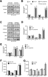
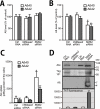
Effect of sphingolipid metabolism on exosome secretion and Aβ amyloidogenesis. A and B, N2a cells or cortical neurons were treated with imipramine, GW4869, D609, or their respective diluent for 24 h. Exosomes were then collected from the medium of each culture (5 × 10⁶ cells) and were subjected to SDS-PAGE, followed by Western blotting (WB) to detect Alix, Tsg101, and GM1 ganglioside. A, representative blots illustrating the amount of Alix in N2a cell lysates (2.5 × 10⁵ cells) and 100,000 × g pellets (Exosome). B, quantification of staining in Western blots. Results shown are the means ± S.E. from two independent experiments (n = 4). *, p < 0.05; **, p < 0.01; t test. C and D, small interfering RNAs (siRNA) active against aSMase, nSMase1, nSMase2, SMS1, and SMS2 were delivered into N2a cells. Exosomes were purified from the media of the siRNA-treated cells as in A, and the amounts of exosome markers in the resulting pellets were determined by Western blotting. C, Alix was detected in the cell lysates and in the exosomes as in A. D, band intensities of exosomal markers were analyzed. Data are presented as the means ± S.E. from two independent experiments (n = 4). *, p < 0.05; **, p < 0.01; t test. E, N2a cells were treated with N-hexanoyl-d-erythrosphingosine (50 μm) or bacterial SMase (100 microunits/ ml) for 24 h. The level of released exosomes were evaluated by Western blotting. Results are expressed as means ± S.E. (n = 3). *, p < 0.05; t test. F, exosomes isolated from the cultures of N2a cells or primary neurons, that had been treated with the indicated inhibitors, were incubated at 37 °C with 25 μm soluble Aβ42. Aβ amyloid fibrils formed in the mixtures were measured by ThT assay. The indicated values for relative fluorescence (AU) are means ± S.E. from two independent experiments (n = 4). *, p < 0.05; **, p < 0.01; ***, p < 0.001; t test. G, exosomes isolated from the cultures of N2a cells that had been treated with the indicated siRNA were mixed with 25 μm Aβ42. After 5 h of incubation, ThT fluorescence was measured. Data are represented as the mean ± S.E. (n = 4). *, p < 0.05; t test. Ctrl, control.
FIGURE 4.
FIGURE 4.
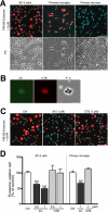
Transfer of exosomes into microglia. A, exosomes purified from N2a cell cultures were labeled with the dye PKH26 (red) and added to BV-2 microglial cells or primary cultures of microglia or cortical neurons. After 3 h of incubation, cells were fixed, stained with DAPI, and analyzed by confocal microscopy. B, N2a-derived exosomes were bound to AlexaFluor-conjugated AV or CTB to detect surface-exposed PS and GM1 ganglioside (GM1), respectively. Fluorescence labeling was visualized by confocal microscopy. The far right panel shows the same field in phase contrast (PC). Scale bar, 200 nm. C and D, exosomes collected from N2a cultures were labeled with the red dye PKH26 and subsequently treated with nothing, AV, or CTB. Labeled exosomes were applied to BV-2 cells and incubated for 3 h. Cells were subsequently fixed and stained with DAPI. C, confocal images of internalized exosomes are shown. D, fluorescence intensities per cell were determined by image analysis. Exosome internalization was quantified from three independent experiments. Values are means ± S.E. ***, p < 0.001; t test. Ctrl, control.
FIGURE 5.
FIGURE 5.
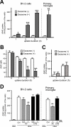
Acceleration of Aβ uptake into microglia by exosome. A and B, N2a-derived exosomes were incubated with 25 μm Aβ42 at 37 °C for 5 h. The preincubated mixtures were then added to BV-2 cells or primary microglia (final concentration of Aβ, 0.5 μm) and further incubated for the indicated times. Levels of Aβ42 in BV-2 cells (A) and in conditioned media (B) were quantified by ELISA. Values are means ± S.E. *, p < 0.05; **, p < 0.01; ***, p < 0.001; t test. C, 50 μm Aβ42 was incubated at 37 °C for 5 days to form amyloid fibrils. The fibrils were incubated with or without N2a-derived exosomes at 37 °C for 5 h. The incubation mixtures were added to BV-2 cells (final concentration of Aβ, 0.5 μm) and incubated for the additional times. The levels of intracellular Aβ in BV-2 cells were measured by ELISA. Values are means ± S.E. D, N2a-derived exosomes (Exo) were incubated with 25 μm Aβ42 at 37 °C for 5 h, then with AV or CTB for an additional 15 min at 37 °C. The mixtures were applied to BV-2 cells or primary microglia and incubated for 3 h. The intracellular levels of Aβ were measured using ELISA. Values represent as the mean ± S.E. **, p < 0.01; t test.
FIGURE 6.
FIGURE 6.
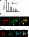
Degradation of Aβ in microglia. A, Aβ42 (25 μm) was incubated in the presence or absence of N2a-derived exosomes at 37 °C for 5 h. The incubation mixtures were subsequently administered to BV-2 cells (final concentration of Aβ, 0.5 μm) for 3 h. After removal of free Aβ and exosomes by washing in medium, the cells were further cultured for up to 48 h. Intracellular levels of Aβ42 were determined at the indicated times by ELISA. B, exosomes were labeled with PKH26 (red) and added to cultures of BV-2 cells. After a 3-h incubation, cells were stained with LysoTracker Green and analyzed by confocal microscopy. Scale bar, 5 μm. C, PKH26-labeled exosomes were mixed with the fluorescent FAM-coupled Aβ42 (25 μm). After a 5-h incubation, the mixtures were administered to cultures of BV-2 cells for 3 h and stained with LysoTracker Blue. Scale bar, 5 μm.
FIGURE 7.
FIGURE 7.
Enhancement of Aβ clearance by SMS2 knockdown. A–C, N2a cells seeded in inserts were transfected with the APP770 and siRNA as indicated. After 24 h, the media were removed and the inserts with the N2a cells were placed into wells with (B) or without (A) BV-2 cells and cultured for another 24 h. The levels of Aβ in the medium (A and B) and in the BV-2 cells (C) were measured by ELISA. Values are means ± S.E. *, p < 0.05; **, p < 0.01; ***, p < 0.001; t test. D, N2a cells were transfected with APP and siRNA for N-SMase2 or SMS2 for 24 h. The media were changed and the cells incubated for an additional 24 h. The exosomes were then collected from the medium of each culture (5 × 10⁶ cells) and were solubilized with guanidine HCl buffer to be analyzed by Western blot or resuspended in TBS to perform ThT assay. Ctrl, control.
FIGURE 8.
FIGURE 8.
Schematic representation of the role of exosome in Aβ metabolism. Both exosomes and Aβs are generated and released from neurons into the extracellular space. Exosome secretion is modulated by the sphingolipid-metabolizing enzymes, N-SMase2 and SMS2, bidirectionally. Exosomes enhance Aβ amyloidogenesis by GSLs on its surface and, subsequently, incorporation of Aβ fibrils into microglia in a PS-dependent manner to degrade Aβ. Neuronal exosomes likely promote Aβ clearance.

Similar articles

Cited by

References

    1. Hardy J., Selkoe D. J. (2002) The amyloid hypothesis of Alzheimer disease. Progress and problems on the road to therapeutics. Science 297, 353–356 - PubMed
    1. Selkoe D. J. (1997) Alzheimer disease. Genotypes, phenotypes, and treatments. Science 275, 630–631 - PubMed
    1. Mawuenyega K. G., Sigurdson W., Ovod V., Munsell L., Kasten T., Morris J. C., Yarasheski K. E., Bateman R. J. (2010) Decreased clearance of CNS β-amyloid in Alzheimer disease. Science 330, 1774. - PMC - PubMed
    1. Bateman R. J., Munsell L. Y., Morris J. C., Swarm R., Yarasheski K. E., Holtzman D. M. (2006) Human amyloid-β synthesis and clearance rates as measured in cerebrospinal fluid in vivo. Nat. Med. 12, 856–861 - PMC - PubMed
    1. Rajendran L., Honsho M., Zahn T. R., Keller P., Geiger K. D., Verkade P., Simons K. (2006) Alzheimer disease β-amyloid peptides are released in association with exosomes. Proc. Natl. Acad. Sci. U.S.A. 103, 11172–11177 - PMC - PubMed

LinkOut - more resources