

Mycobacterium tuberculosis RNA Expression Patterns in Sputum Bacteria Indicate Secreted Esx Factors Contributing to Growth are Highly Expressed in Active Disease
- PMID: 22291682
- PMCID: PMC3254194
- DOI: 10.3389/fmicb.2011.00266
Mycobacterium tuberculosis RNA Expression Patterns in Sputum Bacteria Indicate Secreted Esx Factors Contributing to Growth are Highly Expressed in Active Disease
Abstract
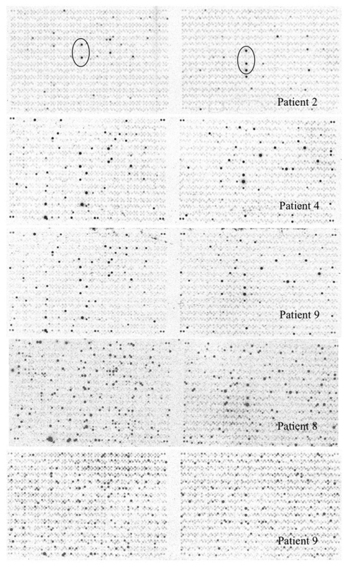
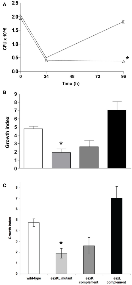
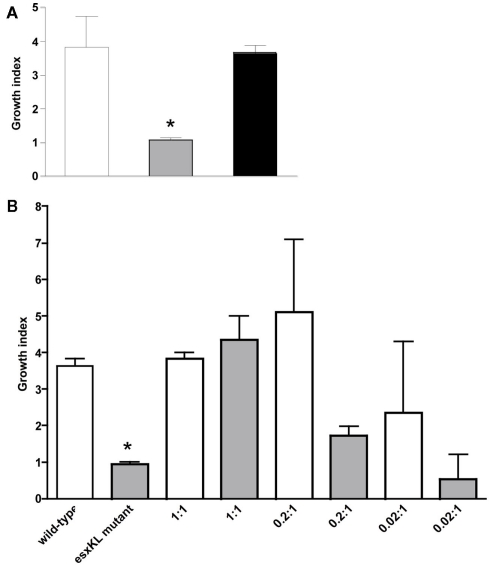
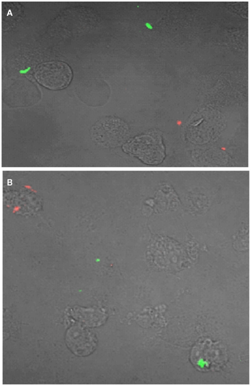
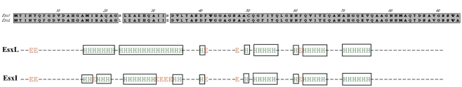
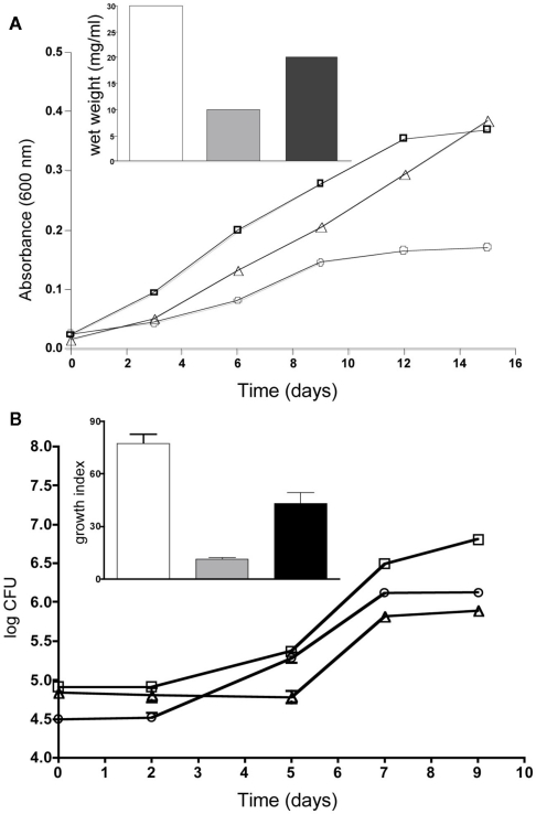
To identify factors contributing to the ability of tubercle bacilli to grow in the lung during active infection, we analyzed RNA expression patterns in bacteria present in patient sputum. Prominent among bacterial transcripts identified were those encoding secreted peptides of the Esat-6 subfamily that includes EsxK and EsxL (Rv1197 and Rv1198). H37Rv esxKL and esxJI transcripts were differentially expressed under different growth conditions, and disruption of these genes altered growth phase kinetics in typical laboratory batch broth cultures. These growth defects, including the reduced intracellular growth of an ΔesxKL mutant in primary human macrophages, were reversed by either low multiplicity co-infection or co-culture with wild-type bacteria, demonstrating the ability of the secreted factors to rescue isogenic mutants. Complementing either only esxL or esxI alone (Rv1198 or Rv1037c) also reduced observed growth defects, indicating these genes encode factors capable of contributing to growth. Our studies indicate that the Mycobacterium tuberculosis Mtb9.9 family secreted factors EsxL and EsxI can act in trans to modulate growth of intracellular bacteria, and are highly expressed during active human lung infection.
Keywords: ESX-5; QILLS-paired secreted factors; WGX100; esxN; esxO; esxV.
Figures

Similar articles
-
Characterization of culture filtrate proteins Rv1197 and Rv1198 of ESAT-6 family from Mycobacterium tuberculosis H37Rv.Biochim Biophys Acta Gen Subj. 2017 Feb;1861(2):396-408. doi: 10.1016/j.bbagen.2016.10.013. Epub 2016 Oct 15. Biochim Biophys Acta Gen Subj. 2017. PMID: 27751956
-
Early secreted antigenic target of 6 kda-like proteins of mycobacterium tuberculosis: Diagnostic and vaccine relevance.Int J Mycobacteriol. 2022 Jan-Mar;11(1):10-15. doi: 10.4103/ijmy.ijmy_232_20. Int J Mycobacteriol. 2022. PMID: 35295018
-
Mycobacterium tuberculosis EsxL inhibits MHC-II expression by promoting hypermethylation in class-II transactivator loci in macrophages.J Biol Chem. 2017 Apr 28;292(17):6855-6868. doi: 10.1074/jbc.M117.775205. Epub 2017 Feb 16. J Biol Chem. 2017. Retraction in: J Biol Chem. 2019 Feb 1;294(5):1632. doi: 10.1074/jbc.W119.007461 PMID: 28209712 Free PMC article. Retracted.
-
Disruption of the ESX-5 system of Mycobacterium tuberculosis causes loss of PPE protein secretion, reduction of cell wall integrity and strong attenuation.Mol Microbiol. 2012 Mar;83(6):1195-209. doi: 10.1111/j.1365-2958.2012.08001.x. Epub 2012 Feb 20. Mol Microbiol. 2012. PMID: 22340629
-
ESX/type VII secretion systems and their role in host-pathogen interaction.Curr Opin Microbiol. 2009 Feb;12(1):4-10. doi: 10.1016/j.mib.2008.11.003. Epub 2009 Jan 18. Curr Opin Microbiol. 2009. PMID: 19155186 Review.
Cited by
-
A Duplicated ESAT-6 Region of ESX-5 Is Involved in Protein Export and Virulence of Mycobacteria.Infect Immun. 2015 Nov;83(11):4349-61. doi: 10.1128/IAI.00827-15. Epub 2015 Aug 24. Infect Immun. 2015. PMID: 26303392 Free PMC article.
-
Draft Genome Sequence of Mycobacterium bovis Strain SP38, a Pathogenic Bacterium Isolated from a Bovine in Brazil.Genome Announc. 2015 May 21;3(3):e00511-15. doi: 10.1128/genomeA.00511-15. Genome Announc. 2015. PMID: 25999553 Free PMC article.
-
The open pan-genome architecture and virulence landscape of Mycobacterium bovis.Microb Genom. 2021 Oct;7(10):000664. doi: 10.1099/mgen.0.000664. Microb Genom. 2021. PMID: 34714230 Free PMC article.
-
Frequent transmission of the Mycobacterium tuberculosis Beijing lineage and positive selection for the EsxW Beijing variant in Vietnam.Nat Genet. 2018 Jun;50(6):849-856. doi: 10.1038/s41588-018-0117-9. Epub 2018 May 21. Nat Genet. 2018. PMID: 29785015 Free PMC article.
-
The efflux pumps Rv1877 and Rv0191 play differential roles in the protection of Mycobacterium tuberculosis against chemical stress.Front Microbiol. 2024 Mar 4;15:1359188. doi: 10.3389/fmicb.2024.1359188. eCollection 2024. Front Microbiol. 2024. PMID: 38516013 Free PMC article.
References
-
- Alderson M. R., Bement T., Day C. H., Zhu L., Molesh D., Skeiky Y. A., Coler R., Lewinsohn D. M., Reed S. G., Dillon D. C. (2000). Expression cloning of an immunodominant family of Mycobacterium tuberculosis antigens using human CD4(+) T cells. J. Exp. Med. 191, 551–56010.1084/jem.191.3.551 - DOI - PMC - PubMed
-
- Bardarov S., Bardarov S., Jr., Pavelka M. S., Jr., Sambandamurthy V., Larsen M., Tufariello J., Chan J., Hatfull G., Jacobs W. R., Jr. (2002). Specialized transduction: an efficient method for generating marked and unmarked targeted gene disruptions in Mycobacterium tuberculosis, M. bovis BCG and M. smegmatis. Microbiology 148, 3007–3017 - PubMed
Referecnes
LinkOut - more resources
Full Text Sources
Other Literature Sources