. 2012 Jan;8(1):e1002502.
doi: 10.1371/journal.ppat.1002502. Epub 2012 Jan 26.

HCMV targets the metabolic stress response through activation of AMPK whose activity is important for viral replication

Affiliations

HCMV targets the metabolic stress response through activation of AMPK whose activity is important for viral replication

Jessica McArdle et al. PLoS Pathog. 2012 Jan.

Abstract

Human Cytomegalovirus (HCMV) infection induces several metabolic activities that have been found to be important for viral replication. The cellular AMP-activated protein kinase (AMPK) is a metabolic stress response kinase that regulates both energy-producing catabolic processes and energy-consuming anabolic processes. Here we explore the role AMPK plays in generating an environment conducive to HCMV replication. We find that HCMV infection induces AMPK activity, resulting in the phosphorylation and increased abundance of several targets downstream of activated AMPK. Pharmacological and RNA-based inhibition of AMPK blocked the glycolytic activation induced by HCMV-infection, but had little impact on the glycolytic pathway of uninfected cells. Furthermore, inhibition of AMPK severely attenuated HCMV replication suggesting that AMPK is an important cellular factor for HCMV replication. Inhibition of AMPK attenuated early and late gene expression as well as viral DNA synthesis, but had no detectable impact on immediate-early gene expression, suggesting that AMPK activity is important at the immediate early to early transition of viral gene expression. Lastly, we find that inhibition of the Ca²⁺-calmodulin-dependent kinase kinase (CaMKK), a kinase known to activate AMPK, blocks HCMV-mediated AMPK activation. The combined data suggest a model in which HCMV activates AMPK through CaMKK, and depends on their activation for high titer replication, likely through induction of a metabolic environment conducive to viral replication.


Conflict of interest statement

The authors have declared that no competing interests exist.

Figures

Figure 1
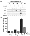
Figure 1. AMPK is activated during HCMV infection.
Serum-starved MRC-5 human fibroblasts were mock- infected or infected with HCMV (MOI = 3). After adsorption, cells were treated with DMSO or the AMPK inhibitor, Compound C (5 µM). At 24, 48 and 72 h post infection cells were harvested in lysis buffer and the resulting lysates were assayed for AMPK activity using the SAMS peptide as a substrate. Values are mean + SE (n = 2).
Figure 2
Figure 2. Impact of AMPK inhibition on AMPK substrate accumulation during HCMV infection.
(A) MRC-5 human fibroblasts were incubated in glucose free media with DMSO (-) or AICAR for 1h or 2.5 h prior to blotting with antibodies specific for pSer79-specific ACC1, total ACC1, or tubulin. (B-D) Serum-starved MRC-5 human fibroblasts were mock infected or infected with HCMV (MOI = 3). After adsorption, cells were treated with the AMPK inhibitor, Compound C (5 µM), or DMSO (-). Cells were harvested at 24, 48 and 72 h post infection and analyzed by Western blot with antibodies specific for pSer79-specific ACC1, total ACC1 and tubulin in (B), TSC1, Glut4 and tubulin in (C), and pThr172-specific AMPK, total AMPK and tubulin (D). Relative pACC/ACC signal ratios in (B) were estimated during HCMV infection for DMSO and Compound C-treated cells using densitometry and subsequently normalized to the ratio of the DMSO control sample.
Figure 3
Figure 3. Inhibition of AMPK blocks HCMV-induced glycolytic activation.
Serum-starved MRC-5 human fibroblasts were mock infected or infected with HCMV (MOI = 3). After adsorption, cells were treated with the AMPK inhibitor Compound C (5 µM) or DMSO. (A) At 48 h post infection, cells were labeled with [14C]-deoxyglucose for 5 min and harvested for scintillation counting as described in the materials and methods. (B) At 48 h post infection, cells were labeled for 1 min with [13C] glucose containing DMEM, quenched with cold methanol, and processed for LC-MS/MS to measure the accumulation of [13C] fructose bisphosphate. All values are means + SE (n = 2). (C) At 40 h post infection, fresh media was added to all cells. At 58 h post infection, media was removed from mock and HCMV-infected fibroblasts and lactate secretion into the media over the 18 hr time frame was analyzed. All values are means + SE (n = 6). (D) Serum-starved MRC-5 human fibroblasts were HCMV infected (MOI = 3) and Compound C (5 µM) was added either immediately after adsorption (0) or 24 h post infection. Lactate excretion was measured from 40–58 hpi or from 58–76 hpi. Values are shown as the percent of the HCMV-infected, DMSO treated control and are means + SE (n≥3).
Figure 4
Figure 4. Pharmaceutical inhibition of AMPK attenuates HCMV viral replication.
(A) Serum-starved MRC-5 human fibroblasts were mock-infected or infected with HCMV (MOI = 3). After adsorption, cells were treated with the AMPK inhibitor, Compound C (CC), at concentrations of 2.5 µM or 5 µM or with DMSO alone. Cells were harvested at 96 h post infection and the production of infectious viral progeny was measured by a standard plaque assay. Values are means + SE (n = 3). (B) Analysis of the potential toxicity of Compound C treatment. Confluent MRC-5 fibroblasts were mock infected or infected with HCMV (MOI = 3). After adsorption, cells were treated with 5 µM Compound C (CC). At 72 h post infection, cell viability was measured via a Live/Dead cell viability assay. Green indicates the presence of esterase activity associated with viable cells, while red indicates loss of cellular membrane integrity associated with cell death. As a positive control for staining of a breakdown in membrane integrity, cells were briefly treated with ethanol. (C) Serum-starved MRC-5 human fibroblasts were HCMV-infected (MOI = 3). Compound C was added at adsorption (0), 24, 48 or 72 h post infection. As a viral growth control, DMSO was added to HCMV infected cells at adsorption. All samples were harvested at 96 h post infection and the production of infectious viral progeny was measured by a standard plaque assay. Values are means + SE (n = 2).
Figure 5
Figure 5. Impact of AMPK pharmaceutical inhibition on viral protein and DNA accumulation.
(A) Analysis of the impact of AMPK inhibition on viral protein accumulation. Serum-starved MRC-5 human fibroblasts were mock infected or infected with HCMV (MOI = 3). After adsorption, cells were treated with the AMPK inhibitor Compound C (CC, 5 µM) or DMSO alone (-). Cells were harvested at 6, 24, 48 and 72 h post infection and analyzed by Western blotting with antibodies specific for IE1, UL44, pp28 and tubulin. (B) Analysis of the impact of AMPK inhibition on viral DNA accumulation. Serum-starved MRC-5 cells were mock infected or infected with HCMV (MOI = 3). After adsorption, cells were treated with the AMPK-specific inhibitor Compound C (CC, 5 µM) or DMSO. Viral DNA was extracted from cells that were harvested at 24, 48, and 72 h post infection and processed for qPCR analysis of viral DNA accumulation. Values are means + SE.
Figure 6
Figure 6. RNAi-mediated targeting of AMPK inhibits HCMV replication.
MRC-5 fibroblasts were transfected with control or AMPK-specific siRNA. Fibroblasts were then serum-starved at 48 h post transfection and subsequently mock or HCMV-infected 24 h later. (A) Samples were harvested for Western blot analysis of AMPK knockdown. The RNAi-mediated percent reduction of AMPK expression from the relevant mock or HCMV-infected control sample was estimated using densitometry. (B) At 48 h post infection, samples were harvested in lysis buffer and cellular lysates were assayed for AMPK activity by measuring phosphorylation of the SAMS peptide by 32P-ATP. Results are shown as mean + SE (n = 3). (C) At 40 h post infection, fresh media was added to all cells. At 58 h post infection, media was removed from mock and HCMV-infected fibroblasts and lactate secretion into the media over the 18 hr time frame was analyzed. All values are means + SE (n = 6). (D) At 96 h post infection, HCMV-infected samples transfected with either control or AMPK-specific siRNA were harvested for viral plaque assay to measure viral replication. Results are shown as mean + SE (n = 2).
Figure 7
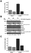
Figure 7. Impact of CaMKK inhibition on AMPK substrates.
Serum-starved MRC-5 human fibroblasts were mock infected or infected with HCMV (MOI = 3). After adsorption, cells were treated with the CaMKK inhibitor STO-609 (10 µg/ml) or DMSO (-). At 24, 48 and 72 h post infection cells harvested in lysis buffer and the resulting lysates were assayed for AMPK activity using the SAMS peptide as a substrate (A) or analyzed by Western blotting with antibodies specific for Ser79-pACC, ACC, TSC1, Glut4,Thr172-pAMPK, AMPK and tubulin (B). Relative pACC/ACC and Thr172-pAMPK/AMPK signal ratios in (B) were estimated during HCMV infection for DMSO and Compound C-treated cells using densitometry and subsequently normalized to the DMSO control. (C) At 40 h post infection, fresh media was added to all cells. At 58 h post infection, media was removed from mock and HCMV-infected fibroblasts and lactate secretion into the media over the 18 hr time frame was analyzed. All values are means + SE (n = 6).
Figure 8
Figure 8. Model of HCMV-mediated manipulation of the AMPK pathway.
HCMV infection results in AMPK activation, likely through activation of CaMKK. AMPK activation results in glycolytic activation to provide energy and building blocks necessary for production of viral progeny. AMPK activation would typically result in inhibition of cellular processes important for viral replication, e.g. protein translation and fatty acid biosynthesis. Viral gene products block these consequences of AMPK activation, e.g. UL38 binds TSC complex and prevents inhibition of mTOR and protein translation. (FP =  fructose-6-phosphate, FBP  =  fructose-1,6-bisphosphate).

Similar articles

Cited by

References

    1. Litman RM, Pardee AB. Production of bacteriophage mutants by a disturbance of deoxyribonucleic acid metabolism. Nature. 1956;178:529–531. - PubMed
    1. Pearson HE, Winzler RJ. Oxidative and glycolytic metabolism of minced day-old mouse brain in relation to propagation of Theiler's GD VII virus. J Biol Chem. 1949;181:577–582. - PubMed
    1. Salzman NP, Lockart RZ, Jr, Sebring ED. Alterations in HeLa cell metabolism resulting from poliovirus infection. Virology. 1959;9:244–259. - PubMed
    1. Kaplan AS, Ben-Porat T. The action of 5-fluorouracil on the nucleic acid metabolism of pseudorabies virus-infected and noninfected rabbit kidney cells. Virology. 1961;13:78–92. - PubMed
    1. Bardell D, Essex M. Glycolysis during early infection of feline and human cells with feline leukemia virus. Infect Immun. 1974;9:824–827. - PMC - PubMed

Publication types

MeSH terms

Substances