. 2012 Mar 16;287(12):8963-73.
doi: 10.1074/jbc.M111.314385. Epub 2012 Jan 30.

Small GTPase Rab17 regulates dendritic morphogenesis and postsynaptic development of hippocampal neurons

Affiliations

Small GTPase Rab17 regulates dendritic morphogenesis and postsynaptic development of hippocampal neurons

Yasunori Mori et al. J Biol Chem. .

Abstract

Neurons are compartmentalized into two morphologically, molecularly, and functionally distinct domains: axons and dendrites, and precise targeting and localization of proteins within these domains are critical for proper neuronal functions. It has been reported that several members of the Rab family small GTPases that are key mediators of membrane trafficking, regulate axon-specific trafficking events, but little has been elucidated regarding the molecular mechanisms that underlie dendrite-specific membrane trafficking. Here we show that Rab17 regulates dendritic morphogenesis and postsynaptic development in mouse hippocampal neurons. Rab17 is localized at dendritic growth cones, shafts, filopodia, and mature spines, but it is mostly absent in axons. We also found that Rab17 mediates dendrite growth and branching and that it does not regulate axon growth or branching. Moreover, shRNA-mediated knockdown of Rab17 expression resulted in a dramatically reduced number of dendritic spines, probably because of impaired filopodia formation. These findings have revealed the first molecular link between membrane trafficking and dendritogenesis.


Figures
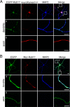
FIGURE 1.
FIGURE 1.
Rab17 is specifically localized at the dendrites of hippocampal neurons. A and B, at 5 DIV mouse hippocampal neurons were transfected with pEGFP-C1-Rab17 (A) or pEGFP-C1 and pEF-Myc-Rab17 (B). The neurons were fixed at 14 DIV and then subjected to immunocytochemistry with antibodies against neurofilament-H (an axon marker; red) and MAP2 (a dendrite marker; blue) (A) or Myc (red) and MAP2 (blue) (B). The middle panels (panels a) and bottom panels (panels b) are magnified views of the boxed areas in the upper right panel. Note that punctate EGFP-Rab17 or Myc-Rab17 signals were specifically observed in the dendrites (MAP2-positive neurites, middle panels in A and B) and were not observed at all in the axons (MAP2-negative neurites, bottom panels in A and B). Bars, 10 μm.
FIGURE 2.
FIGURE 2.
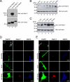
Expression and localization of Rab17 protein in the hippocampal neuron are developmentally regulated. A, specificity of the anti-Rab17 antibody used in this study. COS-7 cells were transfected with a vector encoding FLAG-tagged Rab5A, Rab17, or Rab20, and 2 days after transfection, the cells were lysed and subjected to immunoblot analysis with anti-Rab17 antibody (upper panel) and anti-FLAG tag antibody (lower panel). Note that Rab17 antibody specifically recognized Rab17 (upper panel, lane 2) but did not recognize its related isoforms, Rab5A and Rab20 (upper panel, lanes 1 and 3). B and C, Rab17 expression is developmentally regulated (B) in the mouse hippocampus and (C) in primary mouse hippocampal neuron cultures. Tissue homogenates (50 μg) of mouse hippocampus from embryonic day 18 (E18) to adult mice (B) or lysates of murine hippocampal neurons at 7, 11, 14, 21, or 28 DIV (C) were analyzed by immunoblotting with anti-Rab17 antibody (upper panels) and anti-actin antibody (lower panels). The asterisks indicate nonspecific bands. The positions of the molecular mass markers (in kilodaltons) are shown on the left. D and E, at 2 DIV mouse hippocampal neurons were transfected with a vector encoding EGFP, and the neurons were fixed at 3 DIV (D) or 11 DIV (E) and subjected to immunocytochemistry with antibodies against GFP (green), Rab17 (red), and MAP2 (blue). The bottom three panels (panels a–c) are magnified views of the boxed areas in the upper left panels. Note that at 3 DIV, endogenous Rab17 signals were specifically observed in the cell body, and they were hardly observed in the dendrites and axons, whereas at 11 DIV some Rab17 signals were clearly observed in the dendrites, but they were rarely observed in the axons. Bars, 50 μm (top panels in D and E) and 10 μm (middle and bottom panels in D and E).
FIGURE 3.
FIGURE 3.
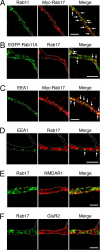
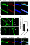
Endosomal localization of Rab17 in developing dendrites of the hippocampal neuron. A–C, at 4 DIV hippocampal neurons were transfected with a vector encoding Myc-Rab17 or EGFP-Rab11A. The neurons were fixed at 16 DIV (A and C) or 18 DIV (B) and subjected to immunocytochemistry with antibodies against Myc (red) and Rab11 (a recycling endosome marker; green) (A), endogenous Rab17 (red) and EGFP-Rab11A (green) (B), and Myc (red) and EEA1 (an early endosome marker; green) (C). D, partial co-localization between endogenous Rab17 and EEA1. Hippocampal neurons were fixed at 16 DIV and subjected to immunocytochemistry with antibodies against Rab17 (red) and EEA1 (green). The arrows indicate the co-localization points. E and F, Rab17 was not co-localized with the NMDA receptor or AMPA receptor in the dendrites of developing neurons. Hippocampal neurons were fixed at 18 DIV and subjected to immunocytochemistry with antibodies against Rab17 (green) and NMDAR1 (E, red) or GluR2 (F, red). The dashed lines indicate dendritic shafts identified as MAP2-positive areas. Bars, 10 μm.
FIGURE 4.
FIGURE 4.
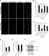
Rab17 regulates dendritic morphogenesis in hippocampal neurons. A–C, at 4 DIV hippocampal neurons were transfected with a vector encoding EGFP and control shRNA (upper panels in A) or Rab17 shRNA (lower panels in A), and the neurons were fixed at 11 DIV and subjected to immunocytochemistry with antibodies against GFP, neurofilament-H (red), and MAP2 (blue). A, typical images of Rab17 knockdown neurons. The arrows and arrowheads indicate axons and dendrites, respectively. Bar, 50 μm. B and C, quantification of total dendrite branching tip numbers (B) and total dendrite length (C) of the control neurons (n = 52), Rab17 knockdown neurons (n = 47), Rab17SR-expressing control neurons (n = 54), and Rab17SR-expressing Rab17 knockdown neurons (n = 62). Note that both the total dendrite length and total dendrite branching tip numbers of Rab17 knockdown neurons were significantly lower than in the control cells. **, p < 0.0025. D and E, quantification of total axon branching tip numbers (D) and total axon length (E) of control neurons (n = 25) and Rab17 knockdown neurons (n = 25). Dendrites (MAP2-positive neurites) and axons (neurofilament-H-positive neurites) were analyzed separately. **, p < 0.0025. F, knockdown efficiency of Rab17 shRNA. COS-7 cells were transfected with a vector encoding control shRNA or with Rab17 shRNA together with EGFP-Rab17 (lanes 1 and 2) or EGFP-Rab17SR (lane 3), and 2 days after transfection the cells were lysed and subjected to immunoblot analysis with anti-GFP antibody (upper panel) and anti-actin antibody (lower panel). The positions of the molecular mass markers (in kilodaltons) are shown on the left.
FIGURE 5.
FIGURE 5.
Overexpression of Rab17-Q77L, a constitutive active Rab17 mutant, promotes dendritic morphogenesis in hippocampal neurons. A–C, at 2 DIV hippocampal neurons were co-transfected with a vector encoding EGFP and pCAG control (top row and second row in A) or pCAG-Myc-Rab17-Q77L (third row and bottom row in A), and the neurons were fixed at 4 DIV and subjected to immunocytochemistry with antibodies against GFP (green), Tau (red), and MAP2 (blue). A, typical images of Rab17-Q77L-expressing neurons. Bars, 20 μm. The second row and bottom row of panels are magnified views of the boxed areas in the far-right panels in the top row and the third row, respectively. B and C, total dendrite branching tip numbers (B) and the total dendrite length (C) of the control neurons (n = 79) and Rab17-Q77L-expressing neurons (n = 80) were analyzed. **, p < 0.0025; *, p < 0.01.
FIGURE 6.
FIGURE 6.
Rab17 is necessary for postsynaptic development. A, hippocampal neurons were fixed at 24 DIV and subjected to immunocytochemistry with antibodies against Rab17 (green), CaMKIIα (a spine marker; red), and MAP2 (blue). Note that the Rab17 dots were localized at dendritic spines. Bar, 2.5 μm. B, typical images of Rab17 knockdown neurons with reduced numbers of spines and synapses. At 4 DIV hippocampal neurons were transfected with a vector encoding EGFP together with control shRNA (upper panels in B) or Rab17 shRNA (lower panels in B), and at 24 DIV the neurons were fixed and subjected to immunocytochemistry with antibodies against GFP (green), CaMKIIα (red), and MAP2 (blue). Bar, 2.5 μm. C, quantification of the numbers of spines in control neurons (n = 21) and Rab17 knockdown neurons (n = 20). **, p < 0.0025. D, typical images of Rab17 knockdown neurons with a reduced number of synapses. At 4 DIV hippocampal neurons were transfected with a vector encoding PSD95-EGFP together with control shRNA (upper panels in D) or Rab17 shRNA (lower panels in D), and the neurons were fixed at 21 DIV and subjected to immunocytochemistry with antibodies against GFP (green), synaptophysin (a presynapse marker; red), and MAP2 (blue). Bar, 2.5 μm. E, quantification of the numbers of synapses in the control neurons (n = 20) and Rab17 knockdown neurons (n = 27). **, p < 0.0025. F, typical images of Rab17 knockdown neurons with reduced numbers of functional synapses. At 4 DIV hippocampal neurons were transfected with a vector encoding EGFP and control shRNA (upper panels in F) or Rab17 shRNA (lower panels in F), and at 21 DIV they were incubated for 10 min with an antibody against Syt I N-terminal domain (anti-Syt I-N) in the presence of 25 mm KCl-containing medium. The neurons were then fixed and subjected to immunocytochemistry with antibodies against rabbit IgG (red) and MAP2 (blue). The arrows indicate the anti-Syt I-N antibody incorporated into neurons (red). Bar, 5 μm. G, quantification of the numbers of anti-Syt I-N antibody-positive dots in the control neurons (n = 20) and Rab17 knockdown neurons (n = 25). *, p < 0.01.
FIGURE 7.
FIGURE 7.
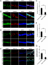
Rab17 is required for filopodia formation in dendrites. A, at 4 DIV hippocampal neurons were transfected with a vector encoding EGFP and control shRNA (upper panels in A) or Rab17 shRNA (lower panels in A), and at 20 DIV the neurons were fixed, subjected to immunocytochemistry with antibodies against EGFP (green) and MAP2 (blue), and stained for F-actin (red) with phalloidin. Bar, 2.5 μm. Note that the control neurons contain a number of protrusions, which represent spines or filopodia, whereas the dendrites of the Rab17 knockdown neurons are devoid of protrusions. B, typical images of Rab17 knockdown neurons with reduced numbers of filopodia. At 4 DIV hippocampal neurons were transfected with a vector encoding gap-Venus together with control shRNA (upper panels in B) or Rab17 shRNA (lower panels in B), and they were examined at 11 DIV. The right panels are magnified views of the boxed areas in the left panels. The arrows indicate filopodia. Bars, 5 μm. C, quantification of the numbers of dendritic filopodia in the control neurons (n = 25) and Rab17 knockdown neurons (n = 20). **, p < 0.0025. D, at 4 DIV hippocampal neurons were co-transfected with a vector encoding gap-Venus and Myc-Rab17, and at 12 DIV the neurons were fixed and subjected to immunocytochemistry with antibodies against Myc (red) and MAP2 (blue). The arrows indicate Rab17-positive dots in filopodia. Bar, 5 μm.

Similar articles

Cited by

References

    1. Urbanska M., Blazejczyk M., Jaworski J. (2008) Molecular basis of dendritic arborization. Acta Neurobiol. Exp. 68, 264–288 - PubMed
    1. Yoshihara Y., De Roo M., Muller D. (2009) Dendritic spine formation and stabilization. Curr. Opin. Neurobiol. 19, 146–153 - PubMed
    1. Perin M. S., Fried V. A., Mignery G. A., Jahn R., Südhof T. C. (1990) Phospholipid binding by a synaptic vesicle protein homologous to the regulatory region of protein kinase C. Nature 345, 260–263 - PubMed
    1. Fei H., Grygoruk A., Brooks E. S., Chen A., Krantz D. E. (2008) Trafficking of vesicular neurotransmitter transporters. Traffic 9, 1425–1436 - PMC - PubMed
    1. Setou M., Nakagawa T., Seog D. H., Hirokawa N. (2000) Kinesin superfamily motor protein KIF17 and mLin-10 in NMDA receptor-containing vesicle transport. Science 288, 1796–1802 - PubMed

Publication types

Substances