. 2012 Mar;23(6):1024-34.
doi: 10.1091/mbc.E11-08-0721. Epub 2012 Jan 25.

Midbody assembly and its regulation during cytokinesis

Affiliations

Midbody assembly and its regulation during cytokinesis

Chi-Kuo Hu et al. Mol Biol Cell. 2012 Mar.

Abstract

The midbody is a transient structure that connects two daughter cells at the end of cytokinesis, with the principal function being to localize the site of abscission, which physically separates two daughter cells. Despite its importance, understanding of midbody assembly and its regulation is still limited. Here we describe how the structural composition of the midbody changes during progression throughout cytokinesis and explore the functional implications of these changes. Deriving from midzones, midbodies are organized by a set of microtubule interacting proteins that colocalize to a zone of microtubule overlap in the center. We found that these proteins split into three subgroups that relocalize to different parts of the midbody: the bulge, the dark zone, and the flanking zone. We characterized these relocalizations and defined domain requirements for three key proteins: MKLP1, KIF4, and PRC1. Two cortical proteins-anillin and RhoA-localized to presumptive abscission sites in mature midbodies, where they may regulate the endosomal sorting complex required for transport machinery. Finally, we characterized the role of Plk1, a key regulator of cytokinesis, in midbody assembly. Our findings represent the most detailed description of midbody assembly and maturation to date and may help elucidate how abscission sites are positioned and regulated.


Figures
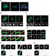
FIGURE 1:
FIGURE 1:
Midzone proteins show distinct relocalization patterns during midbody formation. Cells in different stages of cytokinesis were fixed for immunofluorescence. (A) Midzones between separated sister chromatids gradually converted into the midbody during furrow ingression. Individual midzone bundles were compacted into a single, large, midbody microtubule bundle. A bulge-like structure named the stem body was formed in the center of midbody. The midzone, midbody, and stem body are indicated in yellow. (B) CENPE colocalized with RacGAP1 on midzones in anaphase but relocalized to flank RacGAP1 after furrow ingression onset. (C) MKLP1 and KIF4 colocalized on midzones in anaphase and during furrow ingression but changed to different localizations after furrow ingression. (D) Midbody proteins were categorized into three groups according to their localizations on the midbody in immunofluorescence. All localizations were concluded with observations from a minimal sample size of 50 cells in which >97% showed the same localization patterns. (E) MKLP1 on the bulge localized exclusively to KIF4 on microtubules. (F–H) Localizations of proteins on midbody microtubules were determined and compared by line scans. Peaks of line scans were used as references. PRC1 and KIF4 peaked at the same positions where microtubule signal was low. Aurora B and MKLP2 peaked at the same positions where microtubule signal was high. Line scans of three different kinesins MKLP2, CENPE, and KIF4 peaked at different positions. Bar, 3 μm.
FIGURE 2:
FIGURE 2:
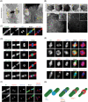
Structure and protein localizations in the dark zone of stem body. (A) Thin-section EM of the midbody. Electron-dense material was enriched in the stem body, and microtubules were compressed at presumptive abscission sites. The area boxed in red was magnified and shown on the right (a′). The midbody and stem body are indicated by yellow lines and the bulge and presumptive abscission site by red and blue arrows, respectively. (B) Series cross-section EM of the stem body. Areas boxed in red (b, c) were magnified (b′, c′). Microtubules within or besides the stem body are boxed in white and magnified (insets in b′ and c′). Vesicles accumulated besides, but not within, the stem body. Vesicles and microtubules are indicated by red and blue arrows, respectively. Electron-dense material was observed to fill the space between microtubules within, but not besides, the stem body. (C) Cells with preabscission or postabscission midbodies were fixed for immunofluorescence. GFP-tubulin (GFP-MT) was observed in the dark zone. KIF4 staining was observed in outer regions of the dark zone. Tubulin staining was masked throughout the whole dark zone. (D) GFP fusion proteins were expressed at low level to reveal their localizations in the dark zone. GFP-MKLP1 localized on the bulge, GFP-KIF4 localized in the dark zone as two bands, and GFP-PRC1 filled the dark zone. GFP-Aurora B was excluded from the stem body. (E) Immunofluorescence of endogenous proteins on midbody remnants. Tubulin staining was continuous in midbody remnant. MKLP1 localized on the bulge as a ring by horizontal views, not on microtubules in the stem body. KIF4 localized as two distinct bands, and PRC1 filled the stem body. Aurora B was excluded from KIF4 localizations on midbody remnants. (F) Cells with midbodies at early (top) and late (bottom) stages of cytokinesis were fixed for immunofluorescence. TSG101 was first concentrated on the bulge and both sides of the stem body and then appeared on the presumptive abscission sites (white arrow) after narrowing onset, exclusive to Aurora B (yellow arrow). (G) Proposed model of midbody structure and formation. Bar, 500 nm (A–B), 100 nM (B, b′–c′), 3 μm (C–E).
FIGURE 3:
FIGURE 3:
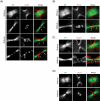
Domain mapping of the stem body protein recruitment. Cells expressing GFP-tagged, full-length or truncated MKLP1, KIF4, and PRC1 were fixed for immunofluorescence. Constructs and domain maps are shown as cartoons, with immunofluorescence below. CC, coiled-coil domain; NLS, nuclear localization signal. (A) MKLP1 C-terminal fragment, which contains a complete coiled-coil domain, was recruited to the stem body. MKLP1 N-terminal fragment contains a motor domain localized on microtubules but was excluded from the stem body. (B) Boxed regions are magnified below. KIF4 missing the C-terminal domain was recruited to the stem body. KIF4 missing the motor domain was recruited to the nucleus, with a small portion localized on the stem body as two distinct bands. (C) PRC1 fragment without the C-terminal microtubule-binding domain could still be recruited to the stem body but was on the bulge. PRC1 missing the N-terminal KIF4- and MKLP1-interacting domain localized on microtubules but was excluded from the stem body. Bar, 3 μm.
FIGURE 4:
FIGURE 4:
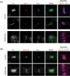
Cortical furrow protein localized on the presumptive abscission sites of the midbody. (A) Furrow protein anillin localized on both bulge and presumptive abscission sites (yellow arrows) of the midbody after furrow ingression completed. This localization was observed in 185 of 200 cytokinetic cells (92.5%). (B) Sharing the same furrow localization with anillin, RhoA also localized on both bulge and presumptive abscission sites of the midbody. (C) Septin (septin 7) was observed on the cortical furrow and two presumptive abscission sites (yellow arrows) but not on the bulge. (D) Citron kinase was observed on the bulge (blue arrow) but was absent from presumptive abscission sites of the midbody. Bar, 3 μm.
FIGURE 5:
FIGURE 5:
Furrow ingression is required for midbody formation. (A, B) Cells were treated with blebbistatin for 30 min and then fixed for immunofluorescence. Furrow ingression was blocked by blebbistatin. Midbody proteins MKLP1, KIF4, PRC1, and Aurora B were still colocalized on microtubules and did not relocalize to distinct parts of the midbody as in control.
FIGURE 6:
FIGURE 6:
Plk1 kinase is required for midbody formation. (A) Cells at different stages of cytokinesis were fixed for immunofluorescence. KIF4 was used as a stem body marker. Line scans were used to measure the relative localizations of KIF4 and Plk1. Plk1 colocalized with KIF4 on midzones and then relocalized to KIF4-flanking regions. After furrow ingression completed, Plk1 relocalized again into the stem body. Plk1 was observed to localize in the core of stem body on midbody remnant. (B) Cells were treated with BI-2536 for 20 min and then fixed for immunofluorescence. In the presence of BI-2536, no immunofluorescence masking was observed on midbodies. Midbody proteins Aurora B, PRC1, KIF4, and MKLP1 all stayed on microtubules, with their localizations biased toward one side of the midbody. Stem body did not form. (C) Cells stably expressing GFP-EB3 were treated with BI-2536 for time-lapse spinning disk microscopy (see Supplemental Movies S1 and S2). Imaging started at time zero. In the non–BI-2536–treated control, the midbody formed and was abscised. In BI-2536–treated cells, the preformed midbody disassembled with time. The GFP-EB3–free zone gradually disappeared. GFP-EB3 eventually was able to move through the intracellular bridge. No abscission occurred. Bar, 3 μm.

Similar articles

Cited by

References

    1. Austin JR, 2nd, Segui-Simarro JM, Staehelin LA. Quantitative analysis of changes in spatial distribution and plus-end geometry of microtubules involved in plant-cell cytokinesis. J Cell Sci. 2005;118:3895–3903. - PubMed
    1. Bastos RN, Barr FA. Plk1 negatively regulates Cep55 recruitment to the midbody to ensure orderly abscission. J Cell Biol. 2010;191:751–760. - PMC - PubMed
    1. Boman AL, Kuai J, Zhu X, Chen J, Kuriyama R, Kahn RA. Arf proteins bind to mitotic kinesin-like protein 1 (MKLP1) in a GTP-dependent fashion. Cell Motil Cytoskeleton. 1999;44:119–132. - PubMed
    1. Buck RC, Tisdale JM. The fine structure of the mid-body of the rat erythroblast. J Cell Biol. 1962;13:109–115. - PMC - PubMed
    1. Canman JC, Cameron LA, Maddox PS, Straight A, Tirnauer JS, Mitchison TJ, Fang G, Kapoor TM, Salmon ED. Determining the position of the cell division plane. Nature. 2003;424:1074–1078. - PubMed

Publication types

Substances

LinkOut - more resources