. 2012 Jan 22;19(2):164-70.
doi: 10.1038/nsmb.2222.

Regulation of Rev1 by the Fanconi anemia core complex

Affiliations

Regulation of Rev1 by the Fanconi anemia core complex

Hyungjin Kim et al. Nat Struct Mol Biol. .

Abstract

The 15 known Fanconi anemia proteins cooperate in a pathway that regulates DNA interstrand cross-link repair. Recent studies indicate that the Fanconi anemia pathway also controls Rev1-mediated translesion DNA synthesis (TLS). We identified Fanconi anemia-associated protein (FAAP20), an integral subunit of the multisubunit Fanconi anemia core complex. FAAP20 binds to FANCA subunit and is required for stability of the complex and monoubiquitination of FANCD2. FAAP20 contains a ubiquitin-binding zinc finger 4 domain and binds to the monoubiquitinated form of Rev1. FAAP20 binding stabilizes Rev1 nuclear foci and promotes interaction of the Fanconi anemia core with PCNA-Rev1 DNA damage bypass complexes. FAAP20 therefore provides a critical link between the Fanconi anemia pathway and TLS polymerase activity. We propose that the Fanconi anemia core complex regulates cross-link repair by channeling lesions to damage bypass pathways and preventing large DNA insertions and deletions.


Conflict of interest statement

COMPETING FINANCIAL INTERESTS

The authors declare no competing financial interests.

Figures

Figure 1
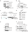
Figure 1. C1orf86 is required for the FA pathway activation
(a) Sequence alignment of the C1orf86 UBZ4 domain with known UBZ4 domains. Stars indicate the conserved residues that form a short mononucleate zinc finger, and arrows point to cysteine residues (Cys147 and 150) important for ubiquitin interaction. (b) FANCA and FANCD2 were analyzed by immunoblot in cell lysates from HeLa cells, transfected with control or C1orf86 siRNA and treated with DNA damage-inducing agents. (c) FACND2 was analyzed by immunoblot in cytosolic (S) and chromatin-containing (P) fractions of HeLa cells, transfected with control or C1orf86 siRNA and treated with 50 ng ml−1 MMC for 17 h. (d) Immunostaining of FANCD2 in HeLa cells, transfected with control or C1orf86 siRNA and treated with 2 mM HU for 6 h. Representative images are shown, and at least 150 cells were counted for quantification. Data shown are mean ± s.d. from three independent experiments. * p < 0.01. (e) Clonogenic survival of HeLa cells transfected with siRNA control, C1orf86, or FANCA treated with increasing doses of MMC and plated for 12 days. (f) Quantification of chromosomal aberrations and radial chromosomes of 293T cells transfected with siRNAs and exposed to 20 ng ml−1 MMC.
Figure 2
Figure 2. FAAP20 is required for the FA core complex stability
(a) Immunoblot of anti-Flag immunoprecipitates (IP) of cell lysates from Flag-FAAP20 expressing 293T cells. (b) Direct interaction between myc-FAAP20 and FANCA in vitro analyzed by anti-myc IP of in vitro translated protein mixture. (c) Anti-Flag IP and immunoblot analysis of 293T cell lysates expressing Flag-tagged FAAP20 (F20) wild-type, ΔN (FAAP2048–180) or ΔC (FAAP201–163). (d) Immunoblot of cell lysates from HeLa cells transfected with siRNA control or FAAP20 for 72 h. (e) FANCA and FANCE were analyzed by immunoblot of HeLa cells, transfected with siRNA oligos and treated with 20 μM MG132 for 4 h. (*) denotes nonspecific band. (f) FANCA was analyzed by immunoblot of HeLa cells, pretreated with siRNA that targets 3′ UTR of FAAP20 mRNA and transfected with Flag-tagged wild-type or indicated mutants for 48 h.
Figure 3
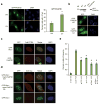
Figure 3. FAAP20 interacts with Rev1 and promotes efficient Rev1 foci formation
(a) Fluorescence microscopy of GFP-Rev1 in U2OS cells, serially transfected with indicated siRNA oligos and GFP-Rev1 followed by treatment with 15 J m−2 UVC for 14 h. Representative images of GFP-Rev1 foci are shown, and the right panel represents the quantification of cells displaying more than five GFP-Rev1 foci. Data shown are mean ± s.d. from three independent experiments. * p < 0.01 compared with UVC-treated siRNA control. The immunoblots show the knockdown efficiency. (**) denotes nonspecific band. (b) Anti-Flag immunoprecipitates were analyzed by immunoblot from 293T cells transiently coexpressing Flag-FAAP20 and GFP-Rev1 or GFP-Polη. (c) Immunoblot of anti-Flag immunoprecipitates of 293T cells transiently coexpressing GFP-Rev1 and Flag-tagged FAAP20 wild-type, UBZ4-point mutant (C147A C150A; CA), or UBZ4-deletion mutant (aa 1–163; Δ).
Figure 4
Figure 4. Monoubiquitination of Rev1 enhances the interaction with FAAP20
(a) Monoubiquitin of Rev1 was visualized by anti-Flag immunoprecipitation and immunoblot of 293T cells transfected with cDNAs encoding Flag-tagged Rev1 with or without HA-tagged ubiquitin. The slower-migrating band, above Flag-Rev1, represents the monoubiquitinated form of Rev1, which was confirmed by anti-HA immunoblot. (b) (Top) schematic of Rev1 mutants. (Bottom) anti-myc immunoprecipitation of 293T cell lysates coexpressing HA-ubiquitin and GFP-tagged Rev1 wild-type, UBM* (L946A P947A L1024A P1025A), or ΔC (aa1–892) mutant, mixed with the lysates expressing myc-tagged FAAP20. (c) Anti-myc immunoprecipitation of 293T cell lysates coexpressing HA-ubiquitin and GFP-tagged Rev1, mixed with lysates expressing myc-tagged FAAP20 wild-type or CA mutant (d) Anti-Flag immunoprecipitation of 293T cell lysates coexpressing HA-ubiquitin and Flag-tagged Rev1, mixed with lysates expressing either myc-tagged FAAP20 wild-type or CA mutant.
Figure 5
Figure 5. FAAP20 and Rev1 colocalize at sites of replication stress
(a) Fluorescence microscopy of U2OS cells, transfected with GFP-tagged FAAP20 and treated with 15 J m−2 UVC for 14 hr. Cells with more than five foci were quantified. Data shown are mean ± s.d. from three independent experiments. * p < 0.01 (b) GFP-fluorescence of undamaged U2OS cells transfected with GFP-FAAP20 wild-type or UBZ4 mutant. Immunoblot shows comparable expression levels. (c) Immunostaining of PCNA in U2OS cells, transfected with GFP-tagged Rev1 or FAAP20 and treated with 15 J m−2 UVC. A representative image is shown, in which 84.1 % of the FAAP20 foci colocalize with PCNA foci. (d) Anti-myc immunostaining in U2OS cells, co-transfected with GFP-Rev1 and myc-tagged FAAP20 wild-type or mutant and treated with 15 J m−2 UVC. Immunostaining of cells transfected with GFP-Rev1 alone served as negative control for antibody specificity. A representative image is shown, in which 83.8 % of the FAAP20 foci colocalize with Rev1 foci. (e) The mutation frequency in damaged (1,000 J m−2 UVC) supF plasmid was determined recovered from siRNA treated 293T cells. Data shown are mean ± s.d. from three independent experiments. * p < 0.05 compared with UVC-treated control.
Figure 6
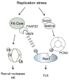
Figure 6. A branched model of the FA pathway
Upon replication stress, the FA core complex monoubiquitinates FANCD2/I to recruit nucleases and promote homologous recombination. FAAP20 stabilizes the FA core complex. The FAAP20-containing FA core complex is also required for efficient Rev1 foci assembly where the ubiquitin-binding property of FAAP20 contributes to stabilizing the PCNA-associated Rev1 complex at the stalled replication fork to direct lesion bypass. FAAP20-Rev1 interaction may be further enhanced by uncharacterized interaction.

Similar articles

Cited by

References

    1. Joenje H, Patel KJ. The emerging genetic and molecular basis of Fanconi anaemia. Nat Rev Genet. 2001;2:446–57. - PubMed
    1. Kee Y, D'Andrea AD. Expanded roles of the Fanconi anemia pathway in preserving genomic stability. Genes Dev. 2010;24:1680–94. - PMC - PubMed
    1. Ciccia A, et al. Identification of FAAP24, a Fanconi anemia core complex protein that interacts with FANCM. Mol Cell. 2007;25:331–343. - PubMed
    1. Smogorzewska A, et al. Identification of the FANCI protein, a monoubiquitinated FANCD2 paralog required for DNA repair. Cell. 2007;129:289–301. - PMC - PubMed
    1. Joo W, et al. Structure of the FANCI-FANCD2 complex: insights into the Fanconi anemia DNA repair pathway. Science. 2011;333:312–6. - PMC - PubMed

Publication types

MeSH terms

Substances