. 2012 Mar 9;287(11):8457-67.
doi: 10.1074/jbc.M111.312561. Epub 2012 Jan 17.

Phosphatidylinositol 4-kinase IIIβ is required for severe acute respiratory syndrome coronavirus spike-mediated cell entry

Affiliations

Phosphatidylinositol 4-kinase IIIβ is required for severe acute respiratory syndrome coronavirus spike-mediated cell entry

Ning Yang et al. J Biol Chem. .

Abstract

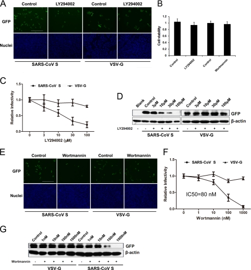
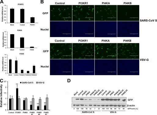
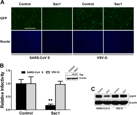
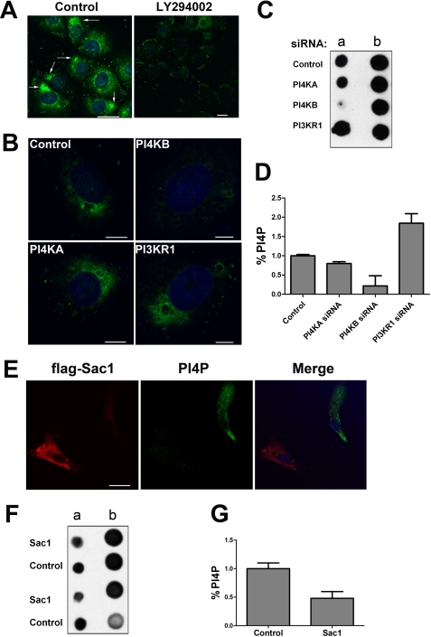
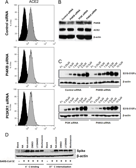
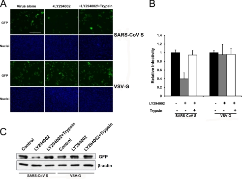
Phosphatidylinositol kinases (PI kinases) play an important role in the life cycle of several viruses after infection. Using gene knockdown technology, we demonstrate that phosphatidylinositol 4-kinase IIIβ (PI4KB) is required for cellular entry by pseudoviruses bearing the severe acute respiratory syndrome-coronavirus (SARS-CoV) spike protein and that the cell entry mediated by SARS-CoV spike protein is strongly inhibited by knockdown of PI4KB. Consistent with this observation, pharmacological inhibitors of PI4KB blocked entry of SARS pseudovirions. Further research suggested that PI4P plays an essential role in SARS-CoV spike-mediated entry, which is regulated by the PI4P lipid microenvironment. We further demonstrate that PI4KB does not affect virus entry at the SARS-CoV S-ACE2 binding interface or at the stage of virus internalization but rather at or before virus fusion. Taken together, these results indicate a new function for PI4KB and suggest a new drug target for preventing SARS-CoV infection.


Figures

FIGURE 1.
FIGURE 1.
LY294002 inhibits SARS-CoV S-mediated entry. A, VeroE6 cells were pretreated with LY294002 at 30 μm or DMSO (control) and then infected with SARS-CoV S or VSV-G pseudovirus. After 48 h, the cells were fixed, and the nuclei were stained with Hoechst 33342. Images were captured on a fluorescence microscope. Scale bar, 400 μm. B, 3-(4,5-dimethylthiazol-2-yl)-2,5-diphenyltetrazolium bromide assay of VeroE6 cells treated with 100 or 1 μm wortmannin for 3 h. C, VeroE6 cells were treated with the indicated concentrations of LY294002 (3–100 μm) or DMSO (control). Statistical analysis was performed on the proportion of GFP-positive cells 48 h after virus infection. For quantification, >1000 cells were scored in three independent experiments. D, effect of LY294002 (3–100 μm) or DMSO (control) treatment or untreated (blank) on SARS pseudovirus entry was determined by assessing the GFP expression level by immunoblotting with the indicated antibodies. Cell lysates were prepared after virus infection for 48 h. E, VeroE6 cells were pretreated with wortmannin at 100 nm or DMSO (control) and then infected with SARS-CoV S or VSV-G pseudovirus. After 48 h, the cells were fixed, and the nuclei were stained with Hoechst 33342. Images were captured on a fluorescence microscope. Scale bar, 400 μm. F, VeroE6 cells were treated with the indicated concentrations of wortmannin or DMSO. Statistical analysis was performed to assess the proportion of GFP-positive cells 48 h after virus infection. For quantification, >1000 cells were scored in three independent experiments. G, effect of wortmannin (1–1000 nm), DMSO, or no treatment (blank) on SARS pseudovirion entry was determined by GFP expression levels by immunoblotting with the indicated antibodies. Cell lysates were prepared after virus infection for 48 h.
FIGURE 2.
FIGURE 2.
PI4KB knockdown inhibits SARS-CoV S-mediated entry, whereas PI3KR1 knockdown increases SARS-CoV S-mediated entry. A, VeroE6 cells were transfected with a nontarget siRNA (control) or siRNAs specific for PI3KR1, PI4KA, or PI4KB. Untreated cells were used as a blank control. The transcripts of target genes were quantified by quantitative RT-PCR at 48 h post-transfection. mRNA levels were normalized to GAPDH levels. Values are presented as the means ± S.D. B, VeroE6 cells were transfected with corresponding siRNAs and then infected with SARS-CoV S or VSV-G pseudovirus. After 48 h, the cells were fixed, and the nuclei were stained with Hoechst 33342. Images were captured on a fluorescence microscope. Scale bar, 400 μm. C, VeroE6 cells were transfected with the corresponding siRNAs and then infected with SARS-CoV S or VSV-G pseudovirus. Statistical analysis was performed on the proportion of GFP-positive cells 48 h after virus infection. For quantification, >1000 cells were scored in three independent experiments. **, p < 0.001 compared with the control. D, siRNA treatment affected SARS-CoV S-mediated entry as determined by GFP expression levels measured by immunoblotting with the indicated antibodies. Cell lysates were prepared after virus infection for 48 h. GFP band intensities were quantitated by densitometric analysis using Quantity One (Bio-Rad) and normalized to β-actin levels.
FIGURE 3.
FIGURE 3.
Overexpression of Sac1 inhibits SARS-CoV S-mediated entry. A, normal VeroE6 cells or cells transiently transfected with FLAG-Sac1 were infected with SARS-CoV S or VSV-G pseudovirus. After 48 h, the cells were fixed, and the nuclei were stained with Hoechst 33342. Images were captured on a fluorescence microscope. Scale bar, 400 μm. B, statistical analysis of the proportion of GFP-positive cells at 48 h post-virus infection in A. Inset, Western blot showing recombinant FLAG-Sac1 expression at 48 h post-transfection. **, p < 0.001. C, GFP expression levels were determined by immunoblotting with the antibodies indicated in A.
FIGURE 4.
FIGURE 4.
PI4P lipid levels are regulated by PI4KB and Sac1 in VeroE6 cells. A, PI4P levels decreased after LY294002 treatment. VeroE6 cells were treated with 100 μm LY294002 or DMSO (control) for 30 min. PI4P lipid content was determined by immunostaining with anti-PI4P antibodies. The white arrows indicate the PI4P distribution (green). Nuclei were stained with Hoechst 33342. Scale bar, 25 μm. B, PI4KB is responsible for a significant fraction of PI4P lipids in VeroE6. The cells were transfected with control, PI4KB, PI4KA, or PI3KR1 siRNAs for 48 h. Then the cells were immunostained with anti-PI4P antibodies. The nuclei were counterstained with Hoechst 33342. Scale bar, 7 μm. C, detection of PI4P from lipid extracts of VeroE6 cells. At 48 h post-transfection of siRNAs, the PI4P lipid levels in VeroE6 cells were determined using PI4P mass strips. Column a consists of PI4P lipid extractions from experimental samples. Column b consists of pre-spotted PI4P standards as follows (top to bottom): 20, 15, 10, and 5 pmol. D, statistical analysis of the relative levels of PI4P in C. Error bars represent the S.E. from two independent experiments. E, recombinant Sac1 expression decreased PI4P lipid levels in VeroE6. The cells were transfected with a FLAG-Sac1 expression plasmid. After 48 h, the transfected cells were detected by indirect immunofluorescence with an anti-FLAG antibody (red). PI4P lipid (green) was detected with anti-PI4P antibody. The nuclei were counterstained with Hoechst 33342. Scale bar, 23 μm. F, detection of PI4P lipids from extracts of VeroE6 cells expressing recombinant Sac1. Column a consists of PI4P lipid extracts from two independent experimental samples. Column b consists of pre-spotted PI4P standards as follows (top to bottom): 4, 2, 1, and 0.5 pmol. G, statistical analysis of the relative levels of PI4P in F. Error bars are S.E. from two independent experiments.
FIGURE 5.
FIGURE 5.
PI4KB facilitates SARS-CoV S-mediated entry after virus internalization into cells. A, flow cytometric analysis of surface levels of ACE2 (gray histograms) in siRNA-transfected VeroE6 cells. Background fluorescence levels (black histograms) were determined by labeling cells with an irrelevant isotype-matched control antibody. B, VeroE6 cells were transfected with the indicated siRNAs, and total cell lysates were analyzed by Western blotting with anti-PI4KB antibodies, anti-ACE2 antibodies, and anti-β-actin antibodies. C, increasing concentrations of recombinant SARS-CoV RBR (S318–510Fc) or control Fc (10 μm) were bound to VeroE6 siRNA cell lines and analyzed by Western blotting. D, VeroE6 cells were pretreated with DMSO (control), LY294002 (30 μm), or wortmannin (100 nm) for 30 min, and SARS-CoV S was added to the cells. After 2 h at 4 °C, the quantity of bound virus was determined by Western blot using anti-SARS spike antibodies or warming to 37 °C to allow the virus to internalize for another 2 h. Uninternalized virus was removed by treatment with 1 mg/ml proteinase K for 10 min. The internalized virus was assayed by Western blot using anti-SARS spike antibodies. For neutralization experiments, SARS-CoV S pseudovirus was preincubated with anti-SARS spike neutralizing antibody (NA) or an irrelevant control antibody (NA control) and then used for binding or internalization assays.
FIGURE 6.
FIGURE 6.
TPCK-trypsin treatment bypasses LY294002 restriction of SARS-CoV S-mediated entry. A, VeroE6 cells were treated with LY294002 (30 μm) or DMSO (control). After 30 min, the cells were spin-infected at 4 °C with the indicated pseudoviruses and then treated with trypsin or PBS. After 30 °C for 13 min, infected cells were maintained in growth medium for 48 h. The cells were fixed, and nuclei were stained with Hoechst 33342. Images were captured with a fluorescence microscope. Scale bar, 400 μm. B, statistical analysis of the proportion of GFP-positive cells in A.
FIGURE 7.
FIGURE 7.
Model of the PI kinases and PI4P involved in SARS-CoV S-mediated entry. After SARS-CoV binds to ACE2, PI4P, the catalytic product of PI4KB, creates a lipid microenvironment or PI4P-enriched organelle required for the steps leading to fusion. Pharmacological inhibitors of PI4KB, such as LY294002 or wortmannin, suppress PI4KB activity and thereby inhibit SARS-CoV S-mediated entry. Cellular factors, such as PI3Ks or Sac1, that negatively regulate PI4P generation can also inhibit SARS-CoV S-mediated entry.

Similar articles

Cited by

References

    1. Vicinanza M., D'Angelo G., Di Campli A., De Matteis M. A. (2008) Function and dysfunction of the PI system in membrane trafficking. EMBO J. 27, 2457–2470 - PMC - PubMed
    1. Saeed M. F., Kolokoltsov A. A., Freiberg A. N., Holbrook M. R., Davey R. A. (2008) Phosphoinositide 3-kinase-Akt pathway controls cellular entry of Ebola virus. PLoS Pathog. 29, e1000141. - PMC - PubMed
    1. Dunn E. F., Connor J. H. (2011) Dominant inhibition of Akt/protein kinase B signaling by the matrix protein of a negative-strand RNA virus. J. Virol. 85, 422–431 - PMC - PubMed
    1. Ehrhardt C., Wolff T., Pleschka S., Planz O., Beermann W., Bode J. G., Schmolke M., Ludwig S. (2007) Influenza A virus NS1 protein activates the PI3K/Akt pathway to mediate antiapoptotic signaling responses. J. Virol. 81, 3058–3067 - PMC - PubMed
    1. Berger K. L., Cooper J. D., Heaton N. S., Yoon R., Oakland T. E., Jordan T. X., Mateu G., Grakoui A., Randall G. (2009) Roles for endocytic trafficking and phosphatidylinositol 4-kinase IIIα in hepatitis C virus replication. Proc. Natl. Acad. Sci. U.S.A. 106, 7577–7582 - PMC - PubMed

Publication types

MeSH terms

Substances