Clinical Trial
. 2012 Jan;8(1):e1002437.
doi: 10.1371/journal.ppat.1002437. Epub 2012 Jan 5.

Lymphoid tissue damage in HIV-1 infection depletes naïve T cells and limits T cell reconstitution after antiretroviral therapy

Affiliations
Clinical Trial

Lymphoid tissue damage in HIV-1 infection depletes naïve T cells and limits T cell reconstitution after antiretroviral therapy

Ming Zeng et al. PLoS Pathog. 2012 Jan.

Abstract

Highly active antiretroviral therapy (HAART) can suppress HIV-1 replication and normalize the chronic immune activation associated with infection, but restoration of naïve CD4+ T cell populations is slow and usually incomplete for reasons that have yet to be determined. We tested the hypothesis that damage to the lymphoid tissue (LT) fibroblastic reticular cell (FRC) network contributes to naïve T cell loss in HIV-1 infection by restricting access to critical factors required for T cell survival. We show that collagen deposition and progressive loss of the FRC network in LTs prior to treatment restrict both access to and a major source of the survival factor interleukin-7 (IL-7). As a consequence, apoptosis within naïve T cell populations increases significantly, resulting in progressive depletion of both naïve CD4+ and CD8+ T cell populations. We further show that the extent of loss of the FRC network and collagen deposition predict the extent of restoration of the naïve T cell population after 6 month of HAART, and that restoration of FRC networks correlates with the stage of disease at which the therapy is initiated. Because restoration of the FRC network and reconstitution of naïve T cell populations are only optimal when therapy is initiated in the early/acute stage of infection, our findings strongly suggest that HAART should be initiated as soon as possible. Moreover, our findings also point to the potential use of adjunctive anti-fibrotic therapies to avert or moderate the pathological consequences of LT fibrosis, thereby improving immune reconstitution.


Conflict of interest statement

The authors have declared that no competing interests exist.

Figures

Figure 1
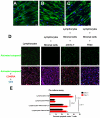
Figure 1. Collagen deposition and loss of the FRC network impede access to and source of IL-7 in HIV-1 infection.
A. FRCs are the major producers of IL-7. LN sections (representative image for one HIV negative subject of 5) stained for desmin (red) and IL-7 (green). Merged confocal image shows co-localization of IL-7 and FRCs in T cell area. Scale bar, 10 µm. B–C. Collagen deposition and loss of the FRC network disrupts interaction between T cells and FRCs. Confocal images of LN sections from an uninfected subject (representative image for one subject of 5) immunofluorescently stained for desmin (green), collagen (red) and CD3 (blue). The merged image shows that FRCs colocalize with collagen and T cells are in contact with the FRC network (B). Confocal images of LN sections from a subject at AIDS stage (representative image for one subject of 6). The merged image shows that the loss of FRCs and associated collagen deposition leads to loss of the contact between FRCs and T cells, which instead contact mainly extra-FRC collagen. Scale bar, 20 µm (C). D. Confocal images of LN sections from HIV negative subjects and from subjects at different stage of HIV infection immunofluorescently stained for desmin (green) and collagen (red), showing the gradual loss of FRCs in the T cell zone within LTs, which is associated with extensive collagen deposition during HIV infection. Scale bar, 20 µm. E. Quantification of average amount of FRCs and collagen deposition at each stage of infection, showing the gradual loss of FRCs and collagen deposition. Error bars represent the s.d. F. Confocal images of LN sections from HIV negative subjects and from subjects at different stage of HIV infection immunofluorescently stained for IL-7 (green), showing that the gradual loss of IL-7 in the T cell zone is associated with gradual depletion of FRCs (E). Scale bar, 20 µm.
Figure 2
Figure 2. Naïve T cells need to contact FRCs to get access to IL-7 for survival.
(A–C) IL-7 is produced and presented on the surface of stromal cells. A-B. Confocal images of monolayer of fixed and permeablized stromal cells isolated from human tonsil immunofluorescently stained for (A) IL-7 (green) or (B) desmin (green) and DAPI (blue) at one-day post passage. Scale bar, 30 µm. C. Confocal image of live stromal cells (DAPI: blue) staining showing the IL-7 (green) on the surface of stromal cells. Scale bar, 10 µm. D-E. FRC-like stromal cells enhance the survival of naïve T cells via IL-7. D. Triple fluorescently stained activated caspase 3+ (green), CD45RA+ (red) and CD3+ (blue) cells in an ex vivo culture system showing that stromal cells enhance the survival of naïve T cell by mechanisms dependent on IL-7 and cell contact. 2x10⁵ lymphocytes from human tonsil were cultured with or without stromal cells for 2–3 days. Naïve T cell apoptosis is reduced in co-cultures with stromal cells (+ stromal cells) compared to cultures without stromal cells. Apoptosis in the naïve T cell population increases with IL-7 blocking antibody (anti-IL-7) or when lymphocytes are separated from stromal cells by a transwell filter (Filter) compared to co-cultures with stromal cells. Scale bar, 60 µm. E. Quantification of the percentages of activated caspase 3+CD45RA+CD3+ naïve T cells in total T cell population at day 2 and day 3 cultures. Values are the mean of the percent apoptotic naïve T cells ± s.d. ANOVA comparison was done on the average percentages of day 2 and day 3.
Figure 3
Figure 3. Loss of FRCs is associated with loss of naïve T cells within LTs.
A. Confocal images of LN sections from subjects at different stage of HIV infection triple immunofluorescently stained for TUNEL (green), CD45RA (red) and CD3 (Blue), showing the gradual loss of CD45RA+CD3+ naïve T cells is associated increased apoptosis in the naïve T cell population within LTs during HIV infection. Scale bar, 10 µm. B. Confocal images of LN sections from subjects at different time points post HIV infection double immunofluorescently stained for CD45RA (green) and CD4 or CD8 (red), showing both naïve CD4 and CD8 T cells are depleted within LTs. Scale bar, 20 µm. C. Quantitative image analysis of the number of apoptotic naïve T cells and the number of naïve T cells (CD45RA+CD3+), showing that increased apoptosis in the naïve T cell population is associated with depletion of naïve T cells (total n = 37, p<0.0001, R2 = 0.5373). D. Quantification of FRCs (the percent area staining positive for desmin in T cell zone) and the number of apoptotic naïve T cells (TUNEL+CD45RA+CD3+), showing that the depletion of FRCs is associated with increased apoptosis in naïve T cell populations (total n = 37, p<0.0001, R2 = 0.5843). E. Quantitative image analysis of FRCs and the number of naïve T cells within LTs, showing that the loss of naïve T cells is associated with loss of FRCs (total n = 37, p<0.0001, R2 = 0.5166). Values are the mean of measurement ± s.d.
Figure 4
Figure 4. The extent of LT destruction before HAART predicts the extent of restoration of naïve T cells after HAART.
A. The area that FRCs occupy before HAART is negatively associated with the number of apoptotic naïve T cells after 6 months of HAART. B. The collagen area before HAART is positively associated with the number of apoptotic naïve T cells after 6 months of HAART.
Figure 5
Figure 5. Restoration of LT structure is slow and incomplete after HAART and is associated with the timing of initiation of HAART.
A. Representative confocal images of immunofluorescent staining for desmin (green) and collagen (red), showing the different extent of restoration of stromal cell network and collagen normalization after 6 months of HAART. Scale bar, 20 µm. B. Quantification of the average area of FRCs and collagen before and after 6 months of HAART in patients receiving the HAART at different stages of infection, showing the different extent of restoration of LTs is associated with the timing of initiation of HAART. Error bars represent the s.d. C. Representative confocal images of immunofluorescent staining for desmin (green) and collagen (red), showing that the different extent of restoration of the FRC network and collagen normalization after 6 months of HAART as represented by the percent area not covered by FRCs. Scale bar, 20 µm. D. Quantification of the percent area covered by FRCs.
Figure 6
Figure 6. Incomplete LT restoration is associated with high level of apoptosis in naïve T cell populations and incomplete restoration of naïve T cells.
A. Confocal images of LN sections from uninfected subjects and patients receiving 6 months of HAART at either acute or AIDS phase of infection, immunofluorescently stained for CD45RA (red) and CD3 (blue), showing the different extent of restoration of naïve T cells. Scale bar, 10 µm. B. Quantification of number of naïve T cells before and after HAART at each stage of infection, showing the different kinetics of restoration of naïve T cells. Error bars represent the s.d. C. Confocal images of LN sections from patients receiving 6 months of HAART at either acute or AIDS phase of infection immunofluorescently stained for CD45RA (red), CD3 (blue) and TUNEL staining of apoptotic cells (green), showing the higher level of apoptosis in naïve T cell populations after 6 months of HAART when HAART was started during chronic phase of infection. Scale bar, 10 µm. D. Quantification of average number of apoptotic naïve T cells before and after HAART at each stage of infection, showing the different kinetics of decrease of apoptotic naïve T cells.
Figure 7
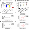
Figure 7. The extent of restoration of naïve T cells after HAART is not associated with viral load or immune activation.
A. Viral load before and after HAART in patients receiving HAART at different stages of infection, showing the inhibition of viral replication is similar for patients at different stage of infection. B. Quantification of the number of Ki67+ cells before and after HAART in patients receiving HAART at different stages of infection, showing the inhibition of immune activation by HAART is similar for patients at different stage of infection (ns, not significant). C, E. Association between the viral load and number of naïve T cells and association between viral load and the number of apoptotic naïve T cells after HAART are not significant. D, F. Association between the number of Ki67+ cells and naïve T cells and association between the number of Ki67+ cells and the number of apoptotic naïve T cells after HAART are not significant.

Similar articles

Cited by

References

    1. Haase AT. Population biology of HIV-1 infection: viral and CD4+ T cell demographics and dynamics in lymphatic tissues. Annu Rev Immunol. 1999;17:625–656. - PubMed
    1. Andersson J, Fehniger TE, Patterson BK, Pottage J, Agnoli M, et al. Early reduction of immune activation in lymphoid tissue following highly active HIV therapy. AIDS. 1998;12:F123–129. - PubMed
    1. Zhang ZQ, Notermans DW, Sedgewick G, Cavert W, Wietgrefe S, et al. Kinetics of CD4+ T cell repopulation of lymphoid tissues after treatment of HIV-1 infection. Proc Natl Acad Sci U S A. 1998;95:1154–1159. - PMC - PubMed
    1. Lewden C, Salmon D, Morlat P, Bevilacqua S, Jougla E, et al. Causes of death among human immunodeficiency virus (HIV)-infected adults in the era of potent antiretroviral therapy: emerging role of hepatitis and cancers, persistent role of AIDS. Int J Epidemiol. 2005;34:121–130. - PubMed
    1. Barbaro G, Barbarini G. HIV infection and cancer in the era of highly active antiretroviral therapy (Review). Oncol Rep. 2007;17:1121–1126. - PubMed

Publication types

MeSH terms