

Phosphoinositide 3-kinase/AKT/mTORC1/2 signaling determines sensitivity of Burkitt's lymphoma cells to BH3 mimetics
- PMID: 22241218
- PMCID: PMC3378513
- DOI: 10.1158/1541-7786.MCR-11-0394
Phosphoinositide 3-kinase/AKT/mTORC1/2 signaling determines sensitivity of Burkitt's lymphoma cells to BH3 mimetics
Abstract
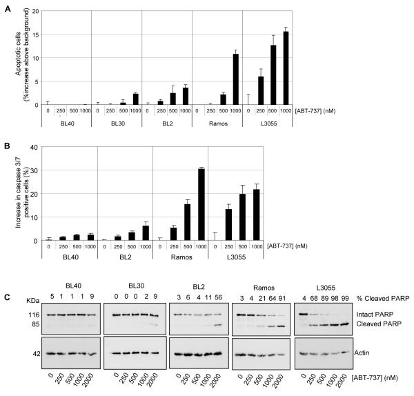
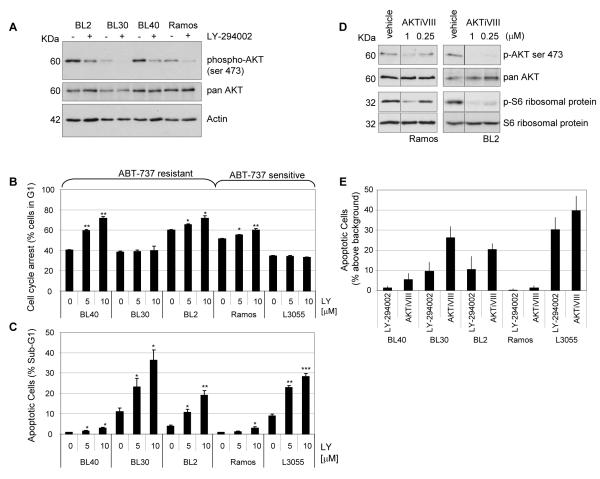
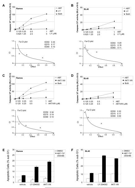
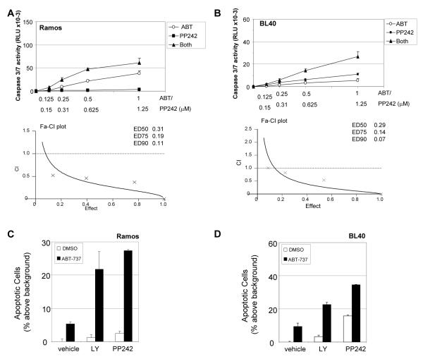
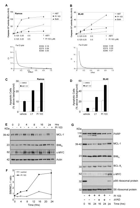
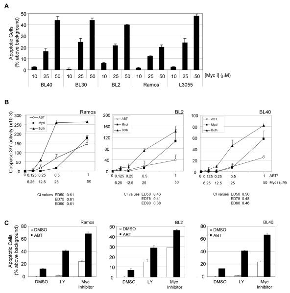
Burkitt's lymphoma (BL), driven by translocation and overexpression of the c-MYC gene, is an aggressive, highly proliferative lymphoma, and novel therapeutic strategies are required to overcome drug resistance following conventional treatments. The importance of the prosurvival BCL-2 family member BCL-X(L) in BL cell survival suggests that antagonistic BH3-mimetic compounds may have therapeutic potential. Here, we show that treatment of BL cell lines with ABT-737 induces caspase-3/7 activation and apoptosis with varying potency. Using selective inhibitors, we identify phosphoinositide 3-kinase (PI3K) as a proproliferative/survival pathway in BL cells and investigate the potential of combined pharmacologic inhibition of both the BCL-2 family and PI3K signaling pathway. PI3K/AKT inhibition and ABT-737 treatment induced synergistic caspase activation, augmented BL cell apoptosis, and rendered chemoresistant cells sensitive. Targeting mTORC1/2 with PP242 was also effective, either as a monotherapy or, more generally, in combination with ABT-737. The combined use of a dual specificity PI3K/mTOR inhibitor (PI 103) with ABT-737 proved highly efficacious. PI 103 treatment of BL cells was associated with an increase in BIM/MCL-1 expression ratios and loss of c-MYC expression. Furthermore, blocking c-MYC function using the inhibitor 10058-F4 also induced apoptosis synergistically with ABT-737, suggesting that maintenance of expression of BCL-2 family members and/or c-MYC by the PI3K/AKT/mTOR pathway could contribute to BL cell survival and resistance to ABT-737. The combined use of BH3 mimetics and selective mTORC1/2 inhibitors may therefore be a useful novel therapeutic approach for the treatment of B-cell malignancy, including chemoresistant lymphomas.
Figures

Similar articles
-
BMX acts downstream of PI3K to promote colorectal cancer cell survival and pathway inhibition sensitizes to the BH3 mimetic ABT-737.Neoplasia. 2014 Feb;16(2):147-57. doi: 10.1593/neo.131376. Neoplasia. 2014. PMID: 24709422 Free PMC article.
-
The mTORC1/2 Inhibitor AZD8055 Strengthens the Efficiency of the MEK Inhibitor Trametinib to Reduce the Mcl-1/[Bim and Puma] ratio and to Sensitize Ovarian Carcinoma Cells to ABT-737.Mol Cancer Ther. 2017 Jan;16(1):102-115. doi: 10.1158/1535-7163.MCT-16-0342. Epub 2016 Dec 15. Mol Cancer Ther. 2017. PMID: 27980105
-
Dual inhibition of Bcl-2 and Bcl-xL strikingly enhances PI3K inhibition-induced apoptosis in human myeloid leukemia cells through a GSK3- and Bim-dependent mechanism.Cancer Res. 2013 Feb 15;73(4):1340-51. doi: 10.1158/0008-5472.CAN-12-1365. Epub 2012 Dec 12. Cancer Res. 2013. PMID: 23243017 Free PMC article.
-
Rational combination of dual PI3K/mTOR blockade and Bcl-2/-xL inhibition in AML.Physiol Genomics. 2014 Jul 1;46(13):448-56. doi: 10.1152/physiolgenomics.00173.2013. Epub 2014 May 13. Physiol Genomics. 2014. PMID: 24824212 Free PMC article. Review.
-
Inhibitors of phosphoinositide 3-kinase (PI3K) and phosphoinositide 3-kinase-related protein kinase family (PIKK).J Enzyme Inhib Med Chem. 2023 Dec;38(1):2237209. doi: 10.1080/14756366.2023.2237209. J Enzyme Inhib Med Chem. 2023. PMID: 37489050 Free PMC article. Review.
Cited by
-
That which does not kill me makes me stronger; combining ERK1/2 pathway inhibitors and BH3 mimetics to kill tumour cells and prevent acquired resistance.Br J Pharmacol. 2013 Aug;169(8):1708-22. doi: 10.1111/bph.12220. Br J Pharmacol. 2013. PMID: 23647573 Free PMC article. Review.
-
Understanding sensitivity to BH3 mimetics: ABT-737 as a case study to foresee the complexities of personalized medicine.J Mol Signal. 2012 Aug 16;7(1):12. doi: 10.1186/1750-2187-7-12. J Mol Signal. 2012. PMID: 22898329 Free PMC article.
-
Transforming growth factor-β directly induces p53-up-regulated modulator of apoptosis (PUMA) during the rapid induction of apoptosis in myc-driven B-cell lymphomas.J Biol Chem. 2013 Feb 15;288(7):5198-209. doi: 10.1074/jbc.M112.410274. Epub 2012 Dec 14. J Biol Chem. 2013. PMID: 23243310 Free PMC article.
-
Blockade of PI3K/AKT pathway enhances sensitivity of Raji cells to chemotherapy through down-regulation of HSP70.Cancer Cell Int. 2013 May 24;13(1):48. doi: 10.1186/1475-2867-13-48. Cancer Cell Int. 2013. PMID: 23706027 Free PMC article.
-
Clinical development of phosphatidylinositol 3-kinase inhibitors for non-Hodgkin lymphoma.Biomark Res. 2013 Nov 15;1(1):30. doi: 10.1186/2050-7771-1-30. Biomark Res. 2013. PMID: 24252186 Free PMC article.
References
-
- Bishop PC, Rao VK, Wilson WH. Burkitt’s lymphoma: molecular pathogenesis and treatment. Cancer Invest. 2000;18(6):574–83. - PubMed
-
- Inman GJ, Allday MJ. Apoptosis induced by TGF-beta 1 in Burkitt’s lymphoma cells is caspase 8 dependent but is death receptor independent. J Immunol. 2000 Sep 1;165(5):2500–10. - PubMed
-
- Spender LC, Inman GJ. Targeting the BCL-2 family in malignancies of germinal centre origin. Expert Opin Ther Targets. 2009 Dec;13(12):1459–72. - PubMed
Publication types
MeSH terms
Substances
Grants and funding
LinkOut - more resources
Full Text Sources
Research Materials
Miscellaneous
