. 2012 Jan 11;481(7382):463-8.
doi: 10.1038/nature10777.

A PGC1-α-dependent myokine that drives brown-fat-like development of white fat and thermogenesis

Affiliations

A PGC1-α-dependent myokine that drives brown-fat-like development of white fat and thermogenesis

Pontus Boström et al. Nature. .

Abstract

Exercise benefits a variety of organ systems in mammals, and some of the best-recognized effects of exercise on muscle are mediated by the transcriptional co-activator PPAR-γ co-activator-1 α (PGC1-α). Here we show in mouse that PGC1-α expression in muscle stimulates an increase in expression of FNDC5, a membrane protein that is cleaved and secreted as a newly identified hormone, irisin. Irisin acts on white adipose cells in culture and in vivo to stimulate UCP1 expression and a broad program of brown-fat-like development. Irisin is induced with exercise in mice and humans, and mildly increased irisin levels in the blood cause an increase in energy expenditure in mice with no changes in movement or food intake. This results in improvements in obesity and glucose homeostasis. Irisin could be therapeutic for human metabolic disease and other disorders that are improved with exercise.


Conflict of interest statement

The authors have no financial interest to disclose.

Figures
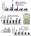
Figure 1
Figure 1. Muscle-specific PGC1α transgenic mice have increased brown/beige fat cells in the subcutaneous depot
QPCR against brown fat and thermogenic genes in epididymal fat, classical brown fat (BAT) (a) and subcutaneous, inguinal (b) fat depots in MCK-PGC1α transgenics or littermate controls. N=7 for each group, repeated in a separate cohort with similar results. c, representative immunohistochemistry against UCP1 in the inguinal depot from indicated mice. d, western blot against UCP1 in the inguinal fat depot (n=3 and repeated in an independent cohort with similar results). e, qPCR against indicated genes in adipocytes differentiated for 6 days from SVF cells. This was done in the presence of conditioned media from primary myocytes with forced expression of GFP or PGC1α (representative for 3 independent experiments). Data is presented as mean +/− SEM, and * p<0.05 compared to control group. Students T-test was used for single comparisons.
Figure 2
Figure 2. Fndc5 is induced with forced PGC1α expression or exercise, and turns on brown/beige fat gene expression
a, qPCR against indicated genes in skeletal muscle from MCK-PGC1α transgenic mice or littermate controls (n=7 from each group). b, qPCR against indicated genes in skeletal muscle from sedentary mice or mice given three weeks of free wheel running (n=10 from each group). Mice were rested for 12 hours prior to euthanization. c, mRNA expression levels from human muscle biopsies before and after 10 weeks of endurance exercise training (8 subjects included). All data points are normalized to baseline levels. d, SVF from the inguinal fat depot, differentiated into adipocytes for 6 days in the presence of saline or recombinant Fndc5 (20 nM), IL-15 (10μM) or VEGFβ (50μM). The graph show normalized mRNA levels of indicated genes. This experiment was repeated three times with similar results. For figure 2d, we performed one-way ANOVA tests where p<0.05 for the effect of Fndc5 on UCP-1 and Cidea expression. All other statistics were performed using students T-test and bar graphs are mean +/− SEM.
Figure 3
Figure 3. Fndc5 is a potent inducer of the brown/beige fat gene program
a, SVF from the inguinal fat depot was differentiated into adipocytes for 6 days in the presence of saline, recombinant Fndc5 (20 nM), or BMP-7 (3.3 μM). The graph show normalized mRNA levels for indicated genes. Similar results were obtained in more than 10 experiments with the fold induction of UCP1 between 7–500 fold. b, mRNA levels of UCP1 from inguinal-derived SVF treated with Fndc5 for 6 days at indicated doses. c, Clark electrode measurements of oxygen consumption in SVF from the inguinal fat depot, differentiated into adipocytes for 6 days in the presence of saline or recombinant Fndc5 (20 nM). Data is representative for three independent experiments and normalized to total cellular protein. d, qPCR of UCP1 mRNA from SVF, differentiated into adipocytes, and treated with Fndc5 or saline for 6 days followed by addition of forskolin for 8 hours. § indicates p<0.05 compared to forskolin treatment. e, qPCR of PPARα after 6 days of Fndc5 treatment (20 nM) during differentiation of primary SVF. f, SVF, differentiated into adipocytes, and treated with Fndc5 and/or GW6471 for 6 days. The graph shows qPCR of indicated genes, and § indicates p<0.05 compared to Fndc5 treatment. For 3d and f; combined one and two-way ANOVA was used. All other statistics were performed using students T-test and bar graphs are mean +/− SEM.
Figure 4
Figure 4. Fndc5 is proteolytically cleaved and secreted from cells
a, Schematic representation of the Fndc5 protein structure (top), flag-tagged Fndc5 protein (middle) and irisin (bottom). SP = signal peptide, H = hydrophobic domain, C = c-terminal domain, Flag = FLAG epiotope. b, 293 cells transfected with a vector expressing the c-terminal flag tagged Fndc5 (CTF-F5, third panel from a), followed by isolation of cell and culture media protein. Samples were adjusted for protein content and western blots were performed against the FLAG antigen (left) or Fndc5 (right). This was repeated in several experiments with similar results. Adjusting for volume (instead of protein content) also gave similar results. C = cell fraction and M = media fraction. c, 293 cells transfected with a vector expressing Fndc5-CTF, followed by isolation of cell and media protein. Respective protein fraction was then treated with PNGase F followed by western blot against Fndc5 after SDS-PAGE. d, representation of the full-length Fndc5 and the irisin fragment mapped by mass spectrometry (bold and underlined).
Figure 5
Figure 5. Detection of irisin in mouse and human plasma
a, Plasma from mice injected intravenously with adenoviral vectors expressing Fndc5 or GFP was subjected to western blot against Fndc5. b, western blot against irisin in plasma from muscle-specific PGC1α knockout (MKO) mice or control littermates (flox/flox). c, western blot against irisin in plasma from wild-type mice or two healthy human subjects (representative for 8 subjects analyzed identically). d, western blot against irisin in serum from control or three weeks exercised mice, followed by 12h rest. Bottom panel shows quantification of the bands. e, western blot analysis of irisin in plasma from human subjects before and after 10 weeks of endurance exercise. 8 subjects in total were analyzed; quantification after internal normalization is displayed in bottom panel. For all plasma analyses, samples were depleted for albumin/IgG, and deglycosylated using PNGase F. Arrow indicated irisin band. Data is presented as mean +/−SEM, and * p<0.05 compared to control group. Students T-test was used for single comparisons.
Figure 6
Figure 6. Irisin induces browning of white adipose tissues in vivo and protects against diet induced obesity and diabetes
a–c, Wild-type BALB/c mice were injected with 1010 GFP or Fndc5-expressing adenoviral particles intravenously (n=7 for each group). Animals were sacrificed after 10 days and inguinal/subcutaneous fat pads were collected and analyzed using qPCR analysis of indicated mRNAs (a) and western blot against UCP1 (b). c, representative images from immunohistochemistry against UCP1 in these mice. All results in a–c were repeated 2 times with similar results. d–g, C57/Bl6 mice fed a 60% kcal high fat diet for 20 weeks were intravenously injected with GFP or Fndc5-expressing adenovirus and all analyses were done 10 days thereafter (n=7 for both groups). d. oxygen consumption at day and night. e, body weights of 10 days after injection with indicated adenovirus in these mice. f, fasting plasma insulin measured using ELISA. g, intraperitoneal glucose tolerance test. h, mice were injected IP with 50μg of rabbit IgG or a rabbit anti-Fndc5 antibody and were either subjected to swimming for 7 days or kept sedentary (n=10 for all groups). Data displays mRNA expression levels from inguinal WAT. All data in d–j were performed at least twice in a separate mouse cohort with similar results. § p<0.05 compared to exercise + IgG. One-way ANOVA was used for statistics in h. All other statistics were performed using students T-test and bar graphs are mean +/− SEM.

Comment in

Similar articles

Cited by

References

    1. Puigserver P, et al. A cold-inducible coactivator of nuclear receptors linked to adaptive thermogenesis. Cell. 1998;92:829–839. - PubMed
    1. Handschin C, Spiegelman BM. The role of exercise and PGC1alpha in inflammation and chronic disease. Nature. 2008;454:463–469. - PMC - PubMed
    1. Sandri M, et al. PGC-1alpha protects skeletal muscle from atrophy by suppressing FoxO3 action and atrophy-specific gene transcription. Proc Natl Acad Sci U S A. 2006;103:16260–16265. - PMC - PubMed
    1. Wenz T, Rossi SG, Rotundo RL, Spiegelman BM, Moraes CT. Increased muscle PGC-1alpha expression protects from sarcopenia and metabolic disease during aging. Proc Natl Acad Sci U S A. 2009;106:20405–20410. - PMC - PubMed
    1. Xu X, et al. Exercise ameliorates high-fat diet-induced metabolic and vascular dysfunction, and increases adipocyte progenitor cell population in brown adipose tissue. Am J Physiol Regul Integr Comp Physiol. 2011;300:R1115–1125. - PMC - PubMed

Publication types

MeSH terms