Rejuvenation of regeneration in the aging central nervous system
- PMID: 22226359
- PMCID: PMC3714794
- DOI: 10.1016/j.stem.2011.11.019
Rejuvenation of regeneration in the aging central nervous system
Abstract
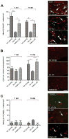
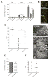
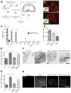
Remyelination is a regenerative process in the central nervous system (CNS) that produces new myelin sheaths from adult stem cells. The decline in remyelination that occurs with advancing age poses a significant barrier to therapy in the CNS, particularly for long-term demyelinating diseases such as multiple sclerosis (MS). Here we show that remyelination of experimentally induced demyelination is enhanced in old mice exposed to a youthful systemic milieu through heterochronic parabiosis. Restored remyelination in old animals involves recruitment to the repairing lesions of blood-derived monocytes from the young parabiotic partner, and preventing this recruitment partially inhibits rejuvenation of remyelination. These data suggest that enhanced remyelinating activity requires both youthful monocytes and other factors, and that remyelination-enhancing therapies targeting endogenous cells can be effective throughout life.
Copyright © 2012 Elsevier Inc. All rights reserved.
Figures


Similar articles
-
Myelin regeneration in multiple sclerosis: targeting endogenous stem cells.Neurotherapeutics. 2011 Oct;8(4):650-8. doi: 10.1007/s13311-011-0065-x. Neurotherapeutics. 2011. PMID: 21904791 Free PMC article. Review.
-
A Subpopulation of Foxj1-Expressing, Nonmyelinating Schwann Cells of the Peripheral Nervous System Contribute to Schwann Cell Remyelination in the Central Nervous System.J Neurosci. 2018 Oct 24;38(43):9228-9239. doi: 10.1523/JNEUROSCI.0585-18.2018. Epub 2018 Sep 18. J Neurosci. 2018. PMID: 30228229 Free PMC article.
-
Small-molecule-induced epigenetic rejuvenation promotes SREBP condensation and overcomes barriers to CNS myelin regeneration.Cell. 2024 May 9;187(10):2465-2484.e22. doi: 10.1016/j.cell.2024.04.005. Epub 2024 May 2. Cell. 2024. PMID: 38701782
-
Retinoid X receptor activation reverses age-related deficiencies in myelin debris phagocytosis and remyelination.Brain. 2015 Dec;138(Pt 12):3581-97. doi: 10.1093/brain/awv289. Epub 2015 Oct 12. Brain. 2015. PMID: 26463675 Free PMC article.
-
The role of oligodendrocytes and oligodendrocyte progenitors in CNS remyelination.Adv Exp Med Biol. 1999;468:183-97. doi: 10.1007/978-1-4615-4685-6_15. Adv Exp Med Biol. 1999. PMID: 10635029 Review.
Cited by
-
[Mechanisms underlying remyelination with special focus on demyelination models of multiple sclerosis].Zhejiang Da Xue Xue Bao Yi Xue Ban. 2020 Aug 25;49(4):524-530. doi: 10.3785/j.issn.1008-9292.2020.08.12. Zhejiang Da Xue Xue Bao Yi Xue Ban. 2020. PMID: 32985167 Free PMC article. Review. Chinese.
-
Astrocyte-derived clusterin disrupts glial physiology to obstruct remyelination in mouse models of demyelinating diseases.Nat Commun. 2024 Sep 6;15(1):7791. doi: 10.1038/s41467-024-52142-7. Nat Commun. 2024. PMID: 39242637 Free PMC article.
-
Tamoxifen accelerates the repair of demyelinated lesions in the central nervous system.Sci Rep. 2016 Aug 24;6:31599. doi: 10.1038/srep31599. Sci Rep. 2016. PMID: 27554391 Free PMC article.
-
Susceptibility to acute cognitive dysfunction in aged mice is underpinned by reduced white matter integrity and microgliosis.Commun Biol. 2024 Jan 16;7(1):105. doi: 10.1038/s42003-023-05662-9. Commun Biol. 2024. PMID: 38228820 Free PMC article.
-
The Immune System in Tissue Environments Regaining Homeostasis after Injury: Is "Inflammation" Always Inflammation?Mediators Inflamm. 2016;2016:2856213. doi: 10.1155/2016/2856213. Epub 2016 Aug 11. Mediators Inflamm. 2016. PMID: 27597803 Free PMC article. Review.
References
-
- Ajami B, Bennett JL, Krieger C, McNagny KM, Rossi FMV. Infiltrating monocytes trigger EAE progression, but do not contribute to the resident microglia pool. Nature Neurosci. 2011;14:1142–1149. - PubMed
-
- Arnett HA, Fancy SPJ, Alberta JA, Zhao C, Plant SR, Raine CS, Rowitch DH, Franklin RJM, Stiles CD. The bHLH transcription factor Olig1 is required for repair of demyelinated lesions in the CNS. Science. 2004;306:2111–2115. - PubMed
-
- Baer AS, Syed YA, Kang SU, Mitteregger D, Vig R, ffrench-Constant C, Franklin RJM, Altmann F, Lubec G, Kotter MR. Myelin-mediated inhibition of oligodendrocyte precursor differentiation can be overcome by pharmacological modulation of Fyn-RhoA and protein kinase C signalling. Brain. 2009;132:465–481. - PMC - PubMed
Publication types
MeSH terms
Grants and funding
LinkOut - more resources
Full Text Sources
Other Literature Sources
Medical
Molecular Biology Databases

