

The role of PAS kinase in PASsing the glucose signal
- PMID: 22219681
- PMCID: PMC3247726
- DOI: 10.3390/s100605668
The role of PAS kinase in PASsing the glucose signal
Abstract
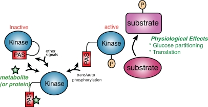
PAS kinase is an evolutionarily conserved nutrient responsive protein kinase that regulates glucose homeostasis. Mammalian PAS kinase is activated by glucose in pancreatic beta cells, and knockout mice are protected from obesity, liver triglyceride accumulation, and insulin resistance when fed a high-fat diet. Yeast PAS kinase is regulated by both carbon source and cell integrity stress and stimulates the partitioning of glucose toward structural carbohydrate biosynthesis. In our current model for PAS kinase regulation, a small molecule metabolite binds the sensory PAS domain and activates the enzyme. Although bona fide PAS kinase substrates are scarce, in vitro substrate searches provide putative targets for exploration.
Keywords: PAS domain; PAS kinase; PASKIN; glucose sensor; metabolic syndrome; protein phosphorylation.
Figures

Similar articles
-
Yeast PAS kinase coordinates glucose partitioning in response to metabolic and cell integrity signaling.EMBO J. 2007 Nov 28;26(23):4824-30. doi: 10.1038/sj.emboj.7601914. Epub 2007 Nov 8. EMBO J. 2007. PMID: 17989693 Free PMC article.
-
PAS kinase: a nutrient sensing regulator of glucose homeostasis.IUBMB Life. 2013 Nov;65(11):921-9. doi: 10.1002/iub.1219. Epub 2013 Nov 7. IUBMB Life. 2013. PMID: 24265199 Free PMC article. Review.
-
Regulation and function of yeast PAS kinase: a role in the maintenance of cellular integrity.Cell Cycle. 2009 Jun 15;8(12):1824-32. doi: 10.4161/cc.8.12.8799. Epub 2009 Jun 20. Cell Cycle. 2009. PMID: 19440050 Free PMC article.
-
The PAS-domain kinase PASKIN: a new sensor in energy homeostasis.Cell Mol Life Sci. 2009 Mar;66(5):876-83. doi: 10.1007/s00018-009-8699-0. Cell Mol Life Sci. 2009. PMID: 19189049 Free PMC article. Review.
-
Per-Arnt-Sim Kinase (PASK): An Emerging Regulator of Mammalian Glucose and Lipid Metabolism.Nutrients. 2015 Sep 7;7(9):7437-50. doi: 10.3390/nu7095347. Nutrients. 2015. PMID: 26371032 Free PMC article. Review.
Cited by
-
Preventing Oxidative Stress in the Liver: An Opportunity for GLP-1 and/or PASK.Antioxidants (Basel). 2021 Dec 20;10(12):2028. doi: 10.3390/antiox10122028. Antioxidants (Basel). 2021. PMID: 34943132 Free PMC article. Review.
-
Energy metabolism regulates clathrin adaptors at the trans-Golgi network and endosomes.Mol Biol Cell. 2013 Mar;24(6):832-47. doi: 10.1091/mbc.E12-10-0750. Epub 2013 Jan 23. Mol Biol Cell. 2013. PMID: 23345590 Free PMC article.
-
Protein Kinases in Obesity, and the Kinase-Targeted Therapy.Adv Exp Med Biol. 2024;1460:199-229. doi: 10.1007/978-3-031-63657-8_7. Adv Exp Med Biol. 2024. PMID: 39287853 Review.
-
Validation of PAS Kinase, a Regulator of Hepatic Fatty Acid and Triglyceride Synthesis, as a Therapeutic Target for Nonalcoholic Steatohepatitis.Hepatol Commun. 2020 Mar 24;4(5):696-707. doi: 10.1002/hep4.1498. eCollection 2020 May. Hepatol Commun. 2020. PMID: 32363320 Free PMC article.
-
A comprehensive protein-protein interactome for yeast PAS kinase 1 reveals direct inhibition of respiration through the phosphorylation of Cbf1.Mol Biol Cell. 2014 Jul 15;25(14):2199-215. doi: 10.1091/mbc.E13-10-0631. Epub 2014 May 21. Mol Biol Cell. 2014. PMID: 24850888 Free PMC article.
References
-
- Warburg O. On the origin of cancer cells. Science. 1956;123:309–314. - PubMed
-
- Hardie D.G. AMP-activated/SNF1 protein kinases: Conserved guardians of cellular energy. Nat. Rev. Mol. Cell Biol. 2007;8:774–785. - PubMed
-
- Steinberg G.R., Kemp B.E. AMPK in health and disease. Physiol. Rev. 2009;89:1025–1078. - PubMed
-
- Martin D.E., Hall M.N. The expanding TOR signaling network. Curr. Opin. Cell Biol. 2005;17:158–166. - PubMed
Publication types
MeSH terms
Substances
LinkOut - more resources
Full Text Sources
Other Literature Sources
Molecular Biology Databases
