. 2011;6(12):e29584.
doi: 10.1371/journal.pone.0029584. Epub 2011 Dec 28.

Oligomerization of ZFYVE27 (Protrudin) is necessary to promote neurite extension

Affiliations

Oligomerization of ZFYVE27 (Protrudin) is necessary to promote neurite extension

D V Krishna Pantakani et al. PLoS One. 2011.

Abstract

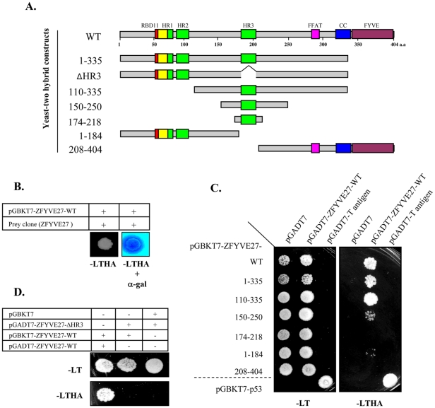
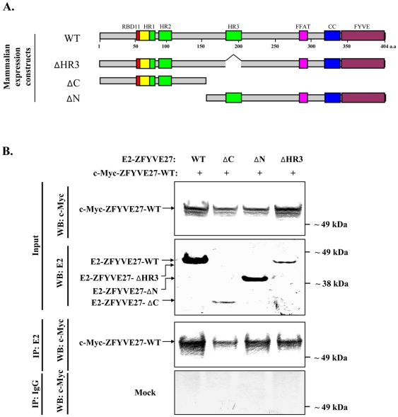
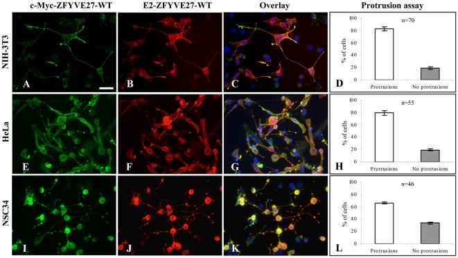
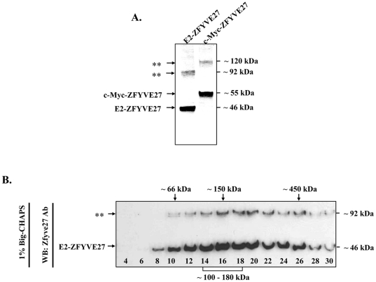
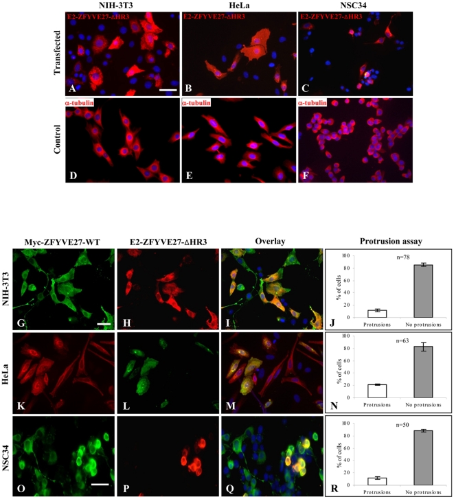
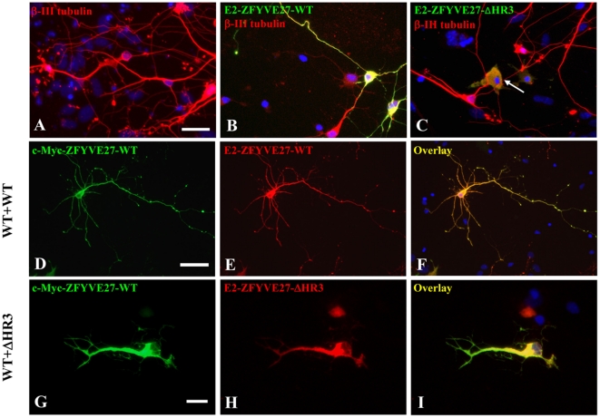
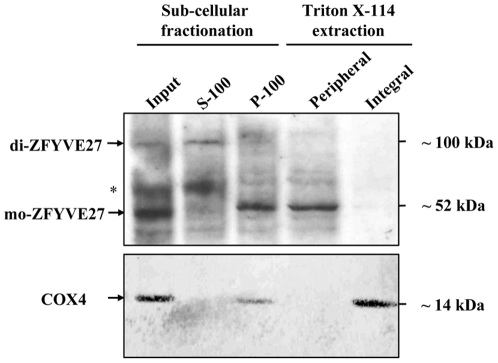
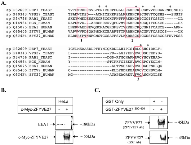
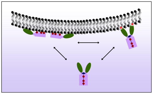
ZFYVE27 (Protrudin) was originally identified as an interacting partner of spastin, which is most frequently mutated in hereditary spastic paraplegia. ZFYVE27 is a novel member of FYVE family, which is implicated in the formation of neurite extensions by promoting directional membrane trafficking in neurons. Now, through a yeast two-hybrid screen, we have identified that ZFYVE27 interacts with itself and the core interaction region resides within the third hydrophobic region (HR3) of the protein. We confirmed the ZFYVE27's self-interaction in the mammalian cells by co-immunoprecipitation and co-localization studies. To decipher the oligomeric nature of ZFYVE27, we performed sucrose gradient centrifugation and showed that ZFYVE27 oligomerizes into dimer/tetramer forms. Sub-cellular fractionation and Triton X-114 membrane phase separation analysis indicated that ZFYVE27 is a peripheral membrane protein. Furthermore, ZFYVE27 also binds to phosphatidylinositol 3-phosphate lipid moiety. Interestingly, cells expressing ZFYVE27(ΔHR3) failed to produce protrusions instead caused swelling of cell soma. When ZFYVE27(ΔHR3) was co-expressed with wild-type ZFYVE27 (ZFYVE27(WT)), it exerted a dominant negative effect on ZFYVE27(WT) as the cells co-expressing both proteins were also unable to induce protrusions and showed cytoplasmic swelling. Altogether, it is evident that a functionally active form of oligomer is crucial for ZFYVE27 ability to promote neurite extensions.


Conflict of interest statement

Competing Interests: The authors have declared that no competing interests exist.

Figures

Figure 1
Figure 1. Yeast two-hybrid (Y2H) screen showing self-interaction of ZFYVE27.
(A) Schematic diagram showing the structural domains of ZFYVE27 and delineation of various deletion constructs of ZFYVE27. ZFYVE27 contains Rab11 binding domain RBD11 (red) at its N-terminus and FYVE domain (purple) at C-terminus. The three hydrophobic regions (HR) are depicted in green and the overlapping region of HR1 region with the RBD11 motif is highlighted in yellow. The FFAT motif (pink) and the coiled-coil region (blue) mediate interaction with VAP-A protein. The generated deletion constructs for Y2H analysis are depicted thereof. (B) Activation of GAL4 reporter genes by interaction of ZFYVE27 with the prey clone (ZFYVE27) in the Y2H screen. A robust growth of yeast strain AH109 was observed on the nutritional selection medium -LTHA (lacking leucine, tryptophan, histidine and adenine) and also was positive for the α-galactosidase (α-gal) activity. (C) Determination of the core interaction region of ZFYVE27, which mediate self-interaction by direct-Y2H. The indicated deletion constructs of ZFYVE27 were fused with DNA binding domain of GAL4 and evaluated for their ability to interact with full-length ZFYVE27 fused to activation domain of GAL4 in Y2H experiments. The interaction between p53 and T-antigen was used as a positive control in the Y2H assay. (D) Evaluation of ZFYVE27-ΔHR3 (deletion of HR3 in ZFYVE271-335 construct) interaction with full-length ZFYVE27.
Figure 2
Figure 2. Co-immunoprecipitation assay to validate interaction of ZFYVE27 in mammalian cells.
(A) Schematic representation of full-length and deletion constructs of ZFYVE27 for mammalian expression studies. (B) The full-length ZFYVE27 fused with c-Myc epitope tag was used for validation of its interaction with E2 tagged ZFYVE27, N-terminus (ΔC), C-terminus (ΔN) as well as with ZFYVE27 lacking the third hydrophobic region (ΔHR3) in NIH-3T3 cells. The cells were transiently transfected with respective constructs and subsequently co-immunoprecipitation was performed. The cell lysates were subjected to immunoprecipitation with E2 tag antibody and the resulting immunoprecipitants were analyzed in the immunoblot with c-Myc tag antibody (Co-IP: E2; WB: c-Myc). A portion of the cell lysates (input) was also subjected to immunoblot with either c-Myc (WB: c-Myc) or E2 (WB: E2) tag antibodies to verify the protein expression of the indicated constructs. For mock experiments, the cell lysates were precipitated with non-specific IgG and subsequently analyzed by immunoblotting (as described above).
Figure 3
Figure 3. Co-localization of ZFYVE27 monomers in various cell lines.
(A–C) The c-Myc-ZFYVE27WT and E2-ZFYVE27WT constructs were used to analyze the co-localization between ZFYVE27 monomers. Overexpression of both constructs in NIH-3T3 cell line revealed a pronounced formation of protrusions from cell soma and showed strong co-localization of ZFYVE27 monomers in the protruded structures. (E–G) A similar protrusions and co-localization was also observed in HeLa cells. (I–K) Overexpression of ZFYVE27WT monomers in the NSC34 (a mouse embryonic spinal cord–neuroblastoma hybrid cell line) resulted in extensive neurite formation with bead-like structures. The ZFYVE27WT monomers showed strongest co-localization in the neurite-beads. (D, H, and L) Protrusion assay → Quantification of proportion of the cells, doubly transfected (E2-ZFYVE27WT and c-Myc-ZFYVE27WT) showing protrusions versus showing no protrusions. Scale bars – 50 µm (A–C, E–G and I–K); n, number of cells analyzed. Error bars indicate the standard deviation (SD) of protrusion assessment by two independent observers.
Figure 4
Figure 4. ZFYVE27 assembles into SDS-sensitive and –resistant dimer/tetramer.
(A) The protein lysates from NIH-3T3 cells overexpressing either E2-ZFYV27WT or c-Myc-ZFYVE27WT were subjected to immunoblot with ZFYVE27 specific antibody. Asterisks denote the formation of SDS-resistant dimers corresponding to E2-ZFYV27WT (∼ 92 kDa) and c-Myc-ZFYVE27WT (∼ 120 kDa). Note: In pCS2-myc vector, six c-Myc epitope tags are fused in tandem therefore resultant c-Myc-ZFYVE27WT protein is larger in size (∼55 kDa) as compared to E2-ZFYVE27WT protein (∼46 kDa). (B) The NIH-3T3 cells transiently transfected with E2-ZFYV27WT were solubilized in 1% Big-CHAPS and size-fractionated by 5-30% sucrose gradient centrifugation. The resultant fractions were analyzed by immunoblot with ZFYVE27 antibody; asterisks denote the formation of SDS-resistant dimers. The mobility of molecular weight markers in the sucrose gradient are indicated on the top.
Figure 5
Figure 5. The truncated ZFYVE27ΔHR3 deters the ability of wild-type ZFYVE27 to promote directional membrane trafficking.
(A–C) Overexpression of the mutant E2-ZFYVE27ΔHR3 (truncated ZFYVE27) failed to induce protrusions in all the analyzed cell lines (NIH-3T3, HeLa and NSC34) and caused cytoplasmic swelling with accumulation of protein either along endoplasmic reticulum or cytoskeleton. (D–F) Staining of control untransfected NIH-3T3, HeLa and NSC34 cells with α-tubulin showed no abnormal morphology. (G–R) Co-expression of both the c-Myc-ZFYVE27WT and mutant E2-ZFYVE27ΔHR3 in various cell lines (NIH-3T3, HeLa and NSC34) showed the co-localization of both forms of ZFYVE27 in the cytosol. Notably, majority of cells co-expressing wild-type as well as ZFYVE27ΔHR3 failed to produce protrusions but rather caused swelling of the cytosol (I, M, and Q). Protrusion assay → Quantification of proportion of the cells, doubly transfected (E2-ZFYVE27ΔHR3 and c-Myc-ZFYVE27WT) showing cytoplasmic swelling (no protrusions) versus cells producing protrusions (J, N and R). Scale bars – 50 µm (A–C, D–F, G–I and K–M), 10 µm (O–Q); n, number of cells analyzed. Error bars indicate the standard deviation (SD) of protrusion assessment by two independent observers.
Figure 6
Figure 6. The truncated ZFYVE27ΔHR3 impairs the ability of wild-type ZFYVE27 to promote neurites in primary neuronal cells.
(A) Control primary neurons stained for β-III tubulin, a neuronal marker. Overexpression of E2-ZFYVE27WT (B) and E2-ZFYVE27ΔHR3 (C) in primary neuronal cells. Arrow in (C) showing the swelling of the cell soma of a neuronal cell expressing E2-ZFYVE27ΔHR3. (D–F) Overexpression of both wild-type ZFYVE27 (c-Myc-ZFYVE27WT and E2-ZFYVE27WT) proteins revealed an enhanced neurites formation from the cell soma and showed co-localization of ZFYVE27 monomers. (G–I) Co-expression of both the c-Myc-ZFYVE27WT and mutant E2-ZFYVE27ΔHR3 in primary neurons showed the co-localization of both forms of ZFYVE27 mostly in cell soma and the neuron failed to produce normal length axon as well as /dendrites. Scale bars – 20 µm (A–C), 50 µm (D–F), 10 µm (G–I).
Figure 7
Figure 7. ZFYVE27 is a peripheral membrane protein.
Sub-cellular fractionation was performed with NSC34 cell extract. The cytosolic fraction (S-100) and the membrane fraction (P-100) were analyzed by immunoblot with ZFYVE27 antibody. Asterisks denote the non-specific band. The monomeric ZFYVE27 (mo-ZFYVE27) and SDS-resistant dimeric ZFYVE27 (di-ZFYVE27) are indicated. The membrane fraction (P-100) was subjected to Triton X-114 phase separation and equal volumes of aqueous phase (peripheral) and detergent phase (integral) fractions were analyzed by immunoblotting. The blot was stripped and re-probed with COX4 antibody, a marker for the integral membrane protein.
Figure 8
Figure 8. ZFYVE27 a novel member of FYVE family, which show binding affinity towards PtdIns3P.
(A) Alignment of the FYVE domain of ZFYVE27 (ZFY27) with other FYVE family proteins; EEA1, ZFYVE9 (ZFYV9), HGS/HRS and yeast FAB1, PEP7, VPS27. The red boxes are highlighting the conserved WXXD motif (1), R(R/K)HHCR motif (2) and RVC motif (3). The conserved cysteine residues which co-ordinates the zinc ion binding were marked with asterisks. (B) The liposomal binding assay shows specific binding of c-Myc-ZFYVE27WT to the PolyPIPosomes consisting of PtdIns3P. As a positive control, we also detected the binding of endogenous EEA1 to the PolyPIPosomes. (C) The recombinant GST-ZFYVE27300-404 protein also showed binding affinity to PtdIns3P. As a negative control, GST (alone) protein was used in the liposomal binding assay.
Figure 9
Figure 9. Schematic diagram illustrating a hypothetical model of ZFYVE27 dimerization.
The monomeric form of ZFYVE27 can associate with the membrane through hydrophobic interactions with its three hydrophobic regions, namely: HR1-3 motifs (maroon filled circles), which might enable the monomers to embed as a peripheral membrane protein. In the cytoplasm, ZFYVE27 can exist as a soluble dimer, which could possibly be formed and stabilized by the self-interaction of three HRs and the coiled-coil domain (blue diamond) between two monomers. The dimerization of ZFYVE27 could most likely occur on the membrane.

Similar articles

Cited by

References

    1. da Silva JS, Dotti CG. Breaking the neuronal sphere: regulation of the actin cytoskeleton in neuritogenesis. Nat Rev Neurosci. 2002;3:694–704. - PubMed
    1. Horton AC, Ehlers MD. Neuronal polarity and trafficking. Neuron. 2003;40:277–295. - PubMed
    1. Dent EW, Callaway JL, Szebenyi G, Baas PW, Kalil K. Reorganization and movement of microtubules in axonal growth cones and developing interstitial branches. J Neurosci. 1999;19:8894–8908. - PMC - PubMed
    1. Wang L, Brown A. Rapid movement of microtubules in axons. Curr Biol. 2002;12:1496–1501. - PubMed
    1. Yu W, Qiang L, Solowska JM, Karabay A, Korulu S, et al. The microtubule-severing proteins spastin and katanin participate differently in the formation of axonal branches. Mol Biol Cell. 2008;19:1485–1498. - PMC - PubMed

Publication types