. 2011 Dec;7(12):e1002455.
doi: 10.1371/journal.ppat.1002455. Epub 2011 Dec 22.

Epstein-Barr virus evades CD4+ T cell responses in lytic cycle through BZLF1-mediated downregulation of CD74 and the cooperation of vBcl-2

Affiliations

Epstein-Barr virus evades CD4+ T cell responses in lytic cycle through BZLF1-mediated downregulation of CD74 and the cooperation of vBcl-2

Jianmin Zuo et al. PLoS Pathog. 2011 Dec.

Abstract

Evasion of immune T cell responses is crucial for viruses to establish persistence in the infected host. Immune evasion mechanisms of Epstein-Barr virus (EBV) in the context of MHC-I antigen presentation have been well studied. In contrast, viral interference with MHC-II antigen presentation is less well understood, not only for EBV but also for other persistent viruses. Here we show that the EBV encoded BZLF1 can interfere with recognition by immune CD4+ effector T cells. This impaired T cell recognition occurred in the absence of a reduction in the expression of surface MHC-II, but correlated with a marked downregulation of surface CD74 on the target cells. Furthermore, impaired CD4+ T cell recognition was also observed with target cells where CD74 expression was downregulated by shRNA-mediated inhibition. BZLF1 downregulated surface CD74 via a post-transcriptional mechanism distinct from its previously reported effect on the CIITA promoter. In addition to being a chaperone for MHC-II αβ dimers, CD74 also functions as a surface receptor for macrophage Migration Inhibitory Factor and enhances cell survival through transcriptional upregulation of Bcl-2 family members. The immune-evasion function of BZLF1 therefore comes at a cost of induced toxicity. However, during EBV lytic cycle induced by BZLF1 expression, this toxicity can be overcome by expression of the vBcl-2, BHRF1, at an early stage of lytic infection. We conclude that by inhibiting apoptosis, the vBcl-2 not only maintains cell viability to allow sufficient time for synthesis and accumulation of infectious virus progeny, but also enables BZLF1 to effect its immune evasion function.


Conflict of interest statement

The authors have declared that no competing interests exist.

Figures

Figure 1
Figure 1. CD4+ T cell recognition is impaired in the BZLF1-expressing LCLs.
(A) The expression plasmid pRTS-CD2-BZLF1 carries a bidirectional DOX regulatable promoter, BI-Tet, which drives the expression of BZLF1 together with a non-functional neuronal growth factor receptor (NGFR) and green fluorescent protein (GFP) as markers of DOX-induced expression. (B) BZLF1KO-LCLs transfected with pRTS-CD2-BZLF1 or pRTS-CD2-control vector were treated with DOX for 24 h. The induced plasmid-containing cells were separated using MACSelect LNGFR MicroBeads. Both the unlabeled cells and the labeled cells were collected, and then analyzed by flow cytometry. The flow-through cells were NGFR/GFP, indicating that they lacked the transfected plasmid and/or DOX treatment failed to induce expression from pRTS-CD2-BZLF1 (dotted line, top histogram). The bound and eluted cells were NGFR+/GFP+, which indicated that expression of BZLF1 from pRTS-CD2-BZLF1 had been induced (dashed line, bottom histogram). (C) These sorted cells were pulsed with or without concentrated EBV B95.8 virus particles and then were co-cultured with ‘LEK’ CD4+ effector T cells (specific for a BXLF2-derived peptide), for a further 18 h before assaying T cell recognition by IFN-γ ELISA. (D) Unpulsed targets were also assayed with ‘SNP’ CD4+ effector T cells (specific for a EBNA1-derived peptide). All results are expressed as IFN-γ release in pg/ml, and error bars indicate standard deviation of triplicate cultures. (E) The pRTS-CD2-control and pRTS-CD2-BZLF1 transfected LCLs were analyzed, before and after DOX induction, by SDS-PAGE and immuno-blotting with antibodies specific for BZLF1, BMRF1, BALF2, EBNA1 or calregulin as a loading control.
Figure 2
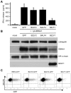
Figure 2. Effect of BZLF1 on CD4+ T cell recognition of MHC-II presented antigen.
MJS-DRB5*01 cells were co-transfected with pCDNA-cytoEBNA1 expression vector together with IRES-GFP (GFP), BZLF2-GFP (BZLF2), BALF4-GFP (BALF4) or BZLF1-GFP (BZLF1) expression plasmids. (A) At 24 h post-transfection, the cells were used as targets for ‘SNP’ CD4+ effector T cells (specific for a EBNA1-derived peptide) as in Figure 1. Error bars indicate standard deviation of five independent experiments. (B) Total cell lysates of target cells were analyzed by immunoblotting using antibodies specific for EBNA1, DRα chain, BZLF1 or calregulin as a loading control. (C) Viable transfected target cells stained with PE-conjugated anti-DR, analyzed by flow cytometry.
Figure 3
Figure 3. BZLF1 downregulates Bcl-2 family members.
(A) EBV-negative Akata-A3 cells transfected with pRTS-CD2-control (solid line) or pRTS-CD2-BZLF1 (dotted line) were induced with DOX and analyzed by flow cytometry at the indicated times for the percentage of cells expressing GFP. Error bars indicate standard deviation of three independent induction experiments. (B) At 48 h after DOX induction, the GFP+ cells from control and BZLF1-transfected cultures were purified on a Mo-Flow cell sorter. The BZLF1 expressing Akata-A3 cell lysate was serially diluted with Akata-A3 cell lysate to give equivalent BZLF1 content of 100%, 50%, 10%, 2%, 1% or 0% and was analyzed by SDS-PAGE alongside a cell lysate from a spontaneous lytic LCL containing 5% BZLF1+ cells. The samples were immunoblotted with antibodies specific for BZLF1 or calregulin as a loading control. (C) Total cell lysates of the purified GFP+ populations from control- and BZLF1-transfected cultures were analyzed by SDS-PAGE and immunoblotting with antibodies specific for BZLF1, Bcl-2, Bcl-xl, or calregulin as a loading control. (D) Bcl-2 and Bcl-xl transcripts in total RNA isolated from induced and sorted cells were measured by QRT-PCR assay, which was normalized to measured GAPDH transcripts.
Figure 4
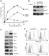
Figure 4. BHRF1 counteracts BZLF1 toxicity.
(A) MJS cells transfected with control IRES-GFP (solid line), BZLF1-GFP (dotted line) or BZLF1-GFP together with BHRF1 plasmids (dashed line) were analyzed by flow cytometry at the indicated times for the percentage of cells expressing GFP. Error bars indicate the standard deviation of three independent transfections. (B) The cell lysates of transfected MJS cells from two independent transfections were analyzed by SDS-PAGE alongside lysates from a spontaneously lytic LCL. The samples were immuno-blotted with antibodies specific for BZLF1, BHRF1 or calregulin as a loading control. The percentage of BZLF1+ cells in each lysate is indicated above each lane of the blot. (C) MJS cells cotransfected with BHRF1 and either IRES-GFP (control) or BZLF1-GFP (BZLF1) expression plasmids were sorted for GFP on a Mo-Flow cell sorter at 48 h post-transfection. Total cell lysates of GFP+ transfected cells were analyzed by immuno-blotting using antibodies specific for BZLF1, Bcl-2, Bcl-xl, DRα chain, CD74, or calregulin as a loading control. (D) MJS cells cotransfected with BHRF1 and either IRES-GFP or BZLF1-GFP expression plasmids were stained with PE-conjugated anti-DR or with CD74 MAb followed by PE conjugated anti-mouse IgG2a antibody, then analyzed by flow cytometry. Histograms show the surface MHC-II DR or CD74 expression on GFP cells (solid line) and GFP+ cells (dashed line). The shaded histogram indicates isotype control staining.
Figure 5
Figure 5. Downregulation of CD74, and not MHC-II DR, is a consistent effect of BZLF1.
(A) EBV-loss Akata-A3 cells transfected with pRTS-CD2-BZLF1 or pRTS-CD2-control were induced with DOX for 24 h, then stained for MHC-II DR or CD74 as in Figure 4D. Histograms show the surface MHC-II DR and CD74 expression on GFP+ cells from pRTS-CD2-control transfection (solid line) and pRTS-CD2-BZLF1 transfection (dashed line). The shaded histogram is the isotype control staining. (B) An LCL with a subpopulation of spontaneously lytic cells was first stained for surface MHC-II DR or CD74 then fixed, permeabilized and stained with anti-BZLF1 followed by FITC-conjugated anti-mouse IgG1. Histograms show the surface MHC-II DR or CD74 expression on latent BZLF1 cells (solid line) and lytic BZLF1+ cells (dashed line). The shaded histogram is the isotype control staining. (C) The same LCL with a subpopulation of spontaneously lytic cells was first stained for surface MHC-II DR or CD74 as before, then were fixed, permeabilized and stained with anti-VCA followed by FITC-conjugated anti-mouse IgG1. Histograms show the surface MHC-II DR or CD74 expression on VCA cells (solid line) and late lytic VCA+ cells (dashed line). The shaded histogram is the isotype control staining.
Figure 6
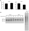
Figure 6. BZLF1 downregulates CD74 post-translationally.
(A) DRα and CD74 transcripts in total RNA isolated from DOX-induced and anti-NGFR bead-sorted control or BZLF1 transfected Akata-A3 cells, were measured by qRT-PCR assay, which was normalized to measured GAPDH transcripts. (B) Dox induced and anti-NGFR bead-sorted control or BZLF1 transfected Akata-A3 cells were metabolically labeled for 15 min with 35S-methionine and chased for the indicated time periods. After lysis in NP-40 detergent buffer, samples were immunoprecipitated with mouse anti-CD74. Samples were separated by 12% SDS/PAGE gel, dried and exposed to autoradiography (left panel). Aliquots of the whole cell lysates before chasing were analyzed (right panel) and served as a loading control.
Figure 7
Figure 7. Inhibition of CD4+ T cell recognition by BZLF1 is retained when toxicity is reversed by BHRF1.
MJS-DRB5*01 cells were co-transfected with a cytoEBNA1 expression vector and IRES-GFP (control) or BZLF1-GFP (BZLF1) expression plasmids, without (A) or with (B) a BHRF1 expression plasmid. At 24 hr post-transfection, the cells were assayed for recognition by ‘SNP’ CD4+ effector T cells (specific for a EBNA1) as in Figure 2A. Error bars for the IFN-γ release in the histograms indicate standard deviation of triplicate cultures. Total cell lysates of the target cell transfections, were analyzed by immunoblotting with antibodies to EBNA1, BZLF1, BHRF1 or calregulin as a loading control.
Figure 8
Figure 8. Downregulation of CD74 is sufficient to impair CD4+ T cell recognition.
(A) MJS-BHRF1 cells were transduced with control shRNA or CD74 shRNA lentivirus and selected with puromycin. The stable cell lines were stained with PE-conjugated anti-DR or with PE-conjugated anti-CD74, then analyzed by flow cytometry. Histograms show the surface MHC-II DR or CD74 expression on control shRNA cells (solid line) and CD74 shRNA cells (dashed line). The shaded histogram indicates isotype control staining. (B) The control shRNA and CD74 shRNA expressing MJS-BHRF1 cells were co-transfected with a cytoEBNA1 expression vector and DRB5*01 β chain expression plasmids. At 24 h post-transfection, the cells were assayed for recognition by ‘SNP’ CD4+ effector T cells specific for a EBNA1 (upper graph). Replicate samples of transfected cells were pulsed with ‘SNP’ peptide to serve as a control (lower graph). Error bars for the IFN-γ release in the histograms indicate standard deviation of triplicate cultures. (C) Total cell lysates of the MJS-BHRF1 target cell transfections, were analyzed by immuno-blotting with antibodies to BHRF1, CD74, DRα, EBNA1, or calregulin as a loading control. (D) LCLs transformed with B95.8 EBV were transduced with control shRNA or CD74 shRNA lentivirus and selected with puromycin. The stable cell lines were stained with PE-conjugated CD74, then analyzed by flow cytometry. Histograms show the surface CD74 expression on control shRNA cells (solid line) and CD74 shRNA cells (dashed line), with isotype control antibody background staining shaded grey. (E) Control shRNA and CD74 shRNA expressing LCLs were assayed for recognition by EBNA1-specific ‘SNP’ CD4+ effector T cells (left graph) and ‘HPV’ CD8+ effector T cells (right graph).

Similar articles

Cited by

References

    1. Rickinson AB, Kieff E. Epstein-Barr virus. In: Knipe DM, Howley PM, editors. Fields Virology. Philadelphia: Walters Kluwer/Lippincott, Williams & Wilkins; 2007. pp. 2655–2700.
    1. Rowe M, Glaunsinger B, van Leeuwen D, Zuo J, Sweetman D, et al. Host shutoff during productive Epstein-Barr virus infection is mediated by BGLF5 and may contribute to immune evasion. Proc Natl Acad Sci U S A. 2007;104:3366–3371. - PMC - PubMed
    1. Zuo J, Thomas W, van Leeuwen D, Middeldorp JM, Wiertz EJ, et al. The DNase of gammaherpesviruses impairs recognition by virus-specific CD8+ T cells through an additional host shutoff function. J Virol. 2008;82:2385–2393. - PMC - PubMed
    1. Hislop AD, Ressing ME, van Leeuwen D, Pudney VA, Horst D, et al. A CD8+ T cell immune evasion protein specific to Epstein-Barr virus and its close relatives in Old World primates. J Exp Med. 2007;204:1863–1873. - PMC - PubMed
    1. Zuo J, Currin A, Griffin BD, Shannon-Lowe C, Thomas WA, et al. The Epstein-Barr virus G-protein-coupled receptor contributes to immune evasion by targeting MHC class I molecules for degradation. PLoS Pathog. 2009;5:e1000255. - PMC - PubMed

Publication types

MeSH terms

Substances