. 2012 Jan 24;109(4):E177-86.
doi: 10.1073/pnas.1119296109. Epub 2011 Dec 27.

TANK-binding kinase 1 (TBK1) controls cell survival through PAI-2/serpinB2 and transglutaminase 2

Affiliations

TANK-binding kinase 1 (TBK1) controls cell survival through PAI-2/serpinB2 and transglutaminase 2

Mireille Delhase et al. Proc Natl Acad Sci U S A. .

Erratum in

  • Proc Natl Acad Sci U S A. 2012 Mar 13;109(11):4332–5

Abstract

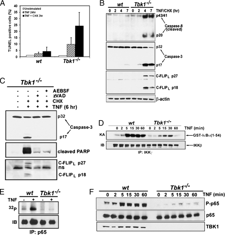
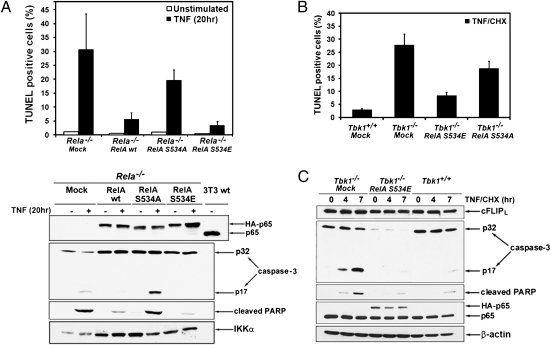
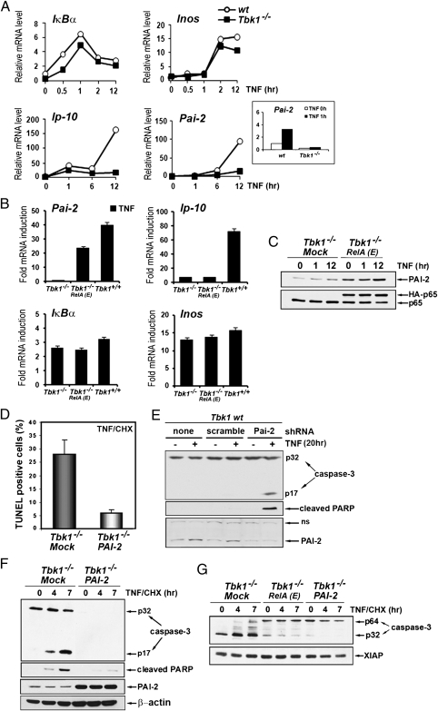
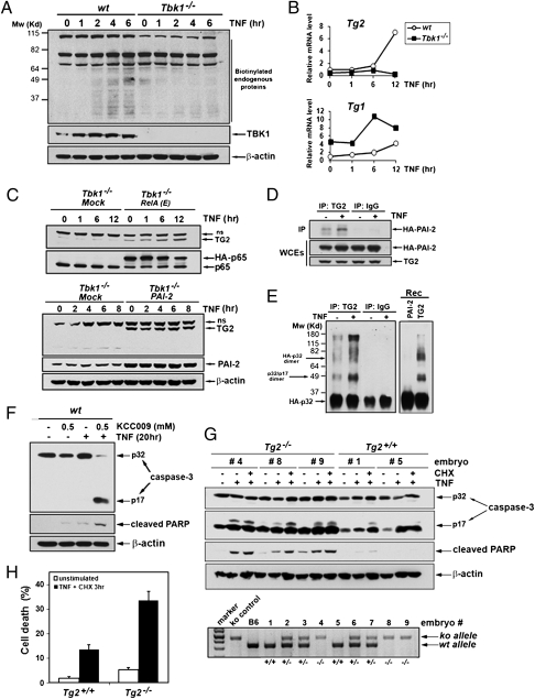
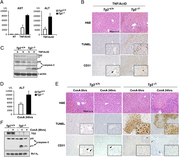
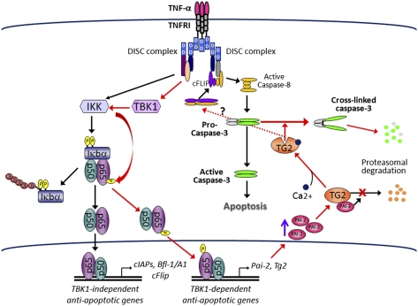
The decision between survival and death in cells exposed to TNF relies on a highly regulated equilibrium between proapoptotic and antiapoptotic factors. The TNF-activated antiapoptotic response depends on several transcription factors, including NF-κB and its RelA/p65 subunit, that are activated through phosphorylation-mediated degradation of IκB inhibitors, a process controlled by the IκB kinase complex. Genetic studies in mice have identified the IκB kinase-related kinase TANK-binding kinase 1 (TBK1; also called NAK or T2K) as an additional regulatory molecule that promotes survival downstream of TNF, but the mechanism through which TBK1 exerts its survival function has remained elusive. Here we show that TBK1 triggers an antiapoptotic response by controlling a specific RelA/p65 phosphorylation event. TBK1-induced RelA phosphorylation results in inducible expression of plasminogen activator inhibitor-2 (PAI-2), a member of the serpin family with known antiapoptotic activity. PAI-2 limits caspase-3 activation through stabilization of transglutaminase 2 (TG2), which cross-links and inactivates procaspase-3. Importantly, Tg2(-/-) mice were found to be more susceptible to apoptotic cell death in two models of TNF-dependent acute liver injury. Our results establish PAI-2 and TG2 as downstream mediators in the antiapoptotic response triggered upon TBK1 activation.


Conflict of interest statement

The authors declare no conflict of interest.

Figures

Fig. 1.
Fig. 1.
TBK1 protects MEFs from TNF-induced apoptosis and modulates IKK-dependent phosphorylation of RelA/p65. (A) Wt and Tbk1−/− MEFs were treated for 24 h with TNF (25 ng/mL) alone or for 3 h with TNF in the presence of CHX (10 μg/mL). Apoptotic cells were detected by TUNEL assays. The bars represent averages ± SD of three different experiments. Approximately 1,200 cells were counted in each experiment. (B) MEFs treated with TNF and CHX for the indicated times were analyzed for caspase-8 and caspase-3 activation by immunodetection of their cleaved forms. (C) Cell extracts prepared from Tbk1−/− MEFs treated for 6 h with TNF and CHX in the presence of z-VAD-FMK (20 μM), AEBSF (0.5 mM), or DMSO (vehicle) were analyzed by immunoblotting for caspase-3 activation and PARP cleavage. (D) IKK activity in TNF-treated MEFs was measured by immunocomplex kinase assay using GST-IκBα(1-54) as a substrate (19). (E) RelA phosphorylation was examined after its immunoprecipitation from [32P]orthophosphate-labeled and TNF-stimulated MEFs. Phospho-RelA was detected by autoradiography and the total amount of RelA by immunoblotting (IB). (F) Phosphorylation of RelA was examined by immunoblotting using a phospho-specific huRelA(Ser536) antibody.
Fig. 2.
Fig. 2.
Phospho-RelA protects MEFs from TNF-induced apoptosis. (A) Rela−/− MEFs infected with an empty retrovirus (mock) or retroviruses expressing HA-tagged wt RelA, RelA(S534A), or RelA(S534E) were left unstimulated or were treated with TNF (20 h). Apoptotic cells were detected by TUNEL staining. Represented are averages ± SD of three separate experiments. Cell lysates were analyzed for caspase-3 activation, PARP cleavage, and RelA expression by immunoblotting. (B) Tbk1−/− MEFs expressing RelA(S534A) or RelA(S534E) were treated for 3 h with TNF in the presence of CHX. Apoptotic cells were detected by TUNEL assay and quantified as described earlier. (C) Lysates were prepared from cells in B that were stimulated with TNF plus CHX for the indicated times. Caspase-3 activation and protein expression were determined by immunoblotting as in A.
Fig. 3.
Fig. 3.
TBK-1 controls NF-κB–dependent expression of the survival factor PAI-2. (A) Induction of NF-κB target genes was determined by qRT-PCR amplification of mRNAs prepared from WT and Tbk1−/− MEFs that were stimulated with TNF for the indicated times. Inset: Short time course (0 and 1 h) of Pai-2 mRNA induction in both cell types. (B) Relative mRNA induction was analyzed by qRT-PCR amplification of RNAs prepared from WT (Tbk1+/+), Tbk1−/−, and Tbk1−/− MEFs expressing RelA(S534E), depicted as RelA(E) that were stimulated with TNF for 2 h (IκBα), 6 h (Pai-2 and Inos), or 14 h (Ip10). (C) Expression of endogenous PAI-2 and RelA was examined by immunoblotting in extracts prepared from cells that were stimulated with TNF for the indicated times. (D) Apoptotic cell death, determined by TUNEL assay as described earlier, was quantified in Tbk1−/− MEFs and in cells stably expressing PAI-2 that were stimulated for 3 h with TNF in the presence of CHX. (E) WT MEFs expressing a Pai-2 shRNA or a scrambled shRNA were treated with TNF for 20 h. Caspase-3 activation, PARP cleavage, and protein expression were determined by immunoblotting. (F and G) Caspase-3 activation, PARP cleavage, and protein expression were determined by immunoblotting in extracts prepared from the indicated cells that were stimulated with TNF in the presence of CHX for the indicated times.
Fig. 4.
Fig. 4.
TG2 is a TBK1-dependent antiapoptotic factor. (A) In vivo transamidation activity was determined in WT and Tbk1−/− MEFs metabolically labeled with BP and stimulated with TNF for the indicated times. Cell extracts were prepared, and biotin-conjugated proteins were detected by immunoblotting using anti–streptavidin-HRP (Pierce). (B) Induction of Tg gene expression was determined by qRT-PCR analysis of mRNAs from WT and Tbk1−/− MEFs stimulated with TNF for the indicated times. (C) Endogenous TG2 protein amounts were determined by immunoblotting in extracts from Tbk1−/− MEFs expressing RelA(E) (Upper), PAI-2 (Lower), or an “empty” retrovirus (mock) and treated with TNF for the indicated times (ns, nonspecific). (D) Coimmunoprecipitation of PAI-2 with TG2 was examined in MEFs stably expressing HA-PAI-2 and human TG2 that were left untreated or were stimulated with TNF for 8 h. Control immunoprecipitations were performed with nonimmune IgGs. (E) In vitro transamidation assay was performed with TG2 or control immunoprecipitates prepared from untreated or TNF-stimulated Tbk1−/− MEFs expressing TG2 and untagged PAI-2 or with recombinant proteins (rec) using HA-procaspase-3 as a substrate. (F) Wt MEFs were treated with TNF (20 h) with or without the transglutaminase-specific inhibitor KCC009 (0.5 mM). Caspase-3 activation, PARP cleavage, and protein expression were determined by immunoblotting. (G) WT and Tg2−/− primary MEFs prepared from littermate embryos were left untreated or treated with TNF alone (20 h) or TNF plus CHX (6 h). Caspase-3 activation and apoptosis were determined as detailed earlier. Genotyping (Lower) was performed by PCR amplification as described (31). (H) The extent of cell death in WT and Tg2−/− MEFs that were stimulated for 3 h with TNF plus CHX was quantified by staining with PI.
Fig. 5.
Fig. 5.
TG2 protects mice from TNF-dependent liver apoptosis. (AC) WT and Tg2−/− mice were injected with TNF and ActD and analyzed 16 h later. (A) Serum AST and ALT levels were determined in untreated mice (NT) or mice treated with TNF and Act D. Data are averages ± SD (n = 3). (B) Histological analysis (H&E staining), TUNEL staining, and anti-CD31 immunostaining were performed on sequential liver sections of WT and Tg2−/− mice 16 h after TNF and Act D injection. Insets: Higher magnification of a selected area. Arrowheads show vascular endothelial cells (CD31-positive), which are TUNEL-negative. CD31 immunostaining in Tg2−/− liver sections displayed some nonspecific background staining as a result of the presence of cell debris. (C) Caspase-3 activation was determined by immunoblot analysis of liver protein extracts prepared 16 h after TNF and Act D injection. (DF) The same analyses were performed on WT and Tg2−/− at 8 h (E and F) or 24 h (D and E) after injection of Con A (35 mg/kg). (D) Serum AST and ALT levels measured after Con A injection. Data are averages ± SD (n = 2). (E) Histological analysis, TUNEL assays, and anti-CD31 staining were performed on sequential liver sections of WT and Tg2−/− mice at 8 h or 24 h after injection of Con A. Insets: Higher magnification of a selected area. TUNEL-positive cells (nuclear staining) in Tg2−/− liver sections are CD31-negative and represent apoptotic hepatocytes. Twenty-four hours of treatment with Con A induced massive liver degeneration in Tg2−/− mice, and dead cells were no longer detectable by TUNEL staining. Arrowheads point to vascular endothelial cells (CD31-positive cells), which are TUNEL-negative. (F) Liver extracts prepared from WT and Tg2−/− mice challenged with Con A (8 h) or PBS solution were analyzed for caspase-3 activation.
Fig. P1.
Fig. P1.
Schematic model illustrating the signaling pathway through which TBK1 inhibits TNF-α–induced apoptosis (red arrows) and the parallel pathway involving IKK-mediated NF-κB activation (black arrows). Engagement of TNFR1 triggers proapoptotic and survival signals through assembly of distinct multiprotein signaling platforms (DISK complexes). Heterodimeric cFLIP:caspase-8 complexes recruited to the DISK prevent cell death unless cFLIP is inactivated or degraded, allowing caspase-8 to induce apoptosis. The survival response is mediated through activation of TBK1 and IKK, both of which are required for NF-κB activation. Although TBK1 is dispensable for IκB phosphorylation and NF-κB–mediated induction of most antiapoptotic genes (i.e., TBK1-independent genes), it regulates the activation of Pai-2 and Tg2 (i.e., TBK1-dependent genes), whose expression requires RelA/p65 phosphorylation. Cytosolic PAI-2 associates with TG2, preventing its degradation. Calcium (Ca2+)-activated TG2 accumulates in the cytosol and cross-links procaspase-3 into inactive dimers that are degraded. This process inhibits apoptosis by depleting the pool of procaspase-3 available for activation by caspase-8.

Similar articles

Cited by

References

    1. Green DR, Evan GI. A matter of life and death. Cancer Cell. 2002;1:19–30. - PubMed
    1. Li J, Yuan J. Caspases in apoptosis and beyond. Oncogene. 2008;27:6194–6206. - PubMed
    1. Chen G, Goeddel DV. TNF-R1 signaling: A beautiful pathway. Science. 2002;296:1634–1635. - PubMed
    1. Wajant H, Scheurich P. TNFR1-induced activation of the classical NF-κB pathway. FEBS J. 2011;278:862–876. - PubMed
    1. Karin M, Lin A. NF-kappaB at the crossroads of life and death. Nat Immunol. 2002;3:221–227. - PubMed

Publication types

MeSH terms

LinkOut - more resources