. 2011 Dec 23;44(6):851-63.
doi: 10.1016/j.molcel.2011.12.005.

The cAMP/PKA pathway rapidly activates SIRT1 to promote fatty acid oxidation independently of changes in NAD(+)

Affiliations

The cAMP/PKA pathway rapidly activates SIRT1 to promote fatty acid oxidation independently of changes in NAD(+)

Zachary Gerhart-Hines et al. Mol Cell. .

Abstract

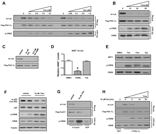
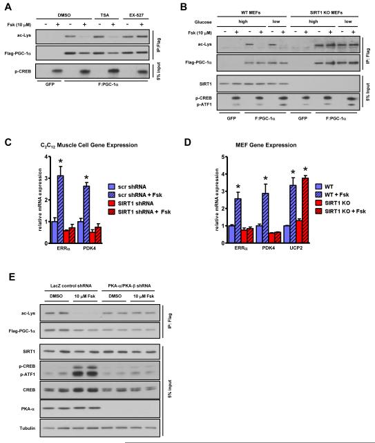
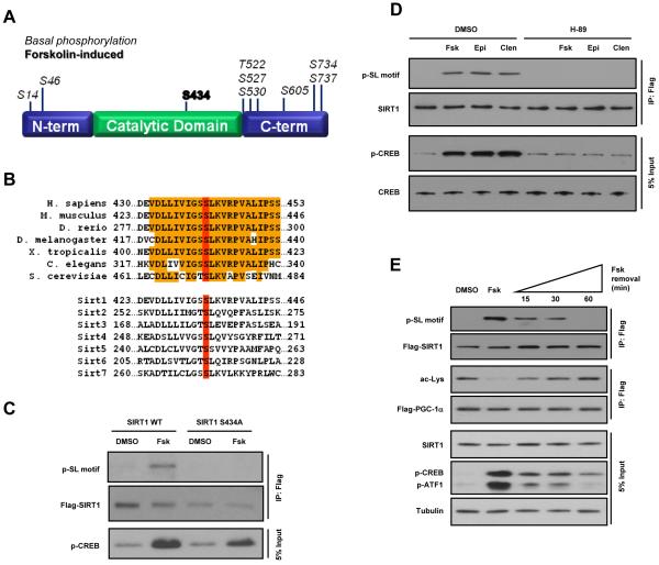
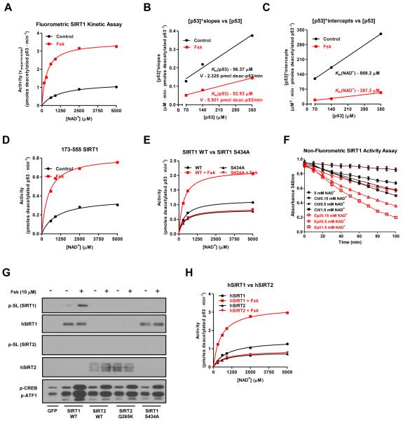
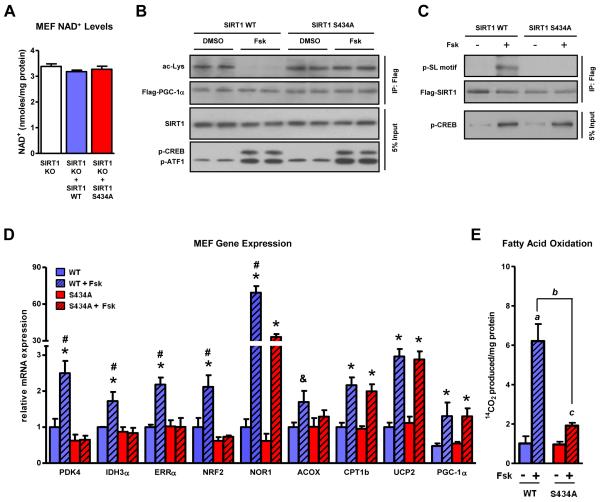
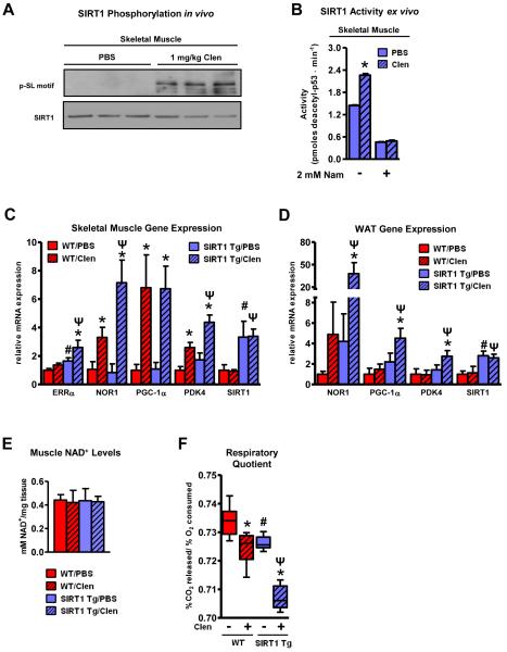
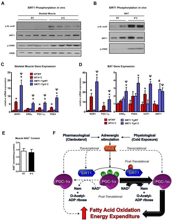
The NAD(+)-dependent deacetylase SIRT1 is an evolutionarily conserved metabolic sensor of the Sirtuin family that mediates homeostatic responses to certain physiological stresses such as nutrient restriction. Previous reports have implicated fluctuations in intracellular NAD(+) concentrations as the principal regulator of SIRT1 activity. However, here we have identified a cAMP-induced phosphorylation of a highly conserved serine (S434) located in the SIRT1 catalytic domain that rapidly enhanced intrinsic deacetylase activity independently of changes in NAD(+) levels. Attenuation of SIRT1 expression or the use of a nonphosphorylatable SIRT1 mutant prevented cAMP-mediated stimulation of fatty acid oxidation and gene expression linked to this pathway. Overexpression of SIRT1 in mice significantly potentiated the increases in fatty acid oxidation and energy expenditure caused by either pharmacological β-adrenergic agonism or cold exposure. These studies support a mechanism of Sirtuin enzymatic control through the cAMP/PKA pathway with important implications for stress responses and maintenance of energy homeostasis.


Figures

Figure 1
Figure 1. Activation of the cAMP signaling pathway induces rapid deacetylation of SIRT1 substrates
PGC-1α is deacetylated following acute stimulation of the cAMP/PKA signaling cascade. Transfected, FLAG-tagged PGC-1α was immunoprecipitated from U2OS cells treated for 30 minutes with, A, forskolin (Fsk) or epinephrine (Epi), B, clenbuterol (clen), and C, Br-cAMP; acetylation status was assessed using a pan-acetyl-lysine antibody (Cell Signaling). D, Intracellular NAD+ and, E, SIRT1 protein levels are not affected by short-term acute PKA activation. NAD+ concentrations were assessed from U2OS cells treated with DMSO (n=4), FK866 (n=4), or Fsk (n=4). Cells were treated with FK866 overnight or with forskolin for 1 h and then harvested in NAD+ extraction buffer. NAD+ levels were measured according to manufacturer’s instructions (BioAssay Systems). F, Endogenous p53 and, G, overexpressed FOXO1 are deacetylated following activation of β-adrenergic pathways. H, Forskolin-mediated PGC-1α deacetylation is rapid. Data are presented as means ± S.D.
Figure 2
Figure 2. cAMP-mediated PGC-1α deacetylation/activation is SIRT1 and PKA-dependent
A, Pharmacological inhibition and, B, genetic ablation of SIRT1 abrogates cAMP-mediated PGC-1α deacetylation. MEFs were subject to cAMP agonism under conditions of high (25 mM) and low (2.5 mM) glucose. C, SIRT1 knockdown by shRNA in skeletal muscle cells or, D, genetic ablation in fibroblasts abolishes the forskolin-mediated increase in fatty acid oxidative genes. PGC-1α was adenovirally expressed in differentiated C2C12 myotubes in addition to either scrambled (scr) control or SIRT1 shRNA for 72 hours to ensure knockdown. Skeletal muscle cells and MEFs were treated with Fsk for 6 h before RNA isolation and mRNA quantification; statistical significance was assessed using one-way ANOVA with a Tukey post-test, *, p<0.01, DMSO vs Fsk. E, cAMP-mediated PGC-1α deacetylation is PKA dependent. PGC-1α was immunoprecipitated from DMSO or Fsk-treated U2OS cell lines expressing either control LacZ shRNA or shRNAs against both PKA-α and β catalytic subunits (Figure S2D). Data are presented as means ± S.D.
Figure 3
Figure 3. PKA activation results in phosphorylation of SIRT1 on serine 434
A, Schematic depicting phosphorylation sites detected using mass spectrometric analysis of SIRT1 protein immunoprecipitated from U2OS cells incubated with Fsk or DMSO. Murine SIRT1 residue, S434 (bold), was found to be differentially phosphorylated following stimulation of the cAMP signaling pathway. B, Phylogenetic alignment shows complete conservation of SIRT1 S434 (red) among orthologs as well as other mammalian Sirtuin family members. C, Mutation of S434 abolishes forskolin-induced SIRT1 phosphorylation. D, Phosphorylation of SIRT1 S434 induced by forskolin, epinephrine, or clenbuterol is blocked by the PKA inhibitor, H-89. E, Removal of forskolin attenuates cAMP signaling and SIRT1 phosphorylation and re-establishes PGC-1α acetylation. Transfected, FLAG-tagged PGC-1α was immunoprecipitated from U2OS cells treated with Fsk for 30 min, washed with PBS, and then incubated in Fsk-free medium for the indicated time points.
Figure 4
Figure 4. The cAMP/PKA pathway increases intrinsic SIRT1 enzymatic activity through serine 434 phosphorylation
A, Forskolin treatment increases SIRT1 activity at multiple concentrations of acetylated substrate and three different NAD+ concentrations (140 μM acetyl-p53 peptide results are shown; data for 70 and 350 μM peptide are found in Figure S4B). Transfected FLAG-tagged SIRT1 was immunoprecipitated from DMSO or Fsk-treated U2OS cells and its activity was quantified using a fluorophore-conjugated acetylated p53 peptide; error bars are present but may be smaller than data point symbols. B-C, Forskolin treatment decreases the SIRT1 Km for NAD+ and increases the reaction rate, V, while leaving the Km for p53 unaffected. Kinetic data from Figure 4A were first plotted reciprocally as [NAD+]/vexperimental vs [NAD+] (Figure S4D) and the slopes and y-intercepts from that graph were used to make secondary plots from which the reaction velocity, V, and the Km’s for both p53 and NAD+ were calculated. V was converted from arbitrary fluorescence units to pmoles of deacetylated p53/min using a deacetylated standard (ENZO). D, The SIRT1 catalytic domain (amino acids 173-555) is sufficient to mediate the effects of forskolin on deacetylase activity. E, S434 mutation abolishes the effect of cAMP on SIRT1 in vitro activity. F, Activation of PKA signaling by epinephrine increases SIRT1 activity in a non-fluorometric based in vitro assay using a H3K14 peptide. G-H, Forskolin induces the phosphorylation and activation of human SIRT1 but not SIRT2 in vitro. Data are presented as means ± S.D.
Figure 5
Figure 5. cAMP-mediated increases in fatty acid oxidation and corresponding transcriptional programs are largely dependent on SIRT1 serine 434 phosphorylation
A, Intracellular NAD+ levels are similar in SIRT1 KO MEFs and MEF retroviral cell lines overexpressing SIRT1 wildtype and the S434A mutant. Mutation of S434 to a non-phosphorylatable residue in cells ablates the forskolin-triggered, B, deacetylation of PGC-1α, C, phosphorylation of SIRT1, D, induction of fatty acid oxidation genes, and significantly blunts, E, utilization of oleic acid. Retroviral cell lines expressing either WT or S434A SIRT1 were established using SIRT1−/− fibroblasts. MEFs were treated with Fsk for 6 h before RNA isolation and mRNA quantification; statistical significance was assessed using one-way ANOVA with a Tukey post-test, *, p<0.05, DMSO vs Fsk; *, p<0.01, DMSO vs Fsk; #, p<0.01, WT/Fsk vs S434A/Fsk. Fatty acid oxidation was quantified by measuring 14CO2 production and statistical significance was assessed using one-way ANOVA with a Tukey post-test; a, p<0.01, WT/DMSO vs WT/Fsk; b, p<0.01, WT/Fsk vs S434A/Fsk; c, p<0.01, S434A/DMSO vs S434A/Fsk. Data are presented as means ± S.D.
Figure 6
Figure 6. Adrenergic signaling induces SIRT1 phosphorylation/activation which promotes lipid oxidation and energy expenditure in vivo
A, SIRT1 is phosphorylated in response to cAMP signaling in vivo. B, SIRT1 catalytic function is enhanced by adrenergic stimulation in vivo. Activity of FLAG-tagged SIRT1 immunoprecipitated from pooled gastrocnemius/soleus samples from Tg mice treated with PBS (n=6) or Clen (n=6) was assessed using the SIRT1 fluorometric activity assay as described in Figure 4 and measured in duplicate; *, p<0.01, PBS vs Clen. Ectopic expression of SIRT1 enhances the clenbuterol-mediated induction of fatty acid oxidative genes in vivo in both, C, skeletal muscle and, D, white adipose tissue (WAT). RNA was isolated from the gastrocnemius and epididymal fat pads of fasted WT (n=5) and SIRT1 transgenic (Tg) (n=6) mice intraperitoneally injected with either saline (PBS) or clenbuterol (Clen-1 mg/kg) overnight for 14 h; statistical significance was assessed using one-way ANOVA with a Tukey post-test, *, p<0.01, PBS vs Clen treatment; #, p<0.01, WT/PBS vs Tg/PBS; Ψ, P<0.01, WT/Clen vs Tg/Clen. Gene expression results are representative of two separate experiments. E, Skeletal muscle NAD+ levels are not affected by clenbuterol administration or SIRT1 overexpression. NAD+ concentrations were assessed from gastrocnemius tissue from treated WT (n=4) and SIRT1 Tg mice (n=4). Muscle samples were homogenized in NAD+ extraction buffer and NAD+ was measured according to the manufacturer’s instructions (BioAssay Systems). F, SIRT1 transgenic mice exhibit lower respiratory quotients (RQ) than WT mice in response to clenbuterol. Metabolic parameters of WT (n=8) and SIRT1 Tg (n=8) mice were measured using CLAMS analysis over the course of a 14 h fast and an initial refeeding (RF). Statistical significance of RQ measurements was determined by one-way ANOVA with a Tukey post-test; *, p<0.01, PBS vs Clen treatment; #, p<0.01, WT/PBS vs Tg/PBS; Ψ, P<0.01, WT/Clen vs Tg/Clen. Data are presented as means ± S.D.
Figure 7
Figure 7. Cold challenge induces SIRT1 phosphorylation/activity which potentiates the thermogenic transcriptional programming in vivo
A, SIRT1 is phosphorylated in skeletal muscle and, B, BAT following cold exposure. C, Ectopic expression of SIRT1 enhances the cold-induction of fatty acid oxidation and energy expenditure genes in skeletal muscle and, D, brown adipose tissue (BAT). RNA was isolated from WT (n=6) and SIRT1 Tg (n=6) mice that were either maintained at room temperature (RT, 25°C) or exposed to cold (4°C) for 6 h; statistical significance was assessed using one-way ANOVA with a Tukey post-test, *, p<0.01, RT vs 4°C treatment; #, p<0.01, WT/RT vs Tg/RT; Ψ, P<0.01, WT/4°C vs Tg/4°C. E, Skeletal muscle NAD+ levels are not affected by cold exposure. F, Stimulation of the cAMP/PKA pathway results in phosphorylation of SIRT1 which enhances the efficiency of NAD+ co-substrate utilization and increases overall catalytic capacity. Adrenergic agonism also induces transcription of both SIRT1 and PGC-1α. Data are presented as means ± S.D.

Comment in

Similar articles

Cited by

References

    1. Asher G, Gatfield D, Stratmann M, Reinke H, Dibner C, Kreppel F, Mostoslavsky R, Alt FW, Schibler U. SIRT1 regulates circadian clock gene expression through PER2 deacetylation. Cell. 2008;134:317–328. - PubMed
    1. Banks AS, Kon N, Knight C, Matsumoto M, Gutiérrez-Juárez R, Rossetti L, Gu W, Accili D. SirT1 gain of function increases energy efficiency and prevents diabetes in mice. Cell Metab. 2008;8:333–341. - PMC - PubMed
    1. Beausoleil SA, Villén J, Gerber SA, Rush J, Gygi SP. A probability-based approach for high-throughput protein phosphorylation analysis and site localization. Nat. Biotechnol. 2006;24:1285–1292. - PubMed
    1. Cantó C, Gerhart-Hines Z, Feige JN, Lagouge M, Noriega L, Milne JC, Elliott PJ, Puigserver P, Auwerx J. AMPK regulates energy expenditure by modulating NAD+ metabolism and SIRT1 activity. Nature. 2009;458:1056–1060. - PMC - PubMed
    1. Donmez G, Guarente L. Aging and disease: connections to sirtuins. Aging Cell. 2010;9:285–290. - PubMed

Publication types

MeSH terms