. 2011 Dec;7(12):e1002433.
doi: 10.1371/journal.ppat.1002433. Epub 2011 Dec 8.

SARS coronavirus nsp1 protein induces template-dependent endonucleolytic cleavage of mRNAs: viral mRNAs are resistant to nsp1-induced RNA cleavage

Affiliations

SARS coronavirus nsp1 protein induces template-dependent endonucleolytic cleavage of mRNAs: viral mRNAs are resistant to nsp1-induced RNA cleavage

Cheng Huang et al. PLoS Pathog. 2011 Dec.

Abstract

SARS coronavirus (SCoV) nonstructural protein (nsp) 1, a potent inhibitor of host gene expression, possesses a unique mode of action: it binds to 40S ribosomes to inactivate their translation functions and induces host mRNA degradation. Our previous study demonstrated that nsp1 induces RNA modification near the 5'-end of a reporter mRNA having a short 5' untranslated region and RNA cleavage in the encephalomyocarditis virus internal ribosome entry site (IRES) region of a dicistronic RNA template, but not in those IRES elements from hepatitis C or cricket paralysis viruses. By using primarily cell-free, in vitro translation systems, the present study revealed that the nsp1 induced endonucleolytic RNA cleavage mainly near the 5' untranslated region of capped mRNA templates. Experiments using dicistronic mRNAs carrying different IRESes showed that nsp1 induced endonucleolytic RNA cleavage within the ribosome loading region of type I and type II picornavirus IRES elements, but not that of classical swine fever virus IRES, which is characterized as a hepatitis C virus-like IRES. The nsp1-induced RNA cleavage of template mRNAs exhibited no apparent preference for a specific nucleotide sequence at the RNA cleavage sites. Remarkably, SCoV mRNAs, which have a 5' cap structure and 3' poly A tail like those of typical host mRNAs, were not susceptible to nsp1-mediated RNA cleavage and importantly, the presence of the 5'-end leader sequence protected the SCoV mRNAs from nsp1-induced endonucleolytic RNA cleavage. The escape of viral mRNAs from nsp1-induced RNA cleavage may be an important strategy by which the virus circumvents the action of nsp1 leading to the efficient accumulation of viral mRNAs and viral proteins during infection.


Conflict of interest statement

The authors have declared that no competing interests exist.

Figures

Figure 1
Figure 1. Schematic diagram of the SCoV genome.
The viral genome consists of a single-stranded, positive-sense RNA of ∼29.7 kb in length. The 5′-proximal gene 1 (∼22-kb) has two ORFs, ORF1a and 1b, which encodes for large polyproteins, polyprotein 1a and polyprotein 1ab. The polyproteins are cleaved into 16 mature non-structural proteins (nsp1-nsp16) by viral proteases PLpro and 3CLpro. Most of the non-structural proteins are involved in viral RNA synthesis and some of the identified functions of the non-structural proteins are: ADRP, ADP-ribose-1”-phosphate phosphatase ; PLpro, papain-like protease ; 3CLpro, 3C-like protease ; Pr, primase ; ssRBP, single-stranded RNA-binding protein ; RDRP, RNA-dependent RNA polymerase ; Hel, helicase ; ExoN, 3′5′ exoribonuclease; N7-MTase, guanine-N7-methyltransferase ; NendoU, poly(U)-specific endoribonuclease ; and 2′OMT, 2′ O-methyltransferase . Major viral structural proteins, including S, M, N and E, and the accessory proteins, including 3a, 3b, 6, 7a, 7b, 8a, 8b, and 9b, are encoded downstream of the ORF1a/1b.
Figure 2
Figure 2. Susceptibilities of IRES-containing dicistronic RNAs to nsp1-mediated RNA cleavage.
(A) Schematic diagram of the structures of full length dicistronic RNA transcripts (Full length), RNA 1 containing the 5′ rluc gene and intercistronic IRES sequence and RNA 2 containing only the 5′ rluc gene. (B) Ren-TMEV-FF was incubated with GST, nsp1 or nsp1-mt or without any protein (mock) in RRL at 30oC for 10 min. RNA samples were extracted and analyzed by Northern blot analysis using the 5′ rluc probe (left panel) and 3′ fluc probe (right panel). Marker is a mixture of in vitro-transcribed, full-length RNA transcripts, RNA 1 and RNA 2. (C) Ren-PV-FF was examined as in (B), except RNA was incubated in RRL+HeLa. RNA 2, full length dicistronic RNA transcripts and RNA 1 were separately applied to the gel and shown in three marker lanes. An asterisk represents a truncated RNA product, which was probably generated by premature transcription termination around the IRES region of full-length RNA and RNA 1. (D) Ren-CSFV-FF was examined as described in (B).
Figure 3
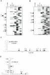
Figure 3. Primer extension analysis of Ren-EMCV-FF and Ren-PV-FF after incubation with GST, nsp1 or nsp1-mt.
(A, B) In vitro-synthesized Ren-EMCV-FF (A) and Ren-PV-FF (B) were incubated with GST, nsp1 or nsp1-mt in RRL (A) and RRL+HeLa (B), respectively. The RNAs were extracted and subjected to primer extension analysis by using the 5′-end 32P labeled primer that binds at ∼100 nt downstream of the fluc translation initiation codon for Ren-EMCV-FF (A) or a labeled primer that binds 6 nt downstream of the fluc initiation codon region for Ren-PV-FF (B). Primer extension products and DNA sequence ladders, which were generated from the plasmid used for RNA synthesis and the primer used for primer extension analysis, were resolved on 8% polyacrylamide/7M Urea DNA sequencing gels. Premature primer extension termination signals are indicated by enumerated arrows and major RNA modification sites are marked with asterisks. IVT RNA, primer extension analysis of in vitro-transcribed RNAs that were not incubated in RRL; AUG, translation initiation AUG (AUG-834) of EMCV IRES. (C, D) RNA modification sites in Ren-EMCV-FF (C) and Ren-PV-FF (D). The structural domain L of the EMCV IRES (C) in Ren-EMCV-FF and the poliovirus IRES stem-loop IV (D) in Ren-PV-FF are also shown. Arrows indicate RNA modification sites and asterisks denote the main ones. The underlined AUG triplet is equivalent to the translation initiation codon AUG-834 in the EMCV genome (C) and the boxed AUG triplet is equivalent to the AUG-586 of the poliovirus genome. AUG-586 of the poliovirus is mapped ∼150 nt upstream of the authentic viral translation initiation codon.
Figure 4
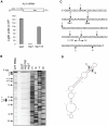
Figure 4. Characterization of nsp1-induced RNA modification in ALA mRNA.
(A) Capped and polyadenylated ALA mRNA was incubated with GST, nsp1 or nsp1-mt in RRL, and rluc reporter activities were determined by an rluc assay (Promega). At the top is a schematic diagram of ALA mRNA, in which 5′ UTR and the rluc gene are not shown to scale. (B) RNAs were extracted after incubation with GST, nsp1 or nsp1-mt and subjected to primer extension analysis by using a 5′-end labeled primer that binds to ∼100 nt downstream of the rluc gene translation initiation codon. Primer extension products were resolved on 8% polyacrylamide/7M Urea DNA sequencing gels, and premature primer extension termination signals are shown by enumerated arrows. Main RNA modification products (sites 3 and 4) are marked with asterisks. 5′-end, full-length primer extension product; IVT RNA, primer extension analysis of in vitro-transcribed ALA mRNA that was not incubated in RRL; AUG, translation initiation codon of rluc gene. (C) RNA modification sites of ALA mRNA. Arrows indicate RNA modification sites with main RNA modification sites marked with asterisks. (D) Predicted secondary structure of human β-actin 5′ UTR. Arrows indicate main RNA modification sites.
Figure 5
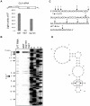
Figure 5. Characterization of nsp1-induced RNA modification in GLA mRNA.
(A-D) Experiments similar to those described in Figure 4 were performed to obtain the results depicted in Figure 5, except that GLA mRNA, instead of ALA mRNA, was used.
Figure 6
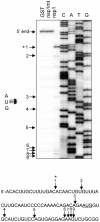
Figure 6. Characterization of nsp1-induced RNA modification in rabbit β-globin mRNA.
Poly A+ RNA was extracted from micrococcal nuclease-untreated RRL (Promega) and incubated with GST, nsp1 or nsp1-mt in micrococcal nuclease-treated RRL. RNA was extracted and subjected to primer extension analysis with a rabbit β-globin mRNA-specific primer. Primer extension products were resolved on 8% polyacrylamide/7M Urea DNA sequencing gels (Top panel). Premature primer extension termination signals are shown by enumerated arrows. The main RNA modification product (site 1) is marked with an asterisk. The 5′-end represents a full-length primer extension product, and the AUG at the top of the figure and underlined AUG represent the translation initiation codon of β-globin mRNA.
Figure 7
Figure 7. Characterization of nsp1-induced RNA fragment of ALA mRNA.
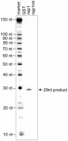
Cap-labeled ALA mRNA was incubated with GST, nsp1 or nsp1-mt in RRL. RNAs were extracted from RRL by using proteinase K digestion and subsequent phenol/chloroform extraction. A cap-radiolabeled RNA fragment was detected in a 10% polyacrylamide/7M Urea DNA sequencing gel electrophoresis. An RNA size marker (ranging from 10 nt to 100 nt in 10 nt increment) was prepared by using the Decade Marker system (Applied Biosystems) and applied to the same gel.
Figure 8
Figure 8. Identification of RNA modification sites of GLA mutant mRNAs.
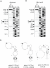
(A) GLA mutant 1 (GLA mt 1) carrying G17U18 and A25C26 mutations was incubated with GST, nsp1 or nsp1-mt in RRL. RNA was extracted from RRL and subjected to primer extension analysis. Premature primer extension termination signals are shown by enumerated arrows and asterisks represent RNA modification products in the 5′ UTR. 5′-end, full-length primer extension product; AUG, translation initiation codon of GLA mutant 1. (B) Similar experiments were performed by using GLA mutant 2 (GLA mt 2) with G17U18 mutations. (C) The predicted RNA secondary structures and the nsp1-induced RNA modification sites at the 5′ UTRs of GLA mutant 1 (left), GLA mRNA (middle) and GLA mutant 2 (right). Arrowheads show the RNA modification sites. Mutated sites in the mutants and the corresponding sites of GAL mRNA are boxed.
Figure 9
Figure 9. Susceptibilities of naturally occurring SCoV mRNAs to nsp1-induced RNA modification.
(A and C) Poly A+ RNA was extracted from SCoV-infected cells and incubated with GST, nsp1 or nsp1-mt in RRL. Extracted RNAs were subjected to primer extension analysis by using primers suitable for detecting RNA modifications in SCoV mRNA 9 (A) or in SCoV mRNA 3 (C). Primer extension products were resolved in 8% polyacrylamide/7M Urea DNA sequencing gels: 5′-end, full-length primer extension product of SCoV mRNA 9 (A) or SCoV mRNA 3 (C). (B) Similar experiments described in (A) were performed, except that RNAs from SCoV-mt-infected cells were used.
Figure 10
Figure 10. Susceptibilities of SCoV-like mRNAs to nsp1-induced RNA modification.
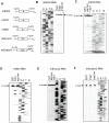
(A) Schematic diagram showing the structures of SCoV mRNA 9-like m9LN3, m9LN, m9N3 and m9Lrluc3 RNAs. N, N ORF; rluc, rluc gene. (B, C) In vitro-synthesized m9LN3 RNA and m9LN RNA were independently incubated with GST, nsp1 or nsp1-mt in RRL. RNAs were extracted and subjected to primer extension analysis by using the same primer described in Figure 9A. 5′-end, full-length primer extension product. (D-F) The experiment was carried out using the same methods as in C, except that the following combinations of RNA and primer were used: (D) m9N3 RNA and a primer that binds ∼76 nt downstream of N gene translation initiation codon; (E) m9Lrluc3 RNA and a primer that binds ∼100 nt downstream of the rluc gene translation initiation codon; and (F) m9Lactin3 RNA template and a primer that binds ∼1 nt downstream of the β-actin gene translation initiation codon.
Figure 11
Figure 11. Susceptibilities of SCoV mRNA 9 mutants to nsp1-induced RNA modification.
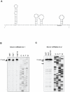
(A) Predicted secondary structure of the 5′ UTR of SCoV mRNA 9. Underlined AUG represents the translation initiation codon. (B, C) In vitro-synthesized SCoV mRNA 9 mt 1 and SCoV mRNA 9 mt 2 RNAs were independently incubated with GST, nsp1 or nsp1-mt in RRL. RNAs were extracted and subjected to primer extension analysis by using the same primer described in Figure 9A. 5′-end, full-length primer extension product. Arrows indicate premature primer extension products.

Similar articles

Cited by

References

    1. Rota PA, Oberste MS, Monroe SS, Nix WA, Campagnoli R, et al. Characterization of a novel coronavirus associated with severe acute respiratory syndrome. Science. 2003;300:1394–1399. - PubMed
    1. Marra MA, Jones SJ, Astell CR, Holt RA, Brooks-Wilson A, et al. The Genome sequence of the SARS-associated coronavirus. Science. 2003;300:1399–1404. - PubMed
    1. Ksiazek TG, Erdman D, Goldsmith CS, Zaki SR, Peret T, et al. A novel coronavirus associated with severe acute respiratory syndrome. N Engl J Med. 2003;348:1953–1966. - PubMed
    1. Lau SK, Woo PC, Li KS, Huang Y, Tsoi HW, et al. Severe acute respiratory syndrome coronavirus-like virus in Chinese horseshoe bats. Proc Natl Acad Sci U S A. 2005;102:14040–14045. - PMC - PubMed
    1. Li W, Shi Z, Yu M, Ren W, Smith C, et al. Bats are natural reservoirs of SARS-like coronaviruses. Science. 2005;310:676–679. - PubMed

Publication types

MeSH terms