. 2011 Dec;7(12):e1002418.
doi: 10.1371/journal.ppat.1002418. Epub 2011 Dec 8.

Epstein-Barr virus nuclear antigen 3C stabilizes Gemin3 to block p53-mediated apoptosis

Affiliations

Epstein-Barr virus nuclear antigen 3C stabilizes Gemin3 to block p53-mediated apoptosis

Qiliang Cai et al. PLoS Pathog. 2011 Dec.

Retraction in

Abstract

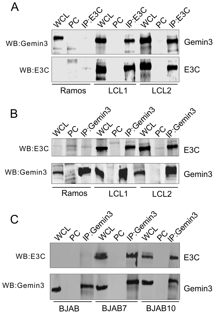
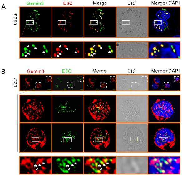
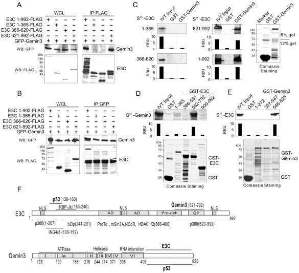
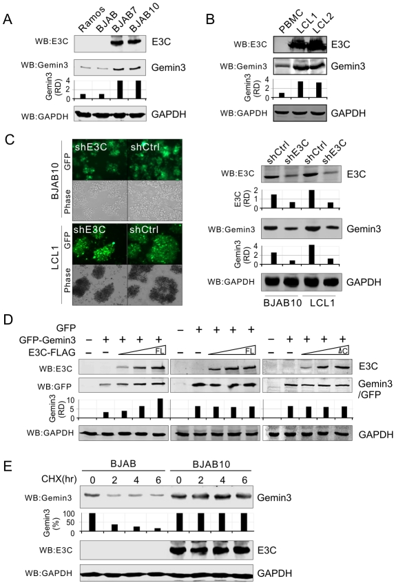
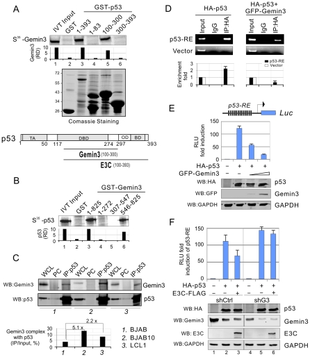
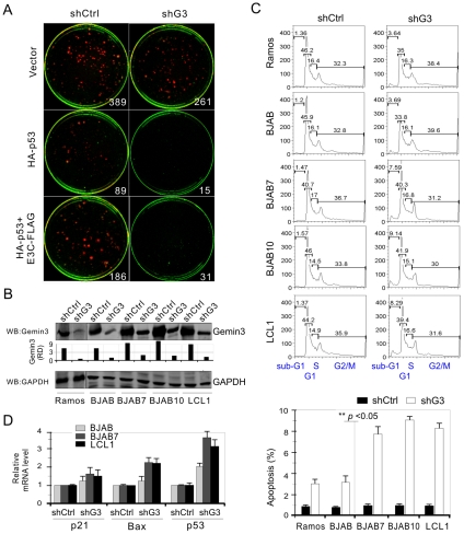
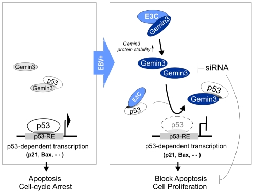
The Epstein-Barr nuclear antigen 3C (EBNA3C), one of the essential latent antigens for Epstein-Barr virus (EBV)-induced immortalization of primary human B lymphocytes in vitro, has been implicated in regulating cell proliferation and anti-apoptosis via interaction with several cellular and viral factors. Gemin3 (also named DDX20 or DP103) is a member of DEAD RNA helicase family which exhibits diverse cellular functions including DNA transcription, recombination and repair, and RNA metabolism. Gemin3 was initially identified as a binding partner to EBNA2 and EBNA3C. However, the mechanism by which EBNA3C regulates Gemin3 function remains unclear. Here, we report that EBNA3C directly interacts with Gemin3 through its C-terminal domains. This interaction results in increased stability of Gemin3 and its accumulation in both B lymphoma cells and EBV transformed lymphoblastoid cell lines (LCLs). Moreover, EBNA3C promotes formation of a complex with p53 and Gemin3 which blocks the DNA-binding affinity of p53. Small hairpin RNA based knockdown of Gemin3 in B lymphoma or LCL cells remarkably attenuates the ability of EBNA3C to inhibit the transcription activity of p53 on its downstream genes p21 and Bax, as well as apoptosis. These findings provide the first evidence that Gemin3 may be a common target of oncogenic viruses for driving cell proliferation and anti-apoptotic activities.


Conflict of interest statement

The authors have declared that no competing interests exist.

Figures

Figure 1
Figure 1. EBNA3C forms a complex with Gemin3 in EBV transformed cells.
(A) and (B) Endogenous Gemin3 associates with EBNA3C. Twenty million of Ramos, LCL1, and LCL2 cells were individually lysed and subjected to immunoprecipitation (IP) with EBNA3C (A10) or Gemin3 (12H12) specific antibody. 5% of whole cell lysates (WCL) were loaded as input. Normal serum IgG was used for preclear (PC). Lysates and IP complexes were resolved by SDS-PAGE and subjected to western blotting (WB) with the indicated antibodies. (C) Exogenous EBNA3C associates with Gemin3. The EBNA3C stable expressing cells (BJAB7 and BJAB10) and parental BJAB cells were individually lysed and subjected to immunoprecipitation (IP) and western blotting as indicated.
Figure 2
Figure 2. EBNA3C colocalizes with Gemin3.
(A) Colocalization of exogenous E3C and Gemin3 in U2OS cells. Cells transfected with E3C-FLAG and GFP-Gemin3 were stained by using M2 antibody, followed by goat anti-mouse antibody conjugated to Alexa Fluor 594 (red). (B) Endogenous Gemin3 colocalizes with E3C in the EBV-transformed cell line LCL1. Endogenous Gemin3 and E3C were individually detected by using mouse anti-Gemin3 (12H12) and rabbit anti-E3C antibodies, followed by Goat anti-mouse antibody conjugated to Alexa Fluor 594 (red) and goat anti-rabbit antibody conjugated to Alexa Fluor 488 (green). Nuclei were counterstained by using DAPI (blue). The images were sequentially captured using an Olympus confocal microscope. All panels are representative pictures from similar repeat experiments. Enlarged sections are shown at the bottom. Arrows show the colocalization of E3C and Gemin3.
Figure 3
Figure 3. EBNA3C and Gemin3 interact through their C-terminal domains.
(A) and (B) Gemin3 associates with the C-terminal domain of EBNA3C. Twenty million of HEK 293 cells were cotransfected with plasmids expressing GFP-tagged Gemin3 and Flag-tagged either full-length or the truncated mutants of EBNA3C. At 36 h post-transfection, cells were harvested and subjected to immunoprecipitation using anti-FLAG (M2) or GFP antibodies followed by western blotting with GFP or M2 antibody. 5% of whole cell lysates (WCL) were used as input. The membrane was stripped and reblotted with M2 or GFP antibody. (C) Gemin3 binds to C-terminal domain of E3C in vitro. The 35S-radiolabeled in vitro-translated proteins of E3C truncated mutants were precleared with GST bead, followed by incubation with GST or GST-Gemin3 beads. The bound protein mixtures were resolved by appropriate SDS-PAGE, and autography. 5% of in vitro translated protein is used as input. The quantification of relative amount of bound protein (RBU) was shown at the bottom. (D) The 35S-radiolabeled in vitro-translated protein full-length Gemin3 was pulled down by truncated mutants of EBNA3C fusion with GST (GST-E3C 1–365, 366–581,582–792, and 900–992). Coomassie blue staining of purified GST-EBNA3C truncated proteins is shown at the bottom panel. (E) E3C binds to C-terminal domain of Gemin3 in vitro. The 35S-radiolabeled in vitro-translated protein full-length EBNA3C was pulled down by truncated mutants of Gemin3 fusion with GST (GST-Gemin3 1–272, 307–547, and 546–825). Coomassie blue staining of purified GST-Gemin3 truncated proteins is shown at the bottom. (F) Schematics illustrate different structural domains of E3C and Gemin3. The respective binding domain(s) of two proteins are indicated by bold.
Figure 4
Figure 4. EBNA3C enhances the protein stability of Gemin3.
(A) and (B) E3C increases the protein production of endogenous Gemin3. Ten million of EBV negative cells (Ramos, BJAB and PBMC), EBNA3C- expressing cells (BJAB7 and BJAB10), and EBV-transformed cells (LCL1 and LCL2) were lysed and subjected to western blot with indicated antibodies. The data show that the protein level of Gemin3 in the EBNA3C or EBV positive cells was significantly upregulated. (C) The protein level of Gemin3 is decreased in BJAB10 and LCL1 cells with EBNA3C stable knockdown. BJAB10 and LCL1 cells with Lentivirus-delivered GFP labeled shRNA against EBNA3C (shE3C) or scramble control (shCtrl) were showed on the left panel. The protein levels of E3C and Gemin3 are detected by western blotting with indicated antibodies. GAPDH immunoblotting was used as the loading control. The relative quantitation of Gemin3 and E3C protein levels are normalized by GAPDH. (D) E3C increases the protein level of exogenous Gemin3. Saos-2 cells were transfected GFP-Gemin3 or GFP with increasing amounts of full length (FL) EBNA3C-FLAG or its C-terminal deleted mutant (ΔC). The transiently transfected cells were harvested at 36 h post-transfection, the protein levels of exogenous GFP-Gemin3 and GFP were detected by western blotting with GFP antibody. The relative quantitation of GFP-Gemin3 or GFP is shown in the middle panel. (E) EBNA3C enhances the protein stability of Gemin3. The cell lysates of BJAB or BJAB10 cells treated with cycloheximide (CHX, 100 µg/ml) for 0, 2, 4 and 6 hours were subjected to immunoblotting as indicated. GAPDH immunoblotting was used as the loading control. The relative quantitation of Gemin3 is shown in the middle panel.
Figure 5
Figure 5. Gemin3 interacts with p53 and contributes to EBNA3C-mediated inhibition of p53 transcriptional activity.
(A) Gemin3 binds with DNA-binding domain of p53 in vitro. 35S-radiolabeled Gemin3 was in-vitro translated and incubated with bacterially purified GST, GST fusion with full length (1–393) or truncated mutants of p53 (1–83, 100–300, and 300–393). The pull-down complexes were resolved by appropriate SDS-PAGE and autography. The amount of bound protein was quantified with ImageQuant software. Coomassie blue staining of purified GST protein is shown in the middle panel. Schematic illustrates the binding domains of p53 with Gemin3 and EBNA3C. (B) C-terminal domain of Gemin3 binds with p53 in vitro. 35S-radiolabeled p53 was in vitro translated and incubated with bacterially purified GST control, full length GST-Gemin3 (1–825) or its truncations (1–272, 307–547, and 546–825). The GST pull down assay was analyzed as the described above. (C) EBNA3C increases the interaction of p53 with Gemin3. Twenty million of BJAB, BJAB10 and LCL1 cells were individually harvested, lysed and subjected to immunoprecipitation (IP) with p53 specific antibody and subsequently western blotted with Gemin3 or p53 antibody. The relative quantitation of Gemin3 complex with p53 was calculated by IP compared with input and shown in bottom panel. (D) Gemin3 reduces DNA-binding affinity of p53. Saos-2 cells were co-transfected HA-p53 with or without GFP-Gemin3 in the presence of p53-RE-Luc containing 13 copies of p53-binding sties or pGL3-basic vector alone. Chromatin immunoprecipitation assays were performed with anti-HA or normal lgG antibodies control. The DNA binding ability of p53 was detected by standard (upper) and quantitative real-time (lower) PCR with primers targeting ampicilin gene of pGL3 plasmid. (E) Gemin3 inhibits the transcriptional activity of p53 in the luciferase reporter assay. Saos-2 cells were co-transfected with 3 µg of the pGL3-p53-RE-Luc, 10 µg HA-p53 in the presence of either vector control or increasing amounts of GFP-Gemin3 (5 µg and 10 µg). Each transfection was performed in triplicate. Error bars indicated standard variations. Bottoms panels show the protein levels detected by western blotting analysis with the indicated antibodies. GAPDH was used as a control for equal loading. The relative luciferase unit (RLU) is showed by normalization with vector alone. (F) Gemin3 knockdown reduces E3C-mediated inhibition of p53 transcriptional activity in the luciferase reporter assays. Saos-2 cells with stable Gemin3 knockdown (shG3) or control (shCtrl) were individually co-transfected p53-RE-Luc with HA-p53, HA-p53 and E3C-FLAG or vector alone. The western blotting data of each protein is shown at the bottom panel. The relative luciferase unit (RLU) is showed by normalization with vector alone. Plasmid expressing RFP was used for normalized transfection efficiency.
Figure 6
Figure 6. Gemin3 knockdown attenuates EBNA3C-mediated inhibition of p53-induced apoptosis.
(A) Saos-2 (p53−/−) cells were electroporated with different combinations of expression plasmids for HA-p53, EBNA3C-FLAG or vector alone in the presence of small hairpin RNA against Gemin3 (shG3) or control (shCtrl) as indicated. 2×10³ transfected cells were cultured in the selection medium (DMEM supplemented with 100 mg/ml G418). After a 2-week selection, cells were fixed on the plates with 4% formaldehyde and stained with 0.1% crystal violet. The area of colonies (pixels) in each dish was calculated by LiCor Odyssey. The number represents the averages of data from two independent experiments. Plasmid expressing RFP was used for normalized transfection efficiency. (B) Western blots showing the protein level of Gemin3 in the lentivirus-mediated Gemin3 or control knockdown cell lines. GAPDH was used as the loading control. (C) Gemin3 knockdown increases apoptosis of EBV negative cells (Ramos and BJAB), EBNA3C positive cells (BJAB7 and BJAB10), and EBV transformed cells (LCL1). Cells were harvested after a 12-h serum starvation and fixed. Levels of cells undergoing apoptosis (sub-G1 phase) in individual PI-stained samples were detected by flow cytometry, and the data were analyzed by FlowJo software. The bar diagram shown at the bottom panel represents the mean of three independent experiments and the results of comparing with each control knockdown sample. (D) Quantitative real-time PCR analysis showed that p53, p21 and Bax genes were upregulated in the Gemin3 knockdown cells. Total RNA was individually isolated from the Gemin3 (shG3) or control (shCtrl) knockdown cells with 12-h serum starvation treatment. A 2 µg total RNA was used to synthesis cDNA. Error bars show standard deviations.
Figure 7
Figure 7. A schematic model depicting the Gemin3-mediated transcriptional regulation of p53 by EBNA3C in the EBV latently infected cells.
Left panel, in response to genotoxic stress, p53 achieves its anti-proliferative properties through its function as a DNA-binding transcriptional activator, and induce expression of downstream target genes (including p21 and BAX) for cell-cycle arrest and apoptosis. Right panel, in the EBV latency III cells, EBNA3C not only stabilizes Gemin3 through the interaction of their C-terminal domains and but also enhances a stable complex of Gemin3 with p53 for blocking p53-mediated transcriptional activity and cell apoptosis. Gemin3 knockdown (siRNA) increases the sensitivity of the EBNA3C-expressing cells to stress-induced apoptosis. p53-RE, p53 responsive element.

Similar articles

Cited by

References

    1. Burkitt D. A sarcoma involving the jaws in African children. Br J Surg. 1958;46:218–223. - PubMed
    1. Epstein MA, Achong BG, Barr YM. Virus Particles In Cultured Lymphoblasts From Burkitt's Lymphoma. Lancet. 1964;1:702–703. - PubMed
    1. Rickinson AB, Kieff E. Epstein-Barr virus. In: Fields BN, Knipe DM, Howley PM, editors. Fields virology, 4th ed. Vol 2, Philadelphia: Lippincott Williams & Wilkins; 2002. pp. 2575–2627.
    1. Henle G, Henle W, Diehl V. Relation of Burkitt's tumor-associated herpes-ytpe virus to infectious mononucleosis. Proc Natl Acad Sci U S A. 1968;59:94–101. - PMC - PubMed
    1. zur Hausen H, Schulte-Holthausen H, Klein G, Henle W, Henle G, et al. EBV DNA in biopsies of Burkitt tumours and anaplastic carcinomas of the nasopharynx. Nature. 1970;228:1056–1058. - PubMed

Publication types

MeSH terms