. 2011 Dec 13;21(6):1156-70.
doi: 10.1016/j.devcel.2011.10.009.

Myosin V transports secretory vesicles via a Rab GTPase cascade and interaction with the exocyst complex

Affiliations

Myosin V transports secretory vesicles via a Rab GTPase cascade and interaction with the exocyst complex

Yui Jin et al. Dev Cell. .

Abstract

Vesicle transport requires four steps: vesicle formation, movement, tethering, and fusion. In yeast, two Rab GTPases, Ypt31/32, are required for post-Golgi vesicle formation. A third Rab GTPase, Sec4, and the exocyst act in tethering and fusion of these vesicles. Vesicle production is coupled to transport via direct interaction between Ypt31/32 and the yeast myosin V, Myo2. Here we show that Myo2 interacts directly with Sec4 and the exocyst subunit Sec15. Disruption of these interactions results in compromised growth and the accumulation of secretory vesicles. We identified the Sec15-binding region on Myo2 and also identified residues on Sec15 required for interaction with Myo2. That Myo2 interacts with Sec15 uncovers additional roles for the exocyst as an adaptor for molecular motors and implies similar roles for structurally related tethering complexes. Moreover, these studies predict that for many pathways, molecular motors attach to vesicles prior to their formation and remain attached until fusion.


Figures

Figure 1
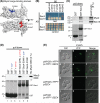
Figure 1. Myo2 directly interacts with Sec4
(A) Surface residues of the Myo2 cargo-binding domain that bind vacuoles (blue) or secretory vesicles (red) (Ishikawa et al., 2003; Lipatova et al., 2008; Pashkova et al., 2006). (B) Sec4 interacts with the secretory vesicle binding region of Myo2. Yeast two-hybrid test of wild-type or mutant Myo2 cargo-binding domain fused with the Gal4 DNA binding domain (BD), versus Sec4 or Vac17 fused with the Gal4 activation domain (AD). Plasmids cotransformed into yeast, grown on SC-LEU-TRP and replica-plated on SCLEU-TRP (control) or SC-ADE-HIS-LEU-TRP + 3AT (test) plates; incubated at 24°C for 9 days. Growth indicates protein-protein interactions. (C) Sec4 and Myo2 are in the same complex. Purified bacterially expressed GST-Sec4 protein pulled down Myo2 and Sec15 from solubilized yeast lysates. Proteins pulled down by GST (lane 1), GST-Sec4 (lane 2), and 8 % of input yeast lysate (lane 3) analyzed by immunoblot with anti-Myo2 cargo-binding domain antibodies (top) Sec15 (middle), or by Coomassie Brilliant Blue (CBB) (bottom). (D) Direct binding of Sec4 to the Myo2 cargo-binding domain requires GTP. Purified GST-Sec4 pulled down purified His-tagged Myo2 cargo-binding domain from E. coli. Proteins pulled down by GST (lane 1), GST+GDP (lane 2), GST+GTPγS (lane 3), GST-Sec4-Q79L+GTPγS (lane 4), GST-Sec4-S34N+GDP (lane 5) or GST-Sec4-N133I (lane 6), and 2 % of input His-tagged Myo2 cargo-binding domain (lane 7) were analyzed by immunoblot with antibody directed against Myo2 cargo-binding domain or by CBB staining. (C and D) CBB stain of proteins bound to glutathione beads shows that the levels of GST (negative control) were higher than the levels of GST-Sec4 (test protein). (E) Sec4 and Myo2 interact in vivo. Myo2-vYFPN and vYFPC-Sec4 coexpressed in myo2Δ cells interact as indicated by the YFP signal (panels a-f). Expression of Myo2- vYFPN with non-tagged Sec4 or vYFPC -Sec4 with non-tagged Myo2 showed no fluorescence (panels g-l).
Figure 2
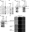
Figure 2. Interaction between Myo2 and Sec4 is essential for yeast viability
(A) Schematic of Rab GTPase fusions of Myo2. Rab GTPases indicated (B) fused in frame to the C-terminus of Myo2. Myo2 motor domain, IQ motifs, coiled-coil region and cargo-binding domain, and mutation site of Y1415R (*) are indicated. (B) (Left panel) Plasmids transformed into a myo2Δ strain (LWY2947), YCp50 [URA3] MYO2: pRS413 [HIS3] MYO2, pRS413 MYO2-SEC4, pMYO2-YPT32, pMYO2-YPT1, pmyo2-Y1415R, pmyo2-Y1415R-SEC4, pmyo2-Y1415R-YPT32 or pmyo2-Y1415R-YPT1. Transformed colonies were re-streaked onto SC+5FOA to counter select against YCp50 [URA3] MYO2 (middle panel) and on SC-His-Ura to show that the cells received a [HIS3] plasmid (right panel). (C) Myo2 fusion proteins expressed at similar levels in SC-His-Ura medium. Cell lysates were analyzed by immunoblot with antibodies directed against Myo2 cargo-binding domain (top) and Pgk1 (loading control). (D) Schematic of Sec4 fused at the C-terminus of Myo2. Sec4 is geranylgeranylated at two cysteine residues of its C-terminus. (E) (Left panel) Plasmids transformed into a myo2Δ strain, YCp50 [URA3] MYO2; pRS413 (mock), pMYO2, pmyo2-Y1415R, pMYO2-SEC4, pmyo2-Y1415R-SEC4, pMYO2-sec4-SS or pmyo2-Y1415R-sec4-SS. Transformed colonies were re-streaked onto SC+5FOA (middle panel) and SC-His-Ura (right panel). (F) Myo2 fusion proteins expressed at similar levels in SC-His-Ura medium. Cell lysates of LWY2947 with the indicated plasmid analyzed by immunoblot with antibodies directed against the Myo2 cargo-binding domain and Pgk1 (loading control).
Figure 3
Figure 3. Myo2 interacts with the exocyst through direct interaction with Sec15
(A) Myo2 cargo-binding domain binds the exocyst complex. Purified bacterially expressed GST-Myo2 cargo-binding domain pulled down Sec3-vYFP, Sec5-vYFP, Sec6, Sec8-vYFP, Sec10-vYFP, Sec15, Exo70-vYFP and Exo84 from yeast lysates. Wild-type strains with vYFP-tag fusions integrated at the endogenous locus were utilized. (B) The amount of Sec3 pulled down with Myo2 was greater than the amount of Sec3 pulled down with Sec15. Pull downs in (A) and (B) were from the same lysates. Asterisks indicate non-specific signals. (C) Sec15 directly binds the Myo2 cargo-binding domain. Purified GST-Sec15 pulled down bacterially expressed, purified His-tagged Myo2 cargo-binding domain. (D) Myo2 cargo-binding domain directly binds Sec15. Purified GSTMyo2 cargo-binding domain pulled down bacterially expressed, purified His-taggedSec15. (E) GST-Sec10 does not bind to His-Myo2 cargo-binding domain. (F) Sec15 and Myo2 interact in vivo. Myo2- vYFPN and Sec15-vYFPC coexpressed in myo2Δ, sec15Δ double knock out cells, interact as indicated by the YFP signal (panels a-f). Expression of Myo2-vYFPN with untagged Sec15 or Sec15-vYFPC with untagged Myo2 did not show a YFP signal (panels g-l).
Figure 4
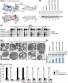
Figure 4. Identification of the potential Sec15 binding site on Myo2
(A) Surface representation indicating mutated residues of the Myo2 cargo-binding domain that show a growth defect (green); Vac17 binding region (blue) (Ishikawa et al., 2003); secretory vesicle/Rab GTPase binding region (red) (Lipatova et al., 2008; Pashkova et al., 2006). (B) Myo2 mutant proteins expressed at similar levels in SC-His-Ura medium. Cell lysates of LWY2947 with indicated plasmids analyzed by immunoblot with antibodies directed against Myo2 cargo-binding domain (top) and Pgk1 (loading control). (C) myo2-R1402E, -Q1472E and –K1473E mutants show a growth defect. pRS413 MYO2, pmyo2-Y1415E, pmyo2-R1402E, pmyo2-Q1472E or pmyo2-K1473E expressed in LWY2947 (myo2Δ cells). Transformed colonies re-streaked onto SC+5FOA to counter select against YCp50 [URA3] MYO2. Colonies from SC + 5FOA were cultured in liquid and serial dilutions spotted on YEPD plates, and incubated at 24, 30 or 37°C for 2 days. (D) myo2 mutants (Y1415E, R1402E and K1372E) accumulate small vesicles. myo2 mutants and corresponding wild-type cells grown to early log phase in YEPD at 24°C. Cells were prepared for electron microscopy and ultrathin sections were stained. Bar = 0.5 μm. (E) Bgl2 secretion is blocked in the myo2 mutants. Accumulation of Bgl2 measured by Western blot using α-Bgl2 antibody (top panel). Bgl2 intensity normalized to an internal loading control (ADH; bottom panel). Averaged units of intensity were plotted (±SEM; n=5), and were significant; p<0.05 for each mutant compared to wild-type (Student's t-test). Strains grown at 24 °C. (F) myo2-R1402E, -Q1472E and -K1473E mutants suppress the vacuole inheritance defect in myo2-2 cells. Indicated plasmids were expressed in the myo2-2 mutant (LWY5516). Quantitative analysis of vacuole inheritance. Vacuole inheritance was assessed as the percent cells with an inherited vacuole in the bud. White bars, normal vacuole inheritance; black bars, bud without detectable FM4-64; gray bars, weaker FM4-64 signal in the bud than in the mother cell. Error bars = S.D. calculated from at least three experiments. Over 170 cells counted.
Figure 5
Figure 5. Myo2-R1402E and -K1473E show a defect in binding to Sec15
(A) myo2-R1402E and –K1473E mutants are defective in binding Sec15 in vitro. Bacterially expressed, purified His-tagged Myo2-R1402E and –K1473E cargo-binding domains have a defect in binding to purified GST-Sec15. (B) Sec15 is mislocalized in myo2 mutants, myo2-R1402E, -Q1472E and –K1473E. Coexpression of vYFP-Sec15 and mCherry-Myo2 (wild-type, R1402E, Q1472E or K1473E) in myo2Δ, sec15Δ double knock out cells (LWY10970). (C) The myo2-R1402E, -Q1472E and –K1473E mutants do not block the ability of Myo2 to bind to Sec4. Yeast two-hybrid test of the Myo2 cargo-binding domain or mutated Myo2 cargo-binding domains, fused with the Gal4 DNA binding domain (BD) and tested for interaction with Sec4 or Vac17 fused with the Gal4 activation domain (AD). (D) In in vitro binding experiments, the Myo2 mutants, R1402E and K1473E, displayed a modest defect in binding Sec4-GTPγS, in contrast with Myo2-Y1415R, which has a major defect in binding Sec4. Purified GST-Sec4 pulled down purified His-tagged Myo2-CBD from E. coli. Proteins pulled down by GST (lanes 1-4), GST-Sec4 (lanes 5-9), and input His-tagged Myo2-CBDs (lanes 10-13) were analyzed by immunoblot with antibody directed against Hisx6 or by Ponceau S staining.
Figure 6
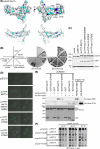
Figure 6. Identification of the Myo2 binding region on Sec15
(A) Model for the structure of yeast Sec15, indicating negatively charged residues (cyan) and conserved negatively charged residues (blue). (B) (Left panel) Plasmids transformed into a sec15Δ strain: pRS415 (mock), pSEC15, pvYFP-SEC15, pvYFP-sec15-D681K/E684K, pvYFP-sec15-D700K, pvYFP-sec15-D705K. Transformed colonies re-streaked on SC+5FOA (middle panel) and SC-Leu-Ura (right panel). (C) Sec15 protein expressed at similar levels in SC-Leu-Ura medium. Cell lysates of LWY10491 with indicated plasmids were analyzed by immunoblot with antibodies directed against Sec15, GFP and Pgk1 (loading control). (D) Lethal Sec15 mutants, D681K/E684K and D705K, are mislocalized. Wild-type yeast (LWY7235) containing the indicated plasmids were visualized by fluorescence microscopy. (E) The Sec15-D705K mutant shows a defect in binding Myo2 in vitro. Bacterially expressed, purified GST-Sec15(577-910)-D705K has a defect in binding purified His-tagged Myo2 cargo-binding domain, but not purified His-tagged Sec4-Q79L. (F) The sec15-D700K mutant suppresses the growth defect of the myo2 mutants, R1402E, Q1472E or K1473E. Plasmids transformed into a myo2Δ, sec15Δ double KO strain: pRS413 MYO2 + pRS415 SEC15, pmyo2-R1402E + pSEC15, pmyo2-R1402E + psec15-D700K, pmyo2-Q1472E + pSEC15, pmyo2-Q1472E + psec15-D700K, pmyo2-K1473E + pSEC15, or pmyo2-K1473E + psec15-D700K. Transformants re-streaked on SC+5FOA to counter select against YCp50 [URA3] SEC15 / MYO2. Colonies from SC + 5FOA were cultured in liquid and serial dilutions spotted on YEPD plates, and incubated at 24, 30 or 37°C for 4 days.
Figure 7
Figure 7. Mammalian myosin-Va interacts with the exocyst complex
(A) Purified GST-mouse myosin Va globular tail domain (GTD) pulled down each exocyst subunit tested, Sec6 and Sec8, from solubilized S100 lysates from NIH 3T3 cells. Proteins pulled down by GST (lane 1) or GST-mMyosin Va-GTD (lane 2), and input S100 (lane 3) analyzed by immunoblot with antibodies directed against Sec6, Sec8 or by CBB stain. (B) mMyosin Va-GTD directly binds to rat Sec15 in vitro. Purified GST-rSec15 pulled down purified His-tagged mMyosin Va-GTD from E. coli. Proteins pulled down by GST (lane 1), GST-rSec15 (lane 2 and 3), and input His-tagged mMyosin Va-GTD (lane 4) analyzed by immunoblot with antibody directed against Hisx6 or by CBB stain.
Figure 8
Figure 8. Model of the direct association of Myo2 with each step in the life of a secretory vesicle
Myosin V is present at each of the exocytic vesicle transport steps. For simplicity, only those vesicle-related molecules that have been demonstrated to interact with the cargo binding domain of Myo2 are shown. Ypt31/32 is required for production of secretory vesicles from the trans-Golgi (Jedd et al., 1997). GTP-bound Ypt31/32 also binds directly to Myo2 (Lipatova et al., 2008). GTP bound Sec4 binds Myo2 (this study, (Santiago-Tirado et al., 2011). Sec15 binds both Myo2 and GTP bound Sec4 (this study). Other exocyst subunits are recruited to the vesicles. Secretory vesicles fuse with the plasma membrane. The precise order of each of these events is not yet established. In this model Myo2 binds to vesicles before they are released from a post Golgi compartment, and remains attached until very late in the fusion pathway.

Similar articles

Cited by

References

    1. Adamo JE, Rossi G, Brennwald P. The Rho GTPase Rho3 has a direct role in exocytosis that is distinct from its role in actin polarity. Mol Biol Cell. 1999;10:4121–4133. - PMC - PubMed
    1. Beronja S, Laprise P, Papoulas O, Pellikka M, Sisson J, Tepass U. Essential function of Drosophila Sec6 in apical exocytosis of epithelial photoreceptor cells. J Cell Biol. 2005;169:635–646. - PMC - PubMed
    1. Boyd C, Hughes T, Pypaert M, Novick P. Vesicles carry most exocyst subunits to exocytic sites marked by the remaining two subunits, Sec3p and Exo70p. J Cell Biol. 2004;167:889–901. - PMC - PubMed
    1. Brennwald P, Kearns B, Champion K, Keranen S, Bankaitis V, Novick P. Sec9 is a SNAP-25-like component of a yeast SNARE complex that may be the effector of Sec4 function in exocytosis. Cell. 1994;79:245–258. - PubMed
    1. Calero M, Chen CZ, Zhu W, Winand N, Havas KA, Gilbert PM, Burd CG, Collins RN. Dual prenylation is required for Rab protein localization and function. Mol Biol Cell. 2003;14:1852–1867. - PMC - PubMed

Publication types

MeSH terms

LinkOut - more resources