. 2011;6(12):e28594.
doi: 10.1371/journal.pone.0028594. Epub 2011 Dec 5.

Inhibition of interferon induction and action by the nairovirus Nairobi sheep disease virus/Ganjam virus

Affiliations

Inhibition of interferon induction and action by the nairovirus Nairobi sheep disease virus/Ganjam virus

Barbara Holzer et al. PLoS One. 2011.

Abstract

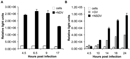
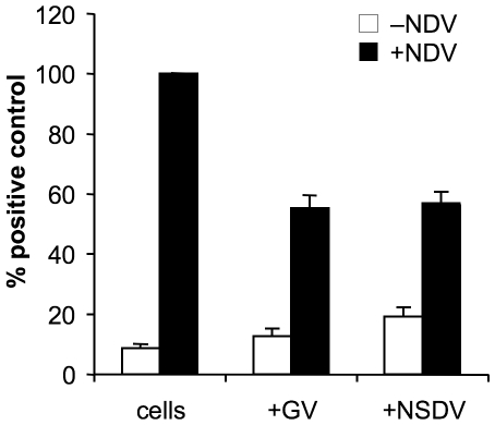
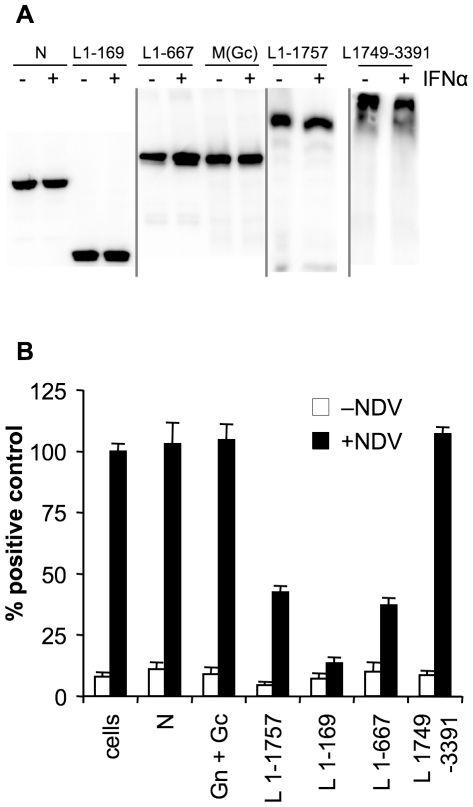
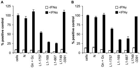
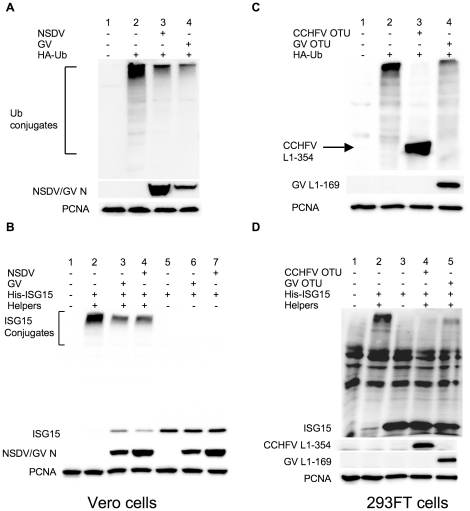
The Nairoviruses are an important group of tick-borne viruses that includes pathogens of man (Crimean Congo hemorrhagic fever virus) and livestock animals (Dugbe virus, Nairobi sheep disease virus (NSDV)). NSDV is found in large parts of East Africa and the Indian subcontinent (where it is known as Ganjam virus). We have investigated the ability of NSDV to antagonise the induction and actions of interferon. Both pathogenic and apathogenic isolates could actively inhibit the induction of type 1 interferon, and also blocked the signalling pathways of both type 1 and type 2 interferons. Using transient expression of viral proteins or sections of viral proteins, these activities all mapped to the ovarian tumour-like protease domain (OTU) found in the viral RNA polymerase. Virus infection, or expression of this OTU domain in transfected cells, led to a great reduction in the incorporation of ubiquitin or ISG15 protein into host cell proteins. Point mutations in the OTU that inhibited the protease activity also prevented it from antagonising interferon induction and action. Interestingly, a mutation at a peripheral site, which had little apparent effect on the ability of the OTU to inhibit ubiquitination and ISG15ylation, removed the ability of the OTU to block the induction of type 1 and the action of type 2 interferons, but had a lesser effect on the ability to block type 1 interferon action, suggesting that targets other than ubiquitin and ISG15 may be involved in the actions of the viral OTU.


Conflict of interest statement

Competing Interests: The authors have declared that no competing interests exist.

Figures

Figure 1
Figure 1. Delayed IFNβ induction in GV and NSDV-infected cells.
Vero cells were transfected with 400 ng of pIFNβ-luc and 200 ng pJATLacZ. After 24 hours of transfection, the cells were infected with (a) NSDV or GV, or (b) NDV at an MOI of 1 TCID50 unit per cell, or left uninfected. At the indicated time points cells were lysed and assayed for luciferase and β-galactosidase activities. The ratio of these two activities was taken as the relative luciferase activity (in RLU). Shown are the data from a representative experiment in triplicates; error bars represent one standard error of the mean.
Figure 2
Figure 2. NSDV and GV suppress transcription from the IFNβ promoter.
Vero cells were transfected with 500 ng each pIFNβ-luc and pJATLacZ. After 24 hours of transfection cells were infected with GV and NSDV at an MOI of 1 TCID50 unit per cell or left uninfected. Eight hours after infection with NSDV or GV, cells were superinfected with Newcastle disease virus (NDV) at an MOI of 1 TCID50 unit per cell or left uninfected, and finally lysed four hours after infection with NDV. The luciferase and β-galactosidase activities of the cell extracts were determined. Results from three separate experiments were combined by setting the RLUs induced by NDV in uninfected cells to 100%; all experiments were performed in triplicate wells. Error bars show standard errors of the normalised data.
Figure 3
Figure 3. GV and NSDV inhibit the induction of expression from IFN-responsive promoters.
Vero cells were transfected with 500 ng pJATLacZ and (a) 500 ng pGL3-Mx-1-luc or (b) pGAS-luc. 30 hours post transfection cells were infected with GV or NSDV at an MOI of 1 or left uninfected. 18 hours post infection cells were treated for 6 hours with IFNα (a) or IFNγ (b), lysed, and the luciferase and β-galactosidase activities determined. Results from two (for IFNα) or three (for IFNγ) separate experiments were combined by setting the RLUs induced by IFNα or IFNγ in uninfected cells to 100%; all experiments were performed in triplicate wells. Error bars show standard errors of the normalised data.
Figure 4
Figure 4. GV and NSDV infection interfere with the tyrosine phosphorylation of STAT1 and STAT2 in response to IFNα or IFNγ.
Vero cells were infected with GV or NSDV at an MOI of 1 TCID50 unit per cell or left uninfected (mock). At the indicated time post-infection cells were treated with or without IFNα (a) or IFNγ (b) for 30 min. The cells were harvested and the levels of STATs and tyrosine-phosphorylated STAT proteins were determined by Western blot analysis with the corresponding specific antibodies. The data shown are from a representative experiment.
Figure 5
Figure 5. Ganjam L protein inhibits transcription from IFNβ-promoter in response to NDV infection.
(a) Vero cells were transfected with 350 ng pIFNβ-luc, 200 ng pJATLacZ and 1.2 µg of a plasmid driving the expression of the indicated viral protein, with the exception of pcDNA6-GV-L1-169 and pcDNA6-GV-N where 700 ng and 300 ng respectively were used in transfection experiments. All transfections were made up to the same amount of DNA using empty vector. After 40 hours of transfection cells were infected with NDV (MOI = 1). After a further 5 hours the cells were (a) lysed with SDS-PAGE sample buffer and the expression of viral proteins detected by Western blot using a monoclonal antibody to the V5 epitope or to the His epitope as required, or (b) lysed with NP40 lysis buffer as described in Methods and the luciferase and β-galactosidase activities determined. Results from two or three separate experiments were combined by setting the RLUs induced by NDV in cells transfected with empty vector to 100%. Error bars show standard errors of normalised data.
Figure 6
Figure 6. GV L protein inhibits transcription from IFN-responsive promoters.
(a) Vero cells were transfected essentially as for Figure 5 except that 100 ng of the reporter gene plasmid pGL3Mx-1-lucwas used. All transfections were made up to the same amount of DNA using empty vector where required. After 40 hours of transfection cells were incubated with or without IFNα. After a further 8 hours the cells were lysed and the luciferase and β-galactosidase activities determined as described in material and methods. (b) Vero cells were transfected essentially as for Figure 5 except that 400 ng of the reporter gene plasmid pGAS-luc was used. 40 hours post-transfection cells were treated with IFNγ. After a further 6 hours cells were lysed and the luciferase and β-galactosidase activities determined. Results from separate experiments were combined by setting the RLUs induced by IFNα in cells transfected with empty vector to 100%. Error bars show standard errors of normalised data.
Figure 7
Figure 7. GV and NSDV inhibit ubiquitination and ISG15ylation.
(a) Vero cells were transfected with 500 ng pHA-Ub. 14 hours post transfection cells were infected with NSDV or GV at an MOI of 1 TCID50 unit per cell. After a further 12 h cells were lysed in SDS PAGE sample buffer and total protein ubiquitination was analysed by Western blot using anti-HA. Lane 1: Untransfected cells; Lanes 2-4:transfected with pUb-HA; Lane 3: GV-infected; Lane 4: NSDV-infected. (b) Vero cells were transfected with 250 ng pHis-mISG15 with 750 ng empty vector (Lanes 5–7) or with 250 ng each HA-mHerc6, mUBE1L-HA and UbcM8 (Lanes 2-4) or left untransfected (Lane 1). After 14 hours of transfection cells were infected with GV (Lane 3, 6) or NSDV (Lane 4, 7) at an MOI of 1 TCID50 unit per cell or left uninfected (Lanes 1, 2, 5). Twelve hours after infection, samples were analysed by Western blot using anti-6His antibody. Samples were also probed for the presence of viral protein (N) to confirm infection. (c) 293FT cells were transfected with 400 ng pUb-HA and with 500 ng empty vector (Lane 2) or with 500 ng of plasmid expressing CCHFV L1-354-HA (Lane 3) or GV L1-169-V5 (Lane 4) or left untransfected (Lane 1). 30 hours post transfection cells were lysed and total protein ubiquitination was determined by Western blot using anti-HA. The expression of GV L1-169 was detected by using anti-V5. (d) 293FT cells were transfected with 250 ng each pHis-mISG15, HA-mHerc6, mUBE1L-HA, UbcM8 alone (Lane 2) or together with 250 ng of plasmid expressing CCHFV L1-354-HA (Lane 4) or GV L1-169-V5 (Lane 5) or left untransfected (Lane 1). As extra negative control cells were transfected with 250 ng pHis-mISG15 and 750 ng empty plasmid (Lane 3). After 30 h of transfection cells were lysed and total ISG15ylation was analyzed by Western blot using anti-6His. The expression level of GV L1-169 and CCHFV L1-354 were assayed by using anti-V5 and anti-HA respectively. PCNA levels served as loading control in all experiments.
Figure 8
Figure 8. The OTU enzymatic activity is required for inhibition of IFN induction and action.
(a) 293FT cells were transfected with 400 ng pHA-Ub and with 500 ng empty vector (Lane 2) or with 500 ng of plasmid expressing GV L1-169-V5 wildtype (Lane 3) or with the mutants C40A (Lane 4), H151A (Lane 5) or Q16R (Lane 6) or left untransfected (Lane 1). Thirty hours post-transfection, cells were lysed and total protein ubiquitination was determined by Western blot using anti-HA. (b) 293FT cells were transfected with 250 ng each pHis-mISG15, HA-mHerc6, mUBE1L-HA, UbcM8 alone (Lane 3) or together with 500 ng of plasmid expressing GV L1-169-V5 (Lane 4) or with one of the following mutants C40A (Lane5), H151A (Lane 6) or L16R (Lane 7), or left untransfected (Lane 1). As an extra negative control, cells were transfected with 250 ng pHis-mISG15 and 1250 ng empty plasmid (Lane 2). After 30 h of transfection cells were lysed and total ISG15ylation was analyzed by Western blot using anti-6His. The expression level of GV L1-169 and its mutants were assayed by using anti-V5. PCNA levels served as loading control in all experiments. (c) Vero cells were transfected with 350 ng of the reporter plasmid pIFNβ-luc plus 200 ng pJATLacZ combined with 700 ng of pcDNA6-GV-L1-169 or of one of the mutants C40A, H151A, or Q16R, or empty vector. After 24 hours of transfection cells were infected with Sendai virus (SV) or left uninfected. Eight hours post-infection the cells were lysed with NP40 lysis buffer and the luciferase and β-galactosidase activities determined as described in material and methods. (d) Vero cells were transfected essentially as in (c) except that 100 ng of the reporter plasmid pGL3Mx-1-luc was used. After 24 hours of transfection cells were incubated with or without IFNα. After a further 8 hours the cells were lysed and the luciferase and β-galactosidase activities determined as described in material and methods. (e) Vero cells were transfected essentially as in (c) except that 400 ng of pGAS-luc was used as the reporter plasmid. After 24 hours of transfection cells were incubated with or without IFNγ. After a further 8 hours the cells were lysed and the luciferase and β-galactosidase activities determined as described in material and methods. Error bars in (c-e) show standard error of the mean of normalised data. Results from three separate experiments were combined by setting the RLUs induced by SV or IFN in cells transfected with empty vector to 100%.

Similar articles

Cited by

References

    1. Montgomery E. On a tick-borne gastro-enteritis of sheep and goats occuring in British East Africa. J Comp Pathol. 1917;30:28–57.
    1. Marczinke BI, Nichol ST. Nairobi sheep disease virus, an important tick-borne pathogen of sheep and goats in Africa, is also present in Asia. Virology. 2002;303:146–151. - PubMed
    1. Daubney R, Hudson JR. Nairobi Sheep Disease. Parasitology. 1931;23:507–524.
    1. Daubney R, Hudson JR. Nairobi Sheep Disease; natural and experimental transmission by ticks other than Rhipicephalus appendiculatus. Parasitology. 1934;26:496–509.
    1. Dandawate CN, Shah KV. Ganjam virus: a new arbovirus isolated from ticks Haemaphysalis intermedia Warburton and Nuttall, 1909 in Orissa, India. Indian J Med Res. 1969;57:799–804. - PubMed

Publication types

MeSH terms