. 2011;6(12):e28308.
doi: 10.1371/journal.pone.0028308. Epub 2011 Dec 5.

PTEN modulates miR-21 processing via RNA-regulatory protein RNH1

Affiliations

PTEN modulates miR-21 processing via RNA-regulatory protein RNH1

Youn-Jae Kim et al. PLoS One. 2011.

Abstract

Aberrant miR-21 expression is closely associated with cell proliferation, anti-apoptosis, migration, invasion, and metastasis in various cancers. However, the regulatory mechanism of miR-21 biogenesis is largely unknown. Here, we demonstrated that the tumor suppressor PTEN negatively regulates the expression of oncogenic miR-21 at the post-transcriptional level. Moreover, our results suggest that PTEN plays such a role through the indirect interaction with the Drosha complex. To elucidate how PTEN regulates pri- to pre-miR-21 processing, we attempted to find PTEN-interacting proteins and identified an RNA-regulatory protein, RNH1. Using the sensor to monitor pri-miR-21 processing, we demonstrated that RNH1 is necessary and sufficient for pri-miR-21 processing. Moreover, our results propose that the nuclear localization of RNH1 is important for this function. Further analysis showed that RNH1 directly interacts with the Drosha complex and that PTEN blocks this interaction. Taken together, these results suggest that the PTEN-mediated miR-21 regulation is achieved by inhibiting the interaction between the Drosha complex and RNH1, revealing previously unidentified role of PTEN in the oncogenic miR-21 biogenesis.


Conflict of interest statement

Competing Interests: The authors have declared that no competing interests exist.

Figures

Figure 1
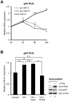
Figure 1. Post-transcriptional regulation of miR-21 expression by PTEN.
(A) Time course of pri- (small dotted line), pre- (large dotted line), or mature miR-21 (single unbroken line) expression in U87MG cells after HA treatment. Expression level of each miRNA was analyzed by qRT-PCR reactions which were normalized to GAPDH for pri- and pre-miR-21, and U6 small nuclear RNA for mature miR-21. (B) Expression level of mature miR-21. U87MG cells pretreated with ActD or overexpressing PTEN were treated with HA for 24 hr. Data represent the mean values of at least three independent experiments performed in triplicate. **P<0.01 and ***P<0.001; n.s., not significant; Student's t test. Error bars indicate s.e.m. PTEN expression and AKT phosphorylation were confirmed by immunoblotting.
Figure 2
Figure 2. PTEN regulates pri-miR-21 processing.
(A) In vitro pri-miR-21 processing with Drosha-expressing cell lysates, Drosha immunoprecipitates, or glioblastoma cell lysates. Lane 1: probe only; lane 2: Drosha-WT overexpressed cell lysates; lane 3: Drosha-TN overexpressed cell lysates; lane 4: Drosha-WT immunoprecipitate; lane 5: Drosha-TN immunoprecipitate; lane 6: Drosha-WT immunoprecipitate; lane 7: LN428 cell lysates; lane 8: U87MG cell lysates. PTEN expression of LN428 (lane 9) and U87MG (lane 10) was confirmed by immunoblotting. (B) In vitro pri-miR-21 processing with parental 293T and PTEN expressing cell lysates. Lane 1: probe only; lane 2: parental 293T cell lysates; lane 3: PTEN expressing cell (clone #25) lysates; lane 4: PTEN expressing cell (clone #33) lysates. PTEN expression of parental 293T (lane 5) and PTEN expressing cells (lane 6: clone #25; lane 7: clone #33) was confirmed by immunoblotting. (C) In vitro pri-let-7a-1 processing with parental 293T and WT-PTEN expressing cell lysates. Lane 1: probe only; lane 2: parental 293T cell lysates; lane 3: PTEN expressing cell (clone #25) lysates; lane 4: PTEN expressing cell (clone #33) lysates. IB; immunoblot.
Figure 3
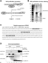
Figure 3. Identification of RNH1 as a novel PTEN-interacting protein.
(A) Schematic diagram of tandem affinity purification (TAP) procedure. A S-tag, double FLAG tag, and streptavidin-binding peptide were fused at the N-terminus of PTEN (SFS-PTEN). After sequential streptavidin and S-protein bead binding, the PTEN-interacting proteins were eluted from S-protein beads. The eluted proteins were analyzed by LC-MS/MS. X and Y represent nonspecifically interacting proteins. (B) The silver staining result of TAP purified proteins. Arrows indicate an RNA-regulatory protein RNH1 (lower) and SFS-PTEN (upper). (C) Peptide sequences (boxes) of RNH1 by LC-MS/MS analysis. (D) Physical interaction between PTEN and RNH1. Plasmids encoding HA-RNH1 and SFS-PTEN were ectopically expressed in 293T cells. SFS-PTEN was precipitated with anti-FLAG M2 affinity agarose gels and then immunoblotting with anti-FLAG, anti-HA, or anti-β-Actin (negative binding control) was performed. IgG (LC): IgG (light chain).
Figure 4
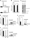
Figure 4. Establishment of pri-miR-21 processing sensors.
(A) Schematic depiction of pri-miR-21 processing sensors. (B) Luciferase assay results of each transfectant. The sensors or control plasmid were ectopically expressed along with the plasmid encoding Drosha (Dros.)-WT in 293T cells. (C) Northern blot analysis of sensor. Total RNAs from U87MG cells were used as positive controls of pre- and mature miR-21 expression. The control plasmid or 5′-sensor was ectopically expressed in 293T cells. Arrows indicate positions of pre- (upper) and mature (lower) miR-21. 5S rRNA was used as the loading control. (D) Luciferase assay results of each transfectant. The sensor was co-transfected into 293T cells along with the plasmid encoding Drosha-WT or Drosha-TN. Drosha expression was confirmed by immunoblotting. (E) Luciferase assay results of each transfectant. The sensor was co-transfected into 293T cells along with siRNA against green fluorescent protein (GFP) or Drosha. Drosha expression was confirmed by immunoblotting. (F) Luciferase assay results of each transfectant. The sensor was co-transfected into 293T cells along with the plasmid encoding FLAG-Lin28-WT or FLAG-Lin28-TN which has no binding activity for the pre-let-7 family. Data represent the mean values of at least three independent experiments performed in triplicate. **P<0.01, and ***P<0.001; Student's t test. Error bars indicate s.e.m.
Figure 5
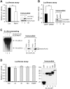
Figure 5. RNH1 modulates pri-miR-21 processing.
(A) Luciferase assay results of each transfectant. The sensor was co-transfected into 293T cells along with the plasmid encoding HA-RNH1. HA-RNH1 expression was confirmed by immunoblotting. (B) Luciferase assay results of each transfectant. The sensor was co-transfected into 293T cells along with siRNA against GFP or RNH1. (C) In vitro pri-miR-21 processing with Drosha or RNH1 immunoprecipitates. The expression of FLAG-RNH1 and Drosha-FLAG was confirmed by immunoblotting. (D) Luciferase assay results of each transfectant. The sensor was co-transfected into 293T cells along with the plasmid encoding GFP, GFP-hnRNP A1 (A1), GFP-hnRNP E1 (E1), GFP-YB1 (YB1), or GFP-FMRP (FMRP). Each expression was confirmed by immunoblotting. Data represent the mean values of at least three independent experiments performed in triplicate. **P<0.01; Student's t test. Error bars indicate s.e.m.
Figure 6
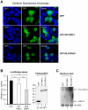
Figure 6. Nuclear localization of RNH1 is essential for the pri-miR-21 processing.
(A) Confocal fluorescence microscopy of each transfectant. The plasmid encoding GFP, GFP-WT-RNH1, or GFP-NLS-RNH1 was ectopically expressed in 293 cells. The experiments were repeated at least three times with similar results. (B) Luciferase assay results of each transfectant. The sensor was co-transfected into 293T cells along with the plasmid encoding GFP, GFP-WT-RNH1 (WT-RNH1), or GFP-NLS-RNH1 (NLS-RNH1). Data represent the mean values of at least three independent experiments performed in triplicate. *P<0.05, **P<0.01; Student's t test. Error bars indicate s.e.m. Each protein expression was confirmed by immunoblotting. (C) Northern blot analysis of each transfectant. The plasmid encoding GFP, WT-RNH1, or NLS-RNH1 was ectopically expressed in 293T cells along with the plasmid encoding the pri-miR-21 minigene. Arrows indicate positions of pre- (upper) and mature (lower) miR-21. 5S rRNA was used as the loading control. The experiments were repeated at least three times with similar results.
Figure 7
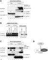
Figure 7. PTEN has inhibitory role for RNH1 interaction to the Drosha complex.
(A) Physical interaction between RNH1 and the Drosha complex. The plasmid encoding Drosha-FLAG and HA-RNH1 were ectopically expressed in 293T cells. Drosha-FLAG was precipitated with anti-FLAG M2 affinity agarose gels and then and then immunoblotting with anti-FLAG (antibody 4C5), anti-HA, or anti-hnRNP A1 (negative binding control) was performed. (B) Physical interaction between RNH1 and pri-miR-21. The plasmid encoding FLAG-RNH1 or Drosha-FLAG was ectopically expressed in HeLa cells. RNA-protein complexes were precipitated with anti-FLAG M2 affinity agarose gels. After IP of RNA-protein complexes, RNAs were isolated and used in RT-PCR reactions with specific oligomers for pri-miR-21 or pri-miR-29a. The PCR products were resolved on 1% agarose gel. Each protein expression was confirmed by immunoblotting. (C) The inhibitory role of PTEN in the interaction of RNH1 with the Drosha complex. The plasmid encoding FLAG-RNH1 and PTEN were ectopically expressed in 293T cells. FLAG-RNH1 was precipitated with anti-FLAG M2 affinity agarose gels and then immunoblotting with anti-Drosha, anti-PTEN, anti-FLAG (anti-DYKDDDDK), or anti-hnRNP A1 (negative binding control) was performed. The experiments were repeated at least three times with similar results. (D) Proposed illustration shows that PTEN negatively modulates the biogenesis of miR-21 by blocking the interaction of the Drosha complex with RNH1, which facilitates pri-miR-21 processing.

Similar articles

Cited by

References

    1. Bartel DP. MicroRNAs: target recognition and regulatory functions. Cell. 2009;136:215–233. - PMC - PubMed
    1. Filipowicz W, Bhattacharyya SN, Sonenberg N. Mechanisms of post-transcriptional regulation by microRNAs: are the answers in sight? Nat Rev Genet. 2008;9:102–114. - PubMed
    1. Garofalo M, Croce CM. microRNAs: Master regulators as potential therapeutics in cancer. Annu Rev Pharmacol Toxicol. 2011;51:25–43. - PubMed
    1. Farazi TA, Spitzer JI, Morozov P, Tuschl T. miRNAs in human cancer. J Pathol. 2011;223:102–115. - PMC - PubMed
    1. Kumar MS, Lu J, Mercer KL, Golub TR, Jacks T. Impaired microRNA processing enhances cellular transformation and tumorigenesis. Nat Genet. 2007;39:673–677. - PubMed

Publication types

MeSH terms