Review
. 2011 Dec 9;12(1):35-48.
doi: 10.1038/nri3111.

The role of ubiquitylation in immune defence and pathogen evasion

Affiliations
Review

The role of ubiquitylation in immune defence and pathogen evasion

Xiaomo Jiang et al. Nat Rev Immunol. .

Abstract

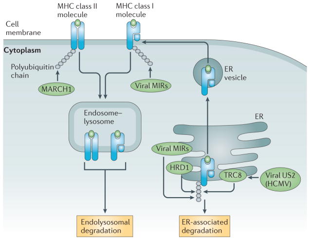
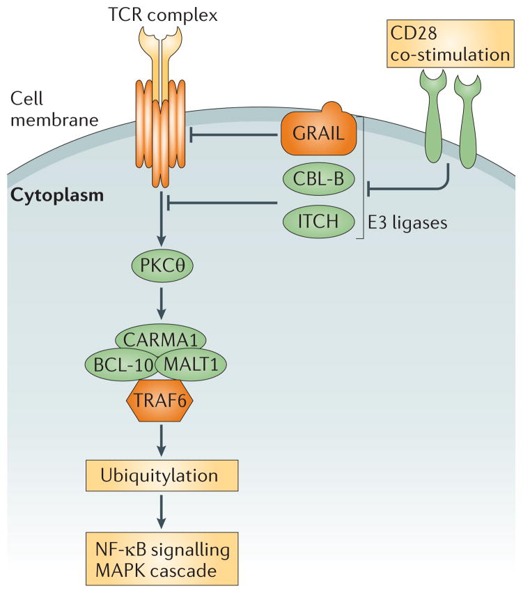
Ubiquitylation is a widely used post-translational protein modification that regulates many biological processes, including immune responses. The role of ubiquitin in immune regulation was originally uncovered through studies of antigen presentation and the nuclear factor-κB family of transcription factors, which orchestrate host defence against microorganisms. Recent studies have revealed crucial roles of ubiquitylation in many aspects of the immune system, including innate and adaptive immunity and antimicrobial autophagy. In addition, mounting evidence indicates that microbial pathogens exploit the ubiquitin pathway to evade the host immune system. Here, we review recent advances on the role of ubiquitylation in host defence and pathogen evasion.


Conflict of interest statement

Competing interests statement

The authors declare no competing financial interests.

Figures

Figure 1
Figure 1. Ubiquitin-mediated signalling in TLR and IL-1R pathways
Stimulation of Toll-like receptors (TLRs) and the interleukin-1 receptor (IL-1R) leads to the recruitment of the adaptor protein myeloid differentiation primary response protein 88 (MYD88), which in turn recruits the kinases IL-1R-associated kinase 4 (IRAK4) and IRAK1, forming the ‘myddosome’. The E3 ligase TNF receptor-associated factor 6 (TRAF6) is then recruited and activated, and synthesizes K63-linked polyubiquitin chains. These polyubiquitin chains recruit kinase complexes containing TGFβ-activated kinase 1 (TAK1) or IκB kinase (IKK) through their ubiquitin-binding subunits — TAK1-associated binding protein 2 (TAB2)–TAB3 and NF-κB essential modulator (NEMO), respectively. Binding of K63-linked polyubiquitin to TAB2 and TAB3 leads to TAK1 activation, which in turn activates the mitogen-activated protein kinase (MAPK) cascade. Binding of K63-linked polyubiquitin to both the IKK and TAK1 complexes facilitates the phosphorylation of IKKβ by TAK1, leading to IKK activation. IKK phosphorylates NF-κB inhibitor (IκB) proteins and targets them for polyubiquitylation by the SCFβTrCP ubiquitin E3 ligase complex. The polyubiquitylated IκB proteins are degraded by the proteasome, allowing nuclear factor-κB (NF-κB) to enter the nucleus to turn on target genes involved in immune and inflammatory responses.
Figure 2
Figure 2. Different types of polyubiquitylation in the TLR4 signalling pathways
Toll-like receptor 4 (TLR4) that is activated at the cell membrane recruits myeloid differentiation primary response protein 88 (MYD88) through TIR domain-containing adaptor protein (TIRAP). Signalling complexes containing MYD88, IL-1 receptor-associated kinases (IRAKs) and TNF receptor-associated factor 6 (TRAF6) are formed at the plasma membrane. TRAF6 activates the E3 ligases cellular inhibitor of apoptosis protein 1 (cIAP1) and cIAP2, which induce the ubiquitylation and subsequent degradation of TRAF3. The degradation of TRAF3 releases TRAF6 signalling complexes from TLR4 into the cytosol, where additional components are recruited and activated. Another branch of signalling pathways is stimulated when TLR4 is endocytosed. The adaptor protein TRAM recruits TIR domain-containing adaptor protein inducing IFNβ (TRIF) to endocytosed TLR4. Through the action of several E3 ligases — such as pellino 1 (PELI1), TRAF3 and TRAF6 — which mediate the ubiquitylation of signalling proteins such as receptor-interacting protein 1 (RIP1) and TRAF3, downstream pathways leading to nuclear factor-κB (NF-κB) activation and type I interferon (IFN) induction are activated. Deubiquitylation enzymes (DUBs), such as A20, provide mechanisms for the downregulation of various pro-inflammatory signalling pathways, by disassembling polyubiquitin chains. IκB, NF-κB inhibitor; IKK, IκB kinase; IRF3, interferon regulatory factor 3; MAPK, mitogen-activated protein kinase; NEMO, NF-κB essential modulator; TAB, TAK1-binding protein; TAK1, TGFβ-activated kinase 1; TBK1, TANK-binding kinase 1.
Figure 3
Figure 3. Roles of ubiquitylation in diverse PRR signalling pathways
a | Nucleotide-binding oligomerization domain protein 2 (NOD2) detects bacterial peptidoglycans and triggers the ubiquitylation of receptor-interacting protein 2 (RIP2), which leads to the activation of the mitogen-activated protein kinase (MAPK) cascade and nuclear factor-κB (NF-κB). b | In the retinoic acid-inducible gene I (RIG-I)-mediated pathway that senses viral RNA, K63-linked polyubiquitylation and the E3 ligases triparite motif-containing protein 25 (TRIM25) and riplet are important for RIG-I activation. RIG-I transduces the signal to mitochondrial antiviral signalling protein (MAVS) on mitochondria, and this induces the activation of the IκB kinase (IKK) and TANK-binding kinase 1 (TBK1) complexes through NF-κB essential modulator (NEMO). Ubiquitylation of NEMO and TBK1 by TRIM23 and mindbomb (MIB) proteins, respectively, has been suggested to mediate TBK1 activation. IKK and TBK1 activate NF-κB and interferon regulatory factors (IRFs) to induce the transcription of pro-inflammatory cytokines and type I interferons (IFNs), respectively. c | Activation of the stimulator of interferon genes (STING)-mediated pathways that sense intracellular DNA also involves ubiquitylation (for example, by TRIM56). cIAP, cellular inhibitor of apoptosis protein; ER, endoplasmic reticulum; TANK, TRAF family member-associated NF-κB activator.
Figure 4
Figure 4. Ubiquitin adaptor proteins mediate selective autophagy of pathogens
Ubiquitylation tags invading bacterial pathogens by targeting bacterial proteins or associated host components. Adaptor proteins, such as p62, NDP52 and optineurin (OPTN), that bind to both ubiquitin and the ubiquitin-like protein LC3, link ubiquitin-tagged bacteria with the autophagic machinery. Antibacterial peptide precursors are targeted in a similar manner by p62 so that the antibacterial peptides are produced in the intracellular compartments that contain the bacteria.
Figure 5
Figure 5. Ubiquitylation controls the expression of MHC molecules
MARCH1 (membrane-associated RING-CH 1)-mediated ubiquitylation of MHC class II molecules promotes their endolysosomal degradation, thus controlling the level of cell-surface expression of MHC class II molecules. MHC class I expression is regulated by the endoplasmic reticulum (ER)-associated degradation pathway and by endolysosomal degradation, both of which are controlled by ubiquitylation. MHC class I ubiquitylation can be mediated by viral E3 ligases, such as modulator of immune response (MIR) proteins, or by cellular E3 enzymes, such as HMG-CoA reductase degradation 1 (HRD1). Other cellular E3 ligases can be exploited by viral proteins; for example, the TRC8-mediated ubiquitylation of MHC class I molecules is induced by the human cytomegalovirus (HCMV) protein US2.
Figure 6
Figure 6. Ubiquitin E3 ligases regulate TCR signalling
Stimulation of the T cell receptor (TCR) leads to the recruitment and activation of protein kinase Cθ (PKCθ), which in turn recruits a protein complex comprising CARMA1, BCL-10 and MALT1. MALT1 binds to and activates TNF receptor-associated factor 6 (TRAF6), which then activates nuclear factor-κB (NF-κB) and mitogen-activated protein kinases (MAPKs) through K63-linked polyubiquitylation. Proper T cell activation requires both TCR stimulation and co-stimulatory signals. In the absence of co-stimulation, several E3 ligases (such as GRAIL, CBL-B and ITCH) keep T cell activation in check by targeting multiple stages of the TCR signalling pathway, including the TCR complex and downstream signalling molecules, such as PKCθ. Co-stimulatory signals release the inhibition of TCR signalling mediated by E3 ligases.

Similar articles

Cited by

References

    1. Pickart CM. Mechanisms underlying ubiquitination. Annu Rev Biochem. 2001;70:503–533. - PubMed
    1. Sun SC. Deubiquitylation and regulation of the immune response. Nature Rev Immunol. 2008;8:501–511. - PMC - PubMed
    1. Harhaj EW, Dixit VM. Deubiquitinases in the regulation of NF-κB signaling. Cell Res. 2011;21:22–39. - PMC - PubMed
    1. Dikic I, Wakatsuki S, Walters KJ. Ubiquitin-binding domains — from structures to functions. Nature Rev Mol Cell Biol. 2009;10:659–671. - PMC - PubMed
    1. Chen ZJ, Sun LJ. Nonproteolytic functions of ubiquitin in cell signaling. Mol Cell. 2009;33:275–286. - PubMed

Publication types