. 2012 Jan 20;287(4):2446-58.
doi: 10.1074/jbc.M111.292748. Epub 2011 Dec 6.

Macromolecular crowding regulates assembly of mRNA stress granules after osmotic stress: new role for compatible osmolytes

Affiliations

Macromolecular crowding regulates assembly of mRNA stress granules after osmotic stress: new role for compatible osmolytes

Ouissame Bounedjah et al. J Biol Chem. .

Abstract

The massive uptake of compatible osmolytes such as betaine, taurine, and myo-inositol is a protective response shared by all eukaryotes exposed to hypertonic stress. Their accumulation results mostly from the expression of specific transporters triggered by the transcriptional factor NFAT5/TonEBP. This allows the recovery of the cell volume without increasing intracellular ionic strength. In this study we consider the assembly and dissociation of mRNA stress granules (SGs) in hypertonic-stressed cells and the role of compatible osmolytes. In agreement with in vitro results obtained on isolated mRNAs, both macromolecular crowding and a high ionic strength favor the assembly of SGs in normal rat kidney epithelial cells. However, after hours of constant hypertonicity, the slow accumulation in the cytoplasm of compatible osmolytes via specific transporters both reduces macromolecular crowding and ionic strength, thus leading to the progressive dissociation of SGs. In line with this, when cells are exposed to hypertonicity to accumulate a large amount of compatible osmolytes, the formation of SGs is severely impaired, and cells increase their chances of survival to another hypertonic episode. Altogether, these results indicate that the impact of compatible osmolytes on the mRNA-associated machineries and especially that associated with SGs may play an important role in cell resistance and adaption to hyperosmolarity in many tissues like kidney and liver.


Figures
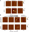
FIGURE 1.
FIGURE 1.
High resolution AFM imaging reveals mRNA assembly into granules and MT bundling under macromolecular crowding environment. A, at moderate ionic strength (20 mm Tris-HCl, pH 7.4, 50 mm KCl), 1% PEG 35K (w/v), a crowding agent, triggers the assembly of MTs into thick bundles, and 20% PEG 35K triggers that of 2Luc mRNA into granules. In contrast, PEG 1K was unable to induce mRNA self-assembly, as expected for excluded volume interactions because of its small size. Incubation time, 30 min. Scale bars, 2.5 μm (AFM images of MTs) and 1 μm (AFM images of mRNAs). B, with 0.5 and 15% PEG 35K at moderate ionic strength, no MT bundles or mRNA aggregates were detected, respectively. However, increasing KCl concentration led to the progressive appearance of MT bundles or mRNA granules. When betaine, a neutral osmolyte, was used instead of KCl to increase the osmolality without increasing the ionic strength, no mRNA granules or MT bundles were observed. Incubation time, 30 min. Scale bars, 2.5 μm (AFM images of MTs) and 1 μm (AFM image of mRNAs).
FIGURE 2.
FIGURE 2.
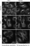
NaCl and sucrose induce SG assembly and MT bundling in NRK cells in contrast with membrane-permeable molecules like urea. NRK cells labeled with anti-YB-1 were exposed to a hypertonic environment (720 mosmol/kg), in which osmolality has been adjusted with urea, NaCl, or sucrose. Scale bar, 15 μm.
FIGURE 3.
FIGURE 3.
Hypertonicity induces SG assembly and MT bundling in cells. A, NRK cells were incubated at the indicated osmolalities for 45 min and then fixed for immunostaining. Anti-YB-1 labeling of NRK cells reveals at 620 mosmol/kg the formation of SGs, which appear even larger at 720 mosmol/kg. Similarly, anti-tubulin labeling indicates a significant tendency for MTs to assemble into bundles, which is especially pronounced at 720 mosmol/kg. Scale bar, 10 μm. B, mean granule area was obtained from A. The results indicate that SG assembly is promoted by increasing the extracellular osmolality above 520 mosmol/kg. Results are the mean ± S.D. (see “Materials and Methods”). C, the rate of cell survival after 16 h of hypertonicity was measured using a hemacytometer with viability determined by trypan blue exclusion. Above 620 mosmol/kg, cells poorly survive to hypertonicity. Results are the mean ± S.D. D, Western blotting of NRK cell extracts show the phosphorylation of the initiation factor eIF2α, triggered after hypertonic treatment for 45 min.
FIGURE 4.
FIGURE 4.
The extracellular osmolality regulates the assembly of SGs in arsenite-stressed cells. A, NRK cells were pretreated at the indicated osmolalities for 45 min before the addition of arsenite for another 45 min. SGs were detected via anti-YB1 labeling. We observed that hypertonicity favors SG assembly in contrast to hypotonic conditions. Scale bar, 15 μm. Statistical analysis clearly indicates a positive correlation between SG formation and extracellular osmolality. B, NRK cells were first pretreated with arsenite for 45 min in iso- or hypotonic conditions and then exposed to various osmolalities in the absence of arsenite. Hypotonic exposure dissociates SGs, in contrast with hypertonicity, which preserves preformed SGs or can make them appear (pretreatment under hypotonic environment). Insets, NRK cells fixed at the end of the 45 min arsenite pretreatment. We observed the presence of SGs in iso- but not hypotonic environment. SGs were detected via anti YB1-labeling. Scale bar, 15 μm. C, NRK cells displayed a homogenous distribution of MTs and YB1 protein under isotonic conditions. SGs and MT bundles appeared after 45 min of hypertonic treatment, and interestingly, upon return to isotonicity for 5 min, both SGs and MT bundles were dissociated. SGs and MTs were observed via anti-YB1 and anti-tubulin staining, respectively. Scale bar, 15 μm.
FIGURE 5.
FIGURE 5.
The accumulation of compatible osmolytes after hypertonic exposure promotes SG disassembly. A, shown is time-lapse immunolabeling of NRK cells labeled with anti-YB1 and anti-tubulin after hypertonic shock. Up to 9 h of hypertonic exposure, SG size grew with time, and MT bundles were thicker. Between 9 and 24 h, SGs and MT bundles dissociated in the few cells that survived. Scale bar, 15 μm. B, we analyzed by NMR the intracellular betaine accumulation in NRK cells after a hypertonic shock with various extracellular concentrations of betaine for 9 h. The results show that betaine transport in NRK cells under hypertonicity is accelerated in the presence of extracellular betaine. Results are the means ± S.D., and 1 a.u. corresponds to 72 fmol of betaine per NRK cell (see “Materials and Methods”). C, two typical NMR spectra were obtained as described in B. In the presence of increasing betaine concentration, the area of cellular betaine peaks (see the asterisks) significantly increases. D, NRK cells were exposed for 9 h to constant hypertonicity in the presence of varying concentrations of betaine. Betaine, above 2.5 mm, promotes the disassemblies of SGs and MT bundles. SGs and MTs were detected with anti-YB1 and anti-tubulin immunostaining, respectively. Scale bar, 15 μm. E, shown are statistical measurements of the mean SG area obtained from the analysis of NRK immunostained with anti-YB1 after the indicated treatment. Although SG size increased between 6 and 9 h for hypertonic-stressed cells in the absence of betaine, a net decrease in SG size was detected in the presence of 10 mm betaine. Results are the means ± S.D.
FIGURE 6.
FIGURE 6.
Hypertonic preconditioning inhibits SG assembly and promotes cell survival. A, NRK cells were pretreated for 24 h as indicated. Then, NaCl osmotic shock was applied for 45 min by increasing the osmolality by 400 mosmol/kg with respect to the osmolality of the pretreatment medium. In the first column, we observed that increasing the osmolality of the pretreatment in the presence of betaine leads to the progressive impairment of SG assembly. In the second column, we observed that not only betaine but also myo-inositol and taurine can also impair SG assembly. These same remarks are also valid for the impairment of MT bundling. SGs and MTs were detected with anti-YB1 and anti-tubulin immunostaining. Scale bar, 10 μm. B, statistical analysis of the SG areas obtained from A and NMR measurement of the betaine accumulation in cells after 24 h of hypertonicity is shown. As expected, the magnitude of the intracellular betaine accumulation is dictated by the extracellular osmolality, and there is a negative correlation between the SG size and the amount of intracellular betaine. Results are the mean ± S.D. (1 a.u. corresponds to 72 fmol of betaine per NRK cell). C, Western blotting of cells extracts from preconditioned NRK cells exposed to an increase of osmolality (+400 mosmol/kg) for 45 min is shown. The phosphorylation of the initiation factor eIF2α is significantly reduced for cells preconditioned in the presence of betaine (10 mm). Control, NRK cells under isotonic condition with 10 mm betaine without hypertonic shock. GAPDH was used as a loading control. D, shown is the rate of cell death after hypertonic shock (+400 mosmol/kg for 6 h) applied to preconditioned NRK cells as determined via trypan blue exclusion. Cell preconditioning under hypertonic environment in the presence of betaine (300 μm) promotes cell resistance to hypertonic aggression. E, shown is phase contrast optical microscopy of NRK cells preconditioned as indicated and exposed to a hypertonic shock (+400 mosmol/kg for 6 h). We observed that without hypertonic preconditioning the integrity of the epithelia was affected, and many cells were detached, in contrast to preconditioned epithelia, which were protected from these damages. Together, these results confirm that hypertonic preconditioning in the presence of betaine promotes cell resistance to hyperosmolarity. See also supplemental Fig. S7 for the effects of other extracellular compatible osmolytes (taurine and myo-inositol).
FIGURE 7.
FIGURE 7.
Betaine efflux in isotonic condition allows the recovery of SGs in preconditioned cells. A, NRK cells were preconditioned for 24 h in the presence of betaine (300 μm) with an extracellular osmolarity of 520 mosmol/kg for 24 h. Cells were then returned to isotonic condition s(320 mosmol/kg) in the absence of betaine for varying times as indicated before being exposed to hypertonic shock (+400 mosmol/kg) for 45 min. In contrast with preconditioned cells, cells returned to isotonic conditions displayed SGs. and their size increased with the time spent in isotonic conditions. Scale bar, 10 μm. B, statistical analysis of the SG areas obtained from A is shown. Results are the means ± S.D. Note that even after long efflux (24 h), SGs remains smaller than those of non preconditioned cells. C, shown is NMR quantification of the intracellular betaine content in preconditioned NRK cells after their return to isotonicity for indicated time. The results show that the rate of betaine efflux is rather rapid during the first hour but then considerably slows down with time. The betaine content is still significant after 24 h. Results are the means ± S.D. (1 a.u. corresponds to 72 fmol of betaine per NRK cell). D, betaine efflux also promotes MT bundling after hypertonic shock in preconditioned NRK cells. SGs and MTs were observed with anti-YB1 and anti-tubulin immunostaining. Scale bar, 10 μm.
FIGURE 8.
FIGURE 8.
Intercellular interactions between preconditioned and non-preconditioned NRK cells prevent SG formation after hypertonic shock in non-preconditioned cells. A, GFP-YB1-transfected NRK cells were loaded onto layers of control or preconditioned NRK cells. Preconditioned cells were returned to isotonic conditions just before the addition of transfected cells. After 4 h, to let the transfected cells incorporate the cell layers, cells were exposed to hypertonic shock (720 mosmol/kg for 45 min) and fixed. GFP fluorescence was used to distinguish transfected cells and anti-YB-1 immunostaining for the detection of SGs in all cells. Isolated transfected cells (control) or transfected cells that had incorporated a non-preconditioned monolayer of cells displayed typical SGs. On the other hand, for transfected cells incorporated into a preconditioned layer, cell-cell interactions led to the inhibition of SG assembly. Scale bar, 10 μm. B, conditions were the same as A with a preconditioned cell layer. Transfected cells not yet fully incorporated in the monolayer of cells or when treated with oleamide, a gap junction inhibitor (50 μm for the 4 h required for transfected cells to incorporate the cell monolayer), displayed large SGs, whereas the assembly of SGs was inhibited in the preconditioned cell monolayer. Scale bar, 10 μm. C, statistical analysis of the SG areas after the treatments indicated in A and B. Results are the means ±S.D.

Similar articles

Cited by

References

    1. Strange K. (2004) Cellular volume homeostasis. Adv. Physiol. Educ 28, 155–159 - PubMed
    1. Wehner F., Olsen H., Tinel H., Kinne-Saffran E., Kinne R. K. (2003) Cell volume regulation. Osmolytes, osmolyte transport, and signal transduction. Rev. Physiol. Biochem. Pharmacol 148, 1–80 - PubMed
    1. Burg M. B., Ferraris J. D. (2008) Intracellular organic osmolytes. Function and regulation. J. Biol. Chem. 283, 7309–7313 - PMC - PubMed
    1. Grinstein S., Woodside M., Sardet C., Pouyssegur J., Rotin D. (1992) Activation of the Na+/H+ antiporter during cell volume regulation. Evidence for a phosphorylation-independent mechanism. J. Biol. Chem. 267, 23823–23828 - PubMed
    1. Strange K., Emma F., Jackson P. S. (1996) Cellular and molecular physiology of volume-sensitive anion channels. Am. J. Physiol. 270, C711–C730 - PubMed

Publication types

LinkOut - more resources