. 2012 Feb;190(2):523-35.
doi: 10.1534/genetics.111.136648. Epub 2011 Nov 30.

Novel transcript truncating function of Rap1p revealed by synthetic codon-optimized Ty1 retrotransposon

Affiliations

Novel transcript truncating function of Rap1p revealed by synthetic codon-optimized Ty1 retrotransposon

Robert M Yarrington et al. Genetics. 2012 Feb.

Abstract

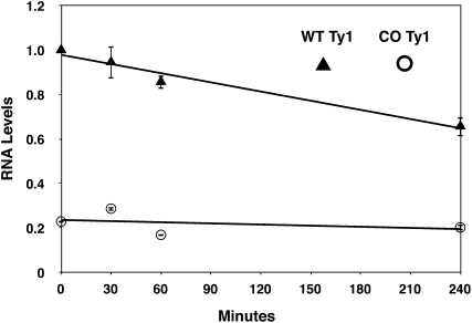
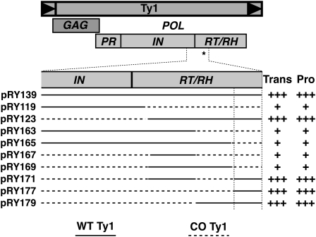
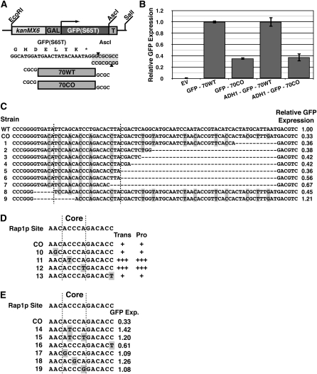
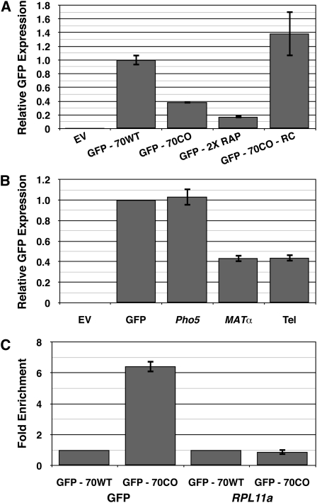
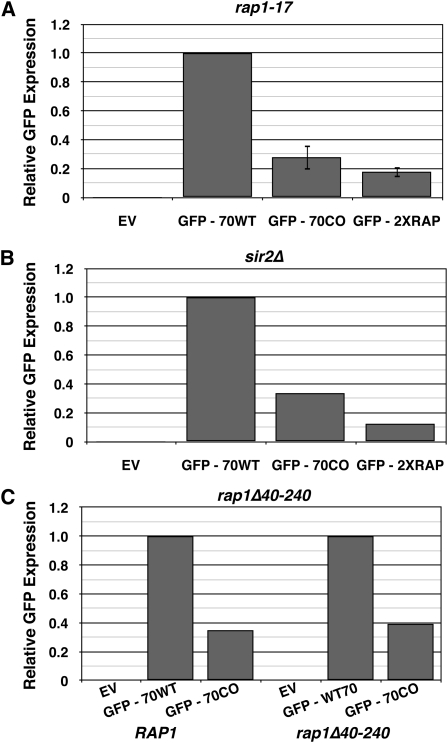
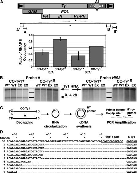
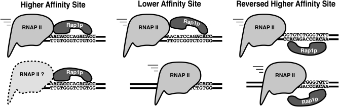
Extensive mutagenesis via massive recoding of retrotransposon Ty1 produced a synthetic codon-optimized retrotransposon (CO-Ty1). CO-Ty1 is defective for retrotransposition, suggesting a sequence capable of down-regulating retrotransposition. We mapped this sequence to a critical ~20-bp region within CO-Ty1 reverse transcriptase (RT) and confirmed that it reduced Ty1 transposition, protein, and RNA levels. Repression was not Ty1 specific; when introduced immediately downstream of the green fluorescent protein (GFP) stop codon, GFP expression was similarly reduced. Rap1p mediated this down-regulation, as shown by mutagenesis and chromatin immunoprecipitation. A regular threefold drop is observed in different contexts, suggesting utility for synthetic circuits. A large reduction of RNAP II occupancy on the CO-Ty1 construct was observed 3' to the identified Rap1p site and a novel 3' truncated RNA species was observed. We propose a novel mechanism of transcriptional regulation by Rap1p whereby it serves as a transcriptional roadblock when bound to transcription unit sequences.


Figures

Figure 1
Figure 1
Functional and biochemical characterization of CO-Ty1. (A) GAL-Ty1-mhis3AI reporter plasmid used for native Ty1 and CO-Ty1 analysis (Curcio and Garfinkel 1991). (B) Transposition assay of pRY022 native Ty1 (WT) and pRY091 CO-Ty1 (CO) grown on galactose and replica plated to SC −His for 2 days. Transposition activity was demonstrated by the appearance of colonies on SC −His plates (see Table S1). (C) Integrase protein (IN) immunoblot analysis of EV, empty vector; WT, pRY022; CO, pRY091. IN is indicated by an arrow and an internal loading control is indicated by *. (D) Above, IN immunoblot analysis of native and CO-Ty1 with and without a protease inactivating in-frame linker insertion (Monokian et al. 1994). EV, empty vector; WT, pRY022; CO, pRY091; WTp, pRY159; COp, pRY160. Protease activity is indicated by a plus or minus sign. Upper arrow indicates GAG-POL; lower arrow indicates IN. Below, RNA blot analysis of samples from above. EV, empty vector; WT, pRY022; CO, pRY091; WTp, pRY159; COp, pRY160. Ty1-HIS3 RNA is indicated by an arrow.
Figure 2
Figure 2
Ty1 RNA stability. Native (WT) and CO-Ty1 (CO) Ty1-HIS3 RNA levels were measured by Q-PCR after addition of glucose to shut off new transcription at the indicated time points. Ty1 RNA levels were normalized to the native Ty1 0 min time point, and error bars are the standard deviation of two biological replicates. Native Ty1-HIS3 RNA is indicated by triangles and CO-Ty1-HIS3 RNA is indicated by circles.
Figure 3
Figure 3
Mapping and characterization of 70-bp retrotransposition critical sequence. Diagram of Ty1 with open reading frames as a mapping reference. Selected and expanded region of IN and RT represents the initially mapped EcoRI–AatII 556-bp region found to contain the critical cis-acting sequence (*). Solid line, native Ty1 sequence; dashed line, CO-Ty1 sequence. Transposition frequency (Trans) and protein levels (Pro) are indicated qualitatively by pluses. Vertical lines define inferred critical sequence affecting Ty1 retrotransposition.
Figure 4
Figure 4
Functional characterization and mapping of critical 70-bp sequence in GFP. (A) GFP reporter construct used for GFP-70WT and GFP-70CO analysis. Tested sequences were inserted into the reporter construct immediately downstream of the GFP stop codon. (B) GFP expression of indicated reporter constructs grown in galactose as measured by fluorescence-activated cell sorting. EV, empty vector; GFP-70WT, construct with 70-bp critical region from native Ty1 inserted immediately after GFP stop codon; GFP-70CO, construct with 70-bp critical region from CO-Ty1 inserted immediately after GFP stop codon; ADH1-GFP-70WT, same as GFP-70WT, but driven by ADH1 promoter; ADH1-GFP-70CO, same as GFP-70CO, but driven by ADH1 promoter (Table S2). (C) Deletion analysis of GFP-70CO reveals 20-bp critical sequence down-regulating GFP expression. GFP expression is normalized to GFP-70WT. Vertical lines enclose minimal critical sequence. Highlighted bases differ between GFP-70WT and GFP-70CO. WT, GFP-70WT; CO, GFP-70CO; 1–9, various mutations in GFP-70CO. (D) Mutation analysis of Rap1p site within CO-Ty1. Transposition frequency (Trans) and protein levels (Pro) are indicated qualitatively by pluses. Vertical lines represent the Rap1p core binding site. Highlighted bases differ between native Ty1 and CO-Ty1. CO, CO-Ty1; 1–4, various mutations in CO-Ty1. (E) Mutation analysis of the identified Rap1p site within GFP-70CO. Vertical lines represent the Rap1p core binding site. GFP expression is normalized to GFP-70WT. Highlighted bases differ between GFP-70WT and GFP-70CO. CO, GFP-70CO; 1–6, various mutations in GFP-70CO.
Figure 5
Figure 5
Rap1p binding sites are capable of down-regulating expression. (A) The orientation and number of Rap1p binding sites affect the ability of Rap1p to down-regulate expression. GFP expression was measured by fluorescence-activated cell sorting. EV, empty vector; GFP-70WT, construct with 70-bp critical region from native Ty1 inserted immediately after GFP stop codon; GFP-70CO, construct with 70-bp critical region from CO-Ty1 inserted immediately after GFP stop codon; GFP-2XRAP, same as GFP-70CO, but with additional Rap1p binding site in the forward orientation; GFP-70CO-RC, same as GFP-70CO, but with the insert inverted. (B) High-affinity Rap1p binding sites from the yeast genome are capable of down-regulating expression. GFP expression was measured by fluorescence-activated cell sorting. EV, empty vector; GFP, empty construct from Figure 4A; Pho5, same as GFP with a documented Rap1p binding site from the PHO5 ORF; MATα, same as GFP with a documented Rap1p binding site from the HMα locus; Tel, as GFP with a documented Rap1p binding site from yeast telomere-like sequence (Buchman et al. 1988). (C) Chromatin immunoprecipitation demonstrates that Rap1p binds the identified Rap1p site in GFP-70CO. DNA was sheared and bound fragments were pulled down by polyclonal antibody to C terminus of Rap1p. Enrichment was measured by Q-PCR and normalized to GFP-70WT. GFP, primers designed to amplify Rap1p binding site within the GFP reporter construct; RPL11a, primers designed to amplify a known Rap1p binding site in the RPL11a promoter (Lieb et al. 2001; Zhao et al. 2006). GFP-70WT, construct with 70-bp critical region from native Ty1 inserted immediately after stop codon of GFP; GFP-70CO, construct with 70-bp critical region from CO-Ty1 inserted immediately after stop codon of GFP.
Figure 6
Figure 6
Neither the N nor the C terminus of the Rap1p protein affect its down-regulation of GFP expression. (A) Constructs were transformed into a yeast strain with a nonsense mutation in Rap1p resulting in a 165-codon deletion of the C-terminal domain (Kyrion et al. 1992). GFP expression was measured by fluorescence-activated cell sorting. EV, empty vector; GFP-70WT, construct with 70 bp of critical region from native Ty1 inserted immediately after stop codon of GFP; GFP-70CO, construct with 70 bp of critical region from CO-Ty1 inserted immediately after stop codon of GFP; GFP-2XRAP, as GFP-70CO, but with additional Rap1p binding site in the forward orientation. (B) Constructs were transformed into a yeast strain with a sir2 deletion genotype. GFP expression was measured by fluorescence-activated cell sorting. EV, empty vector; GFP-70WT, construct with 70 bp of critical region from native Ty1 inserted immediately after stop codon of GFP; GFP-70CO, construct with 70 bp of critical region from CO-Ty1 inserted immediately after stop codon of GFP; GFP-2XRAP, as GFP-70CO, but with additional Rap1p binding site in the forward orientation. (C) Constructs were transformed into a yeast strain with a 200-amino-acid deletion within the N terminus of Rap1p. GRF167 was used as a wild-type control. GFP expression was measured by fluorescence-activated cell sorting. EV, empty vector; GFP-70WT, construct with 70 bp of critical region from native Ty1 inserted immediately after stop codon of GFP; GFP-70CO, construct with 70 bp of critical region from CO-Ty1 inserted immediately after stop codon of GFP.
Figure 7
Figure 7
Rap1p blocks elongation and produces a novel transcript. (A) Diagram of Ty1; Rap1p binding site in CO-Ty1 is indicated by *. Chromatin immunoprecipitation demonstrates RNAP II occupancy; DNA was sheared and bound fragments were pulled down by monoclonal antibody to RPB3, a core component of RNAP II. Occupancy was determined by Q-PCR and the ratio of RNAP II at the downstream primer in a pair to the upstream primer in a pair is shown for CO-Ty1 (CO-Ty1+) and for a variant in which the identified Rap1p binding site has been replaced with corresponding sequence from the native Ty1 (CO-Ty10). A, primers designed to amplify a 109-bp sequence 1086 bp 5′ of the identified Rap1p binding site; A′, primers designed to amplify a 103-bp sequence 1286 bp 5′ of the identified Rap1p binding site; B, primers designed to amplify a 115-bp sequence 1015 bp 3′ of the identified Rap1p binding site; B′, primers designed to amplify a 103-bp sequence 1191 bp 3′ of the identified Rap1p binding site. (B) RNA blot analysis of CO-Ty1+ and CO-Ty10 with probes amplified from a 109-bp sequence 1086 bp 5′ of the identified Rap1p binding site (A) and HIS3. Two different strains (WT, GRF167; EX, rrp6Δ) were used for analysis. Ty1-HIS3 RNA is indicated by an arrow and is the full-length RNA. X represents novel CO-Ty1–specific RNA species. Bands from the 0.5- to 10-kb ladder by Invitrogen are marked. From top to bottom, the sizes are 8, 6, and 4 kb. (C) cRT–PCR strategy used for mapping the 3′ end of the X RNA. Messenger RNA molecules are represented as solid black lines and cDNA as broken lines. The CAP and the “?” represent the 5′ cap and unknown 3′ end, respectively. The “P” indicates the 5′-phosphate group and “P-?” indicates the ligated 5′–3′ junction. (D) Sequence analysis of the 5′–3′ junction of representative X RNA molecules. The Rap1p site and the 5′ start of the Ty1 message are underlined. CO represents the sequence of the intact, nontruncated synthetic Ty1 element and the numbers 1–9 label the sequences of nine X RNA molecules after undergoing the cRT–PCR protocol. The sequence shown for the nine clones before the Rap1p site is immediately joined to the GAGGAG of the 5′ end of the Ty1 message. Dashes are used as a spacer to illustrate the distance the polymerase stalled before the Rap1p site. * and corresponding numbers indicate the distance before the start of the Rap1p site. The shaded box indicates a mutation observed in one of our clones. A few additional RNAs (not pictured) truncated further upstream. The differences in stalling position observed may be the result of polymerase backtracking.
Figure 8
Figure 8
Model of Rap1p-mediated down-regulation of gene expression. (Left) High-affinity site in the sense orientation impedes RNAP II’s progress. (Middle) Lower-affinity site may not bind Rap1p avidly enough to prevent displacement by RNAP II. (Right) High-affinity site in inverted orientation may make Rap1p susceptible to displacement by RNAP II because the second Myb domain lacks the additional contacts provided by the C terminus of the Rap1p DNA binding domain.

Similar articles

Cited by

References

    1. Ashfield R., Patel A. J., Bossone S. A., Brown H., Campbell R. D., et al. , 1994. MAZ-dependent termination between closely spaced human complement genes. EMBO J. 13: 5656–5667 - PMC - PubMed
    1. Belcourt M. F., Farabaugh P. J., 1990. Ribosomal frameshifting in the yeast retrotransposon Ty: tRNAs induce slippage on a 7 nucleotide minimal site. Cell 62: 339–352 - PMC - PubMed
    1. Boeke J. D., Trueheart J., Natsoulis G., Fink G. R., 1987. 5-Fluoroorotic acid as a selective agent in yeast molecular genetics. Methods Enzymol. 154: 164–175 - PubMed
    1. Bolton E. C., Coombes C., Eby Y., Cardell M., Boeke J. D., 2005. Identification and characterization of critical cis-acting sequences within the yeast Ty1 retrotransposon. RNA 11: 308–322 - PMC - PubMed
    1. Braiterman L. T., Monokian G. M., Eichinger D. J., Merbs S. L., Gabriel A., et al. , 1994. In-frame linker insertion mutagenesis of yeast transposon Ty1: phenotypic analysis. Gene 139: 19–26 - PubMed

Publication types

MeSH terms