. 2012 Jan 3;109(1):E23-31.
doi: 10.1073/pnas.1116932108. Epub 2011 Nov 28.

Wolbachia induces reactive oxygen species (ROS)-dependent activation of the Toll pathway to control dengue virus in the mosquito Aedes aegypti

Affiliations

Wolbachia induces reactive oxygen species (ROS)-dependent activation of the Toll pathway to control dengue virus in the mosquito Aedes aegypti

Xiaoling Pan et al. Proc Natl Acad Sci U S A. .

Abstract

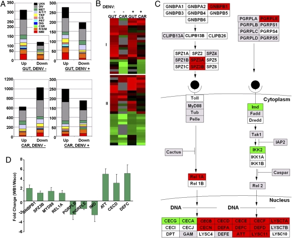
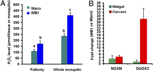
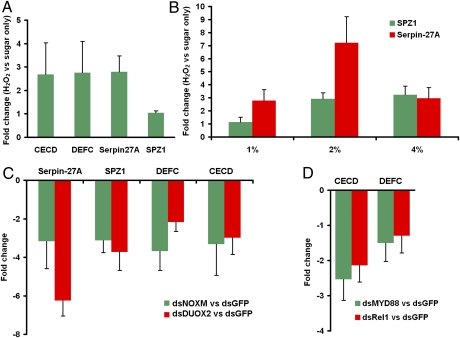
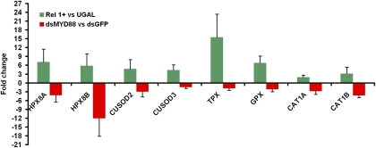
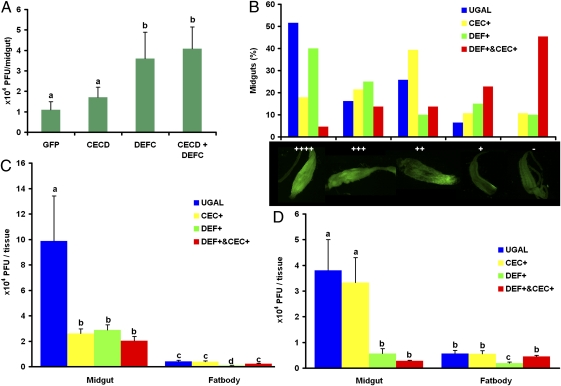
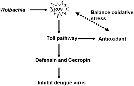
Wolbachia are maternally transmitted symbiotic bacteria that can spread within insect populations because of their unique ability to manipulate host reproduction. When introduced to nonnative mosquito hosts, Wolbachia induce resistance to a number of human pathogens, including dengue virus (DENV), Plasmodium, and filarial nematodes, but the molecular mechanism involved is unclear. In this study, we have deciphered how Wolbachia infection affects the Aedes aegypti host in inducing resistance to DENV. The microarray assay indicates that transcripts of genes with functions related to immunity and reduction-oxidation (redox) reactions are up-regulated in Ae. aegypti infected with Wolbachia. Infection with this bacterium leads to induction of oxidative stress and an increased level of reactive oxygen species in its mosquito host. Reactive oxygen species elevation is linked to the activation of the Toll pathway, which is essential in mediating the expression of antioxidants to counterbalance oxidative stress. This immune pathway also is responsible for activation of antimicrobial peptides-defensins and cecropins. We provide evidence that these antimicrobial peptides are involved in inhibition of DENV proliferation in Wolbachia-infected mosquitoes. Utilization of transgenic Ae. aegypti and the RNAi depletion approach has been instrumental in proving the role of defensins and cecropins in the resistance of Wolbachia-infected Ae. aegypti to DENV. These results indicate that a symbiotic bacterium can manipulate the host defense system to facilitate its own persistent infection, resulting in a compromise of the mosquito's ability to host human pathogens. Our discoveries will aid in the development of control strategies for mosquito-transmitted diseases.


Conflict of interest statement

The authors declare no conflict of interest.

Figures

Fig. 1.
Fig. 1.
Wolbachia-regulated genes in Ae. aegypti. (A) Functional classification of differentially expressed genes in the midgut (GUT) and carcass (CAR) of 7-d-old mosquitoes before a blood meal (DENV−) and 12 d after infection with DENV serotype 2 (DENV+). The graph shows the functional class distributions in real numbers of genes that are regulated by Wolbachia (“Up” indicates induced, and “Down” indicates repressed). The Wolbachia-regulated gene expression data are presented in Tables S1, S2, S3, and S4. Functional group abbreviations: C/S, cytoskeletal and structural; CSR, chemosensory reception; DIG, blood and sugar food digestive; DIV, diverse functions; IMM, immunity; MET, metabolism; PRT, proteolysis; R/S/M, redox, stress, and mitochondrion; R/T/T, replication, transcription, and translation; TRP, transport; UNK, unknown functions. (B) Cluster analysis of 23 immune genes (I) and 27 R/S/M genes (II) that were regulated in midguts and carcasses of Wolbachia-infected female mosquitoes in at least two of four combinations: midgut with (GUT+) or without (GUT−) DENV infection; carcass with (CAR+) or without (CAR−) DENV infection. All genes presented in the cluster are listed in Tables S5 and S6. (C) Regulation of putative Toll signaling pathway genes by Wolbachia in the mosquito midgut 12 d after feeding on DENV-infected blood. Red, green, and gray colors indicate Wolbachia-responsive up-, down- and nonregulated genes, respectively. White indicates unfound or filtered signal. The pathway was built with GenMAPP software based on the immunogenomics prediction (49, 50). CECB, Cecropin B; CECE, Cecropin E; CECF, Cecropin F; CECG, Cecropin G; CECI, Cecropin I; CECJ, Cecropin J; CECN, Cecropin N; CLIPB13A, Clip-domain serine protease 13A; CLIPB13B, Clip-domain serine protease 13B; CLIPB26, Clip-domain serine protease 26; DEFD, Defensin D; DEFE, Defensin E; DPT, Diptericin; Dredd, Death-related ced-3/Nedd2-like protein; Fadd, Fas-Associated Death Domain; GAM, Gambicin; GNBPA1, Gram-negative binding protein A1; GNBPA2, Gram-negative binding protein A2; GNBPB3, Gram-negative binding protein B3; GNBPB4, Gram-negative binding protein B4; GNBPB5, Gram-negative binding protein B5; GNBPB6, Gram-negative binding protein B6; IAP2, Inhibitor of apoptosis 2; IKK1A, I-Kappa-B Kinase 1 A; IKK1B, I-Kappa-B Kinase 1 B; IKK2, I-Kappa-B Kinase 2; LYSC4, C-Type lysozyme 4; LYSC7A, C-Type Lysozyme 7A; LYSC7B, C-Type Lysozyme 7B; LYSC10, C-Type lysozyme 10; LYSC11, C-Type lysozyme 11; Pelle, PGRPLA, Peptidoglycan Recognition Protein (Long) A; PGRPS1, Peptidoglycan Recognition Protein (Short) 1; PGRPLC, Peptidoglycan Recognition Protein (Long) C; PGRPS4, Peptidoglycan Recognition Protein (Short) 4; PGRPLD, Peptidoglycan Recognition Protein (Long) D; PGRPS5, Peptidoglycan Recognition Protein (Short) 5; Rel2, Relish-like protein 2; SPZ2, Spaetzle-likecytokine 2; SPZ4, Spaetzle-like cytokine 4; SPZ5, Spaetzle-like cytokine 5; SPZ1C, Spaetzle-like cytokine 1C; SPZ6, Spaetzle-like cytokine 6; Tak1, TGF-Beta-Activated Kinase-1; Tub, Tube. (D) Real-time PCR validation of Wolbachia-regulated genes identified in the microarray assays. Data are shown as fold change in WB1 midguts compared with Waco midguts 12 d after feeding on DENV-infected blood.
Fig. 2.
Fig. 2.
Wolbachia induces ROS formation in the mosquito Ae. aegypti. (A) Comparison of H2O2 levels in the fat body and whole mosquito in 7-d-old WB1 and Waco mosquitoes before a blood meal. The data shown are means of six replicates, with five fat bodies or three whole mosquitoes for each. The level of H2O2 was significantly higher in both fat bodies and whole bodies of WB1 mosquitoes than in those of Waco mosquitoes. Statistical significance is represented by letters above each column, with different letters signifying distinct statistical groups. Student's t test: a vs. b, P < 0.05; b vs. c, P < 0.001. (B) Fold changes in expression of NADPH oxidase (NOXM) and dual oxidase (DUOX2) in the midguts and carcasses of WB1 and Waco mosquitoes. Real-time PCR analysis was performed using midguts and carcasses of 7-d-old, non–blood-fed, female mosquitoes.
Fig. 3.
Fig. 3.
Wolbachia-induced ROS-dependent activation of the Toll pathway. (A) Fold changes in expression of Toll pathway-related genes in midguts of Waco females after H2O2 treatment. Two-day-old Waco females were fed with a sugar solution containing 1% H2O2 (vol/vol) for 5 d. The total RNA of midguts was extracted to measure the fold change in gene expression compared with a control group fed only a sugar solution. (B) Fold changes in the expression of Toll pathway marker genes in fat bodies of Waco females after H2O2 treatment at three different doses—1%, 2%, and 4% (vol/vol)—compared with a control group fed a sugar solution only. (C) Fold changes in the expression of Toll pathway-related genes in WB1 females after silencing of NOXM and DUOX2. Three days after injection with dsRNA, midguts were collected to extract total RNA and measure the fold change in gene expression compared with that in the control group injected with GFP dsRNA. (D) Fold changes in the expression of CECD and DEFC after silencing of the Toll pathway in WB1. Three days after injection with MYD88 or REL1 dsRNA, midguts were collected to extract the total RNA and measure the fold change in gene expression compared with that in the control group injected with GFP dsRNA.
Fig. 4.
Fig. 4.
The Toll pathway controls the expression of antioxidant proteins. Fat bodies of REL1-overexpressing transgenic mosquitoes (Rel1+) were collected 24 h after a blood meal, and the expression of eight antioxidants was measured using real-time PCR and compared with that of wild-type UGAL mosquitoes. Midguts of WB1 females injected with MYD88 dsRNA were collected to measure the fold changes in gene expression compared with that of the control group injected with GFP dsRNA. For all experiments, the gene expression data were normalized with RPS6. The primer sequences are listed in Tables S8 and S9. Error bars indicate SE.
Fig. 5.
Fig. 5.
Roles of defensins and cecropins in the inhibition of DENV proliferation. Viral infection was detected by plaque assay in C6/36 cells (A, C, and D) or by immunofluorescence staining of the DENV-2 envelope protein (B). (A) DENV loads were measured in the midguts of mosquitoes after RNAi depletion of CECD, DEFC, a double depletion (CECD + DEFC), or in GFP dsRNA-treated control mosquitoes. a vs. b, P < 0.05 (Mann\x{2013}Whitney U test). (B) Midgut infection rates were measured at 7 d after viral infection in transgenic Ae. aegypti with fat body ectopic expression of either defensin A (DEF+) or cecropin A (CEC+) and in their hybrid strain (DEF+ and CEC+). Midgut infection levels were recorded as uninfected (−) or infected in one of four increasing levels (+, ++, +++, ++++). There were significant differences in the levels of midgut DENV infection in the hybrid strain and UGAL controls (χ2 test, P < 0.0001) and in levels in CECA transgenic mosquitoes and UGAL controls (χ2 test, P < 0.05). DENV loads were measured in the midguts and fat bodies of three transgenic mosquitoes and the wild-type UGAL strain 10 d after infection with DENV-2 during the first blood feeding (C) or 10 d after infection with DENV-2 during the second blood feeding (the first blood meal was blood without DENV to activate the transgenes) (D). For all figures, error bars represent SE. Statistical significance is represented by letters above each column, with different letters signifying distinct statistical groups. For C and D, P < 0.05 for a vs b, b vs c, and c vs d in Student's t test.
Fig. 6.
Fig. 6.
A current model of Wolbachia-mediated resistance to DENV in Ae. aegypti.
Fig. P1.
Fig. P1.
A model of Wolbachia-mediated resistance to DENV in Ae. aegypti.

Similar articles

Cited by

References

    1. Hill CA, Kafatos FC, Stansfield SK, Collins FH. Arthropod-borne diseases: Vector control in the genomics era. Nat Rev Microbiol. 2005;3:262–268. - PubMed
    1. Halstead SB. Dengue. Lancet. 2007;370:1644–1652. - PubMed
    1. Kyle JL, Harris E. Global spread and persistence of dengue. Annu Rev Microbiol. 2008;62:71–92. - PubMed
    1. Serbus LR, Casper-Lindley C, Landmann F, Sullivan W. The genetics and cell biology of Wolbachia-host interactions. Annu Rev Genet. 2008;42:683–707. - PubMed
    1. Iturbe-Ormaetxe I, Walker T, O'Neill SL. Wolbachia and the biological control of mosquito-borne disease. EMBO Rep. 2011;12:508–518. - PMC - PubMed

Publication types

MeSH terms

LinkOut - more resources