

Lack of recognition by global-genome nucleotide excision repair accounts for the high mutagenicity and persistence of aristolactam-DNA adducts
- PMID: 22121226
- PMCID: PMC3315299
- DOI: 10.1093/nar/gkr1095
Lack of recognition by global-genome nucleotide excision repair accounts for the high mutagenicity and persistence of aristolactam-DNA adducts
Abstract
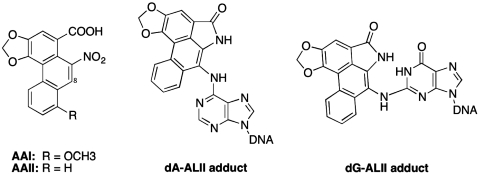
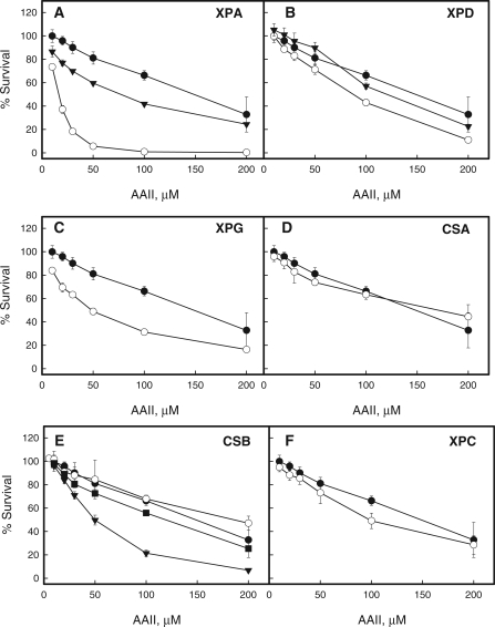
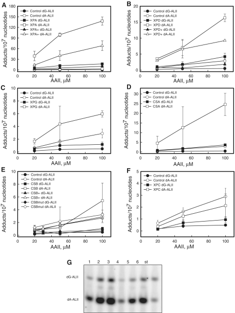
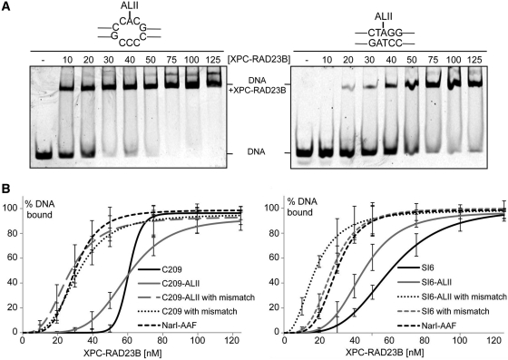
Exposure to aristolochic acid (AA), a component of Aristolochia plants used in herbal remedies, is associated with chronic kidney disease and urothelial carcinomas of the upper urinary tract. Following metabolic activation, AA reacts with dA and dG residues in DNA to form aristolactam (AL)-DNA adducts. These mutagenic lesions generate a unique TP53 mutation spectrum, dominated by A:T to T:A transversions with mutations at dA residues located almost exclusively on the non-transcribed strand. We determined the level of AL-dA adducts in human fibroblasts treated with AA to determine if this marked strand bias could be accounted for by selective resistance to global-genome nucleotide excision repair (GG-NER). AL-dA adduct levels were elevated in cells deficient in GG-NER and transcription-coupled NER, but not in XPC cell lines lacking GG-NER only. In vitro, plasmids containing a single AL-dA adduct were resistant to the early recognition and incision steps of NER. Additionally, the NER damage sensor, XPC-RAD23B, failed to specifically bind to AL-DNA adducts. However, placing AL-dA in mismatched sequences promotes XPC-RAD23B binding and renders this adduct susceptible to NER, suggesting that specific structural features of this adduct prevent processing by NER. We conclude that AL-dA adducts are not recognized by GG-NER, explaining their high mutagenicity and persistence in target tissues.
Figures
Similar articles
-
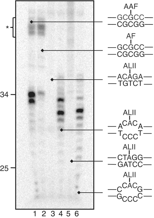
Structure and stability of DNA containing an aristolactam II-dA lesion: implications for the NER recognition of bulky adducts.Nucleic Acids Res. 2012 Mar;40(6):2759-70. doi: 10.1093/nar/gkr1094. Epub 2011 Nov 25. Nucleic Acids Res. 2012. PMID: 22121223 Free PMC article.
-
Aristolochic acid-induced upper tract urothelial carcinoma in Taiwan: clinical characteristics and outcomes.Int J Cancer. 2013 Jul;133(1):14-20. doi: 10.1002/ijc.28013. Epub 2013 Feb 12. Int J Cancer. 2013. PMID: 23292929
-
The relationships between XPC binding to conformationally diverse DNA adducts and their excision by the human NER system: is there a correlation?DNA Repair (Amst). 2014 Jul;19:55-63. doi: 10.1016/j.dnarep.2014.03.026. Epub 2014 Apr 29. DNA Repair (Amst). 2014. PMID: 24784728 Free PMC article.
-
Molecular basis for damage recognition and verification by XPC-RAD23B and TFIIH in nucleotide excision repair.DNA Repair (Amst). 2018 Nov;71:33-42. doi: 10.1016/j.dnarep.2018.08.005. Epub 2018 Aug 23. DNA Repair (Amst). 2018. PMID: 30174301 Free PMC article. Review.
-
Aristolochic acid as a probable human cancer hazard in herbal remedies: a review.Mutagenesis. 2002 Jul;17(4):265-77. doi: 10.1093/mutage/17.4.265. Mutagenesis. 2002. PMID: 12110620 Review.
Cited by
-
Nucleotide excision repair efficiencies of bulky carcinogen-DNA adducts are governed by a balance between stabilizing and destabilizing interactions.Biochemistry. 2012 Feb 21;51(7):1486-99. doi: 10.1021/bi201794x. Epub 2012 Feb 9. Biochemistry. 2012. PMID: 22242833 Free PMC article.
-
Comparative Research of Chemical Profiling in Different Parts of Fissistigma oldhamii by Ultra-High-Performance Liquid Chromatography Coupled with Hybrid Quadrupole-Orbitrap Mass Spectrometry.Molecules. 2021 Feb 11;26(4):960. doi: 10.3390/molecules26040960. Molecules. 2021. PMID: 33670350 Free PMC article.
-
Adenine-DNA adducts derived from the highly tumorigenic Dibenzo[a,l]pyrene are resistant to nucleotide excision repair while guanine adducts are not.Chem Res Toxicol. 2013 May 20;26(5):783-93. doi: 10.1021/tx400080k. Epub 2013 Apr 24. Chem Res Toxicol. 2013. PMID: 23570232 Free PMC article.
-
Analysis of TP53 mutation spectra reveals the fingerprint of the potent environmental carcinogen, aristolochic acid.Mutat Res. 2013 Jul-Sep;753(1):41-49. doi: 10.1016/j.mrrev.2013.02.003. Epub 2013 Feb 17. Mutat Res. 2013. PMID: 23422071 Free PMC article. Review.
-
Hypermutation in human cancer genomes: footprints and mechanisms.Nat Rev Cancer. 2014 Dec;14(12):786-800. doi: 10.1038/nrc3816. Nat Rev Cancer. 2014. PMID: 25568919 Free PMC article. Review.
References
-
- Debelle FD, Vanherweghem JL, Nortier JL. Aristolochic acid nephropathy: a worldwide problem. Kidney Int. 2008;74:158–169. - PubMed
-
- National Toxicology Program. Aristolochic Acids. Rep. Carcinogens. 2011;12:45–49. - PubMed
-
- Kumar V, Poonam PAK, Parmar VS. Naturally occurring aristolactams, aristolochic acids and dioxoaporphines and their biological activities. Nat. Product Rep. 2003;20:565–583. - PubMed
-
- Vanherweghem JL, Debelle F, Muniz Martinez MC, Nortier J. In: Clinical Nephrotoxins. 2nd edn. De Broe ME, Porter GA, Bennet WM, Verpooten GA, editors. Netherlands: Dordrecht Kluwer; 2003. pp. 588–601.
-
- Schmeiser HH, Bieler CA, Wiessler M, van Ypersele de Strihou C, Cosyns JP. Detection of DNA adducts formed by aristolochic acid in renal tissue from patients with Chinese herbs nephropathy. Cancer Res. 1996;56:2025–2028. - PubMed
Publication types
MeSH terms
Substances
Grants and funding
LinkOut - more resources
Full Text Sources
Research Materials
Miscellaneous
