. 2011 Nov 23;147(5):1066-79.
doi: 10.1016/j.cell.2011.10.039.

Lin28A and Lin28B inhibit let-7 microRNA biogenesis by distinct mechanisms

Affiliations

Lin28A and Lin28B inhibit let-7 microRNA biogenesis by distinct mechanisms

Elena Piskounova et al. Cell. .

Abstract

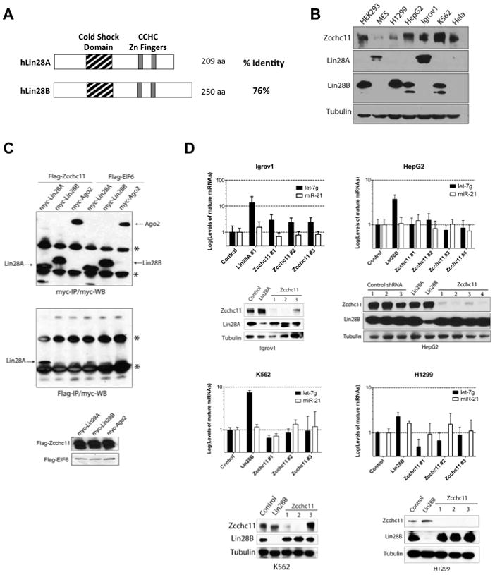
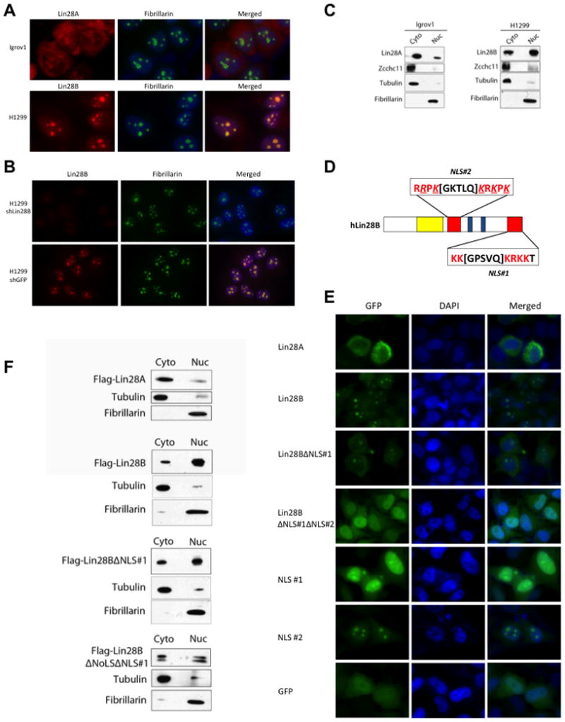
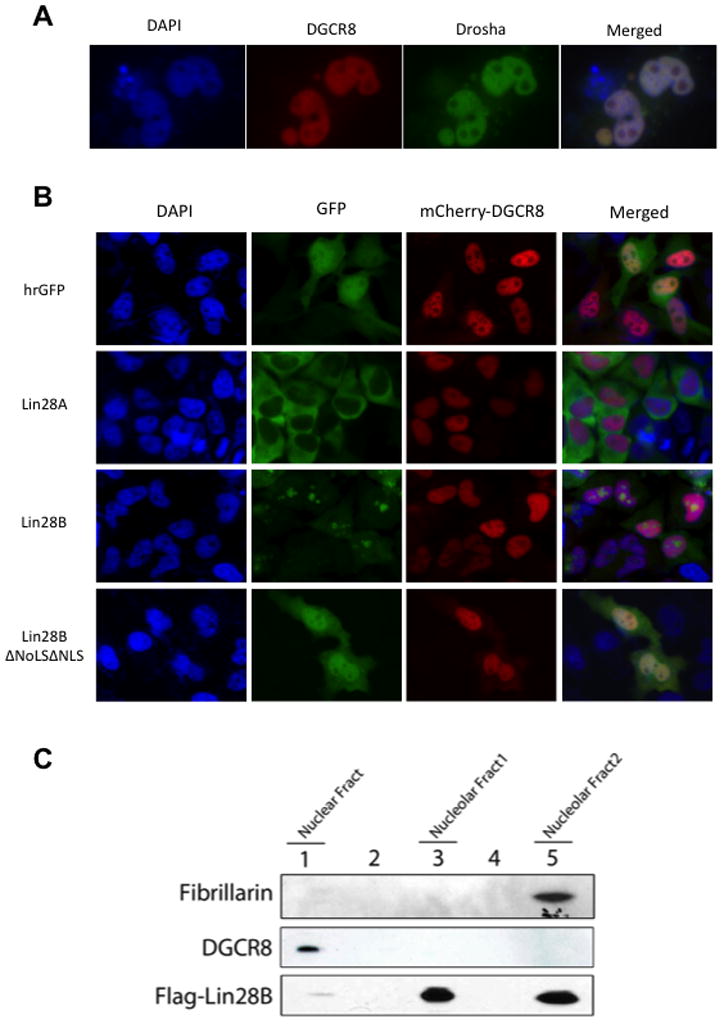
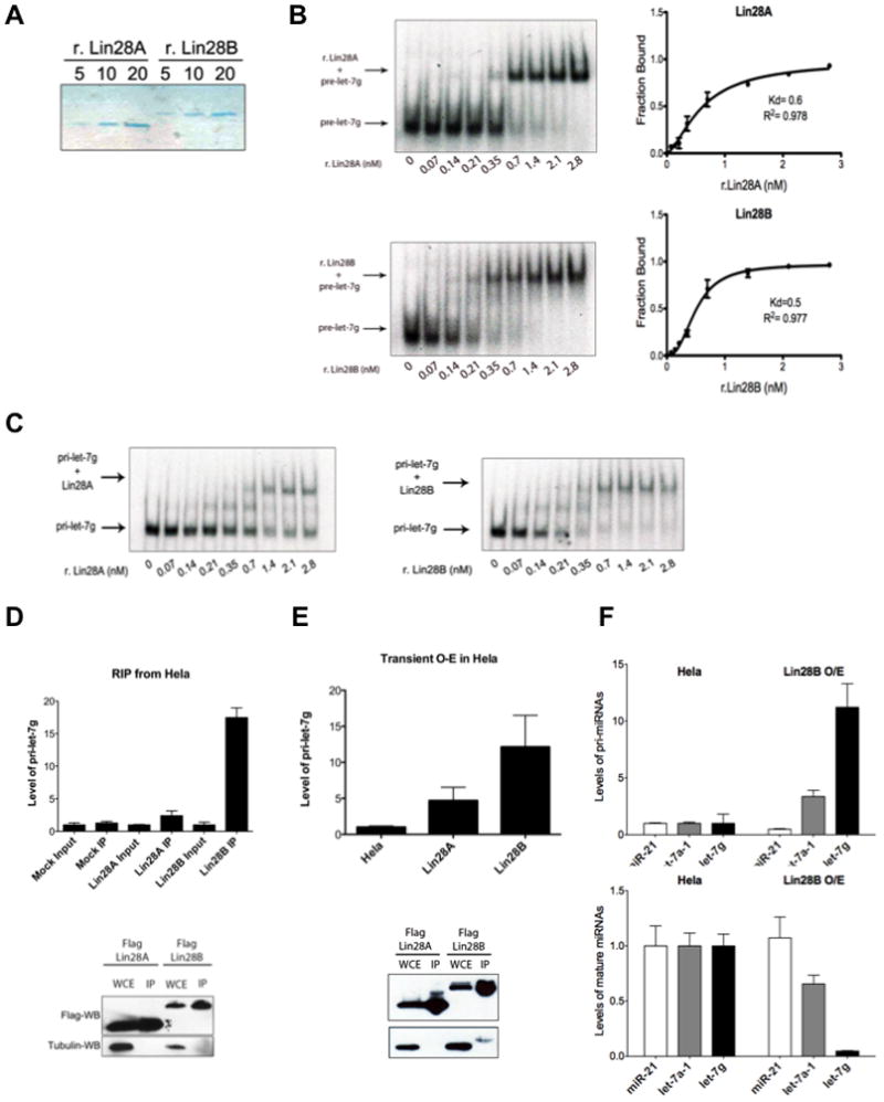
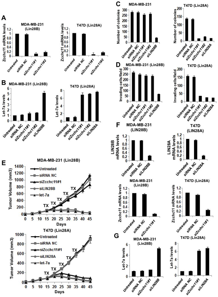
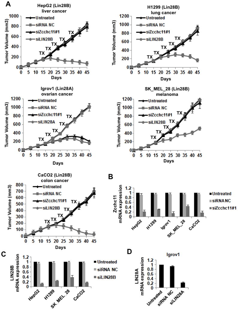
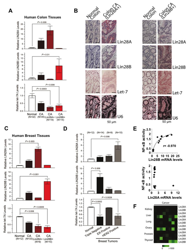
Lin28A and Lin28B selectively block the expression of let-7 microRNAs and function as oncogenes in a variety of human cancers. Lin28A recruits a TUTase (Zcchc11/TUT4) to let-7 precursors to block processing by Dicer in the cell cytoplasm. Here we find that unlike Lin28A, Lin28B represses let-7 processing through a Zcchc11-independent mechanism. Lin28B functions in the nucleus by sequestering primary let-7 transcripts and inhibiting their processing by the Microprocessor. The inhibitory effects of Zcchc11 depletion on the tumorigenic capacity and metastatic potential of human cancer cells and xenografts are restricted to Lin28A-expressing tumors. Furthermore, the majority of human colon and breast tumors analyzed exclusively express either Lin28A or Lin28B. Lin28A is expressed in HER2-overexpressing breast tumors, whereas Lin28B expression characterizes triple-negative breast tumors. Overall our results illuminate the distinct mechanisms by which Lin28A and Lin28B function and have implications for the development of new strategies for cancer therapy.


Figures

Figure 1
Figure 1. Lin28B regulates let-7 biogenesis through a TUTase-independent mechanism
(A) Schematic representation of human Lin28A and Lin28B (B) Western Blot analysis of Zcchc11, Lin28A, and Lin28B in extracts prepared from human cancer cell lines. (C) Co-immunoprecipitation (co-IP): Hela cells were co-transfected with Human myc-Lin28A, myc-Lin28B, or myc-Ago2 with either Flag-Zcchc11 or Flag-EIF6. Flag-IP and Flag- and Myc-western blots were performed to detect expression and interaction respectively. See Also Figure S1. (D) Stable knockdown of Zcchc11 leads to upregulation of mature let-7g levels in Lin28A-expressing cells but not Lin28B-expressing cell lines. miRNA levels were measured by q.RT-PCR. Error bars represent SEM (n=3). Protein knockdown was monitored by Western blot.
Figure 2
Figure 2. Lin28A and Lin28B are differentially localized within the cell
(A) Immunofluorescence detection of endogenous Lin28A in Igrov1 and Lin28B in H1299 cell lines. Fibrillarin, a known nucleolar protein, was used as a positive control. (B) Immunofluorescence analysis of control- and Lin28B-knockdown H1299 cell lines. (C) Biochemical fractionation of Igrov1 and H1299 cell lines. Endogenous Lin28A, Lin28B, and Zcchc11 in each fraction were detected by western blot. Fibrillarin was used as a nuclear marker; Tubulin was used as a cytoplasmic marker. (D) Schematic of nuclear localization signals (NLS) in the Lin28B protein. An Arginine as well as several Lysines that were replaced by Glycines are underlined and italicized (E) Localization of GFP-Lin28 fusion proteins in Hela cells. (F) Fractionation of Flag-Lin28 proteins, exogenously expressed in Hela cells. Proteins were detected by Flag western blot.
Figure 3
Figure 3. Lin28B localizes to nucleoli where Microprocessor is absent
(A) Co-localization of Microprocessor components GFP-Drosha and mCherry-DGCR8 in Hela cells reveals their distribution throughout the nucleus and exclusion from nucleoli. (B) Localization of GFP-Lin28A, Lin28B, or mutant Lin28B proteins with mCherry-DGCR8 in Hela cells reveals non-overlapping localization of Lin28B and DGCR8. (B) Fractionation of a Flag-Lin28B Hela stable cell line. Flag-Lin28B and endogenous DGCR8 were detected western blot and shows a non-overlapping subcellular localization of Lin28B and the Microprocessor. Fibrillarin was used as a control for nucleolar localization.
Figure 4
Figure 4. Lin28B directly binds and sequesters pri-let-7
(A) Colloidal Blue staining of purified recombinant His-Lin28A and His-Lin28B proteins. (B) Binding of r.Lin28A and r.Lin28B to pre-let-7g was assessed by EMSA performed with 0.5 nM 5′-end labeled pre-let-7g RNA and the indicated concentration of recombinant protein. Band intensities were quantitated from three independent experiments and represented as the fraction of bound pre-let-7g RNA in the plots. Values are given as average ±S.E.M. (n=3). See also Figure S2. (C) EMSA performed indicated concentration of r.Lin28A and r.Lin28B with in vitro transcribed uniformly labeled pri-let-7g. (D) RNA-Immunoprecipitation (RIP) analysis of RNA associated with immunopurified Flag-Lin28A and Flag-Lin28B from Hela cells. RNA was extracted from IP material and analyzed by q.RT-PCR. Error bars ±S.E.M. (n=3). Lower panel indicates relative Lin28A and Lin28B expression levels by Flag-Western blot. (E) Accumulation of pri-let-7 by transient Lin28B expression in Hela cell detected by q.RT-PCR. Error bars ±S.E.M. (n=3). Lower panel indicates relative expression levels of Lin28A and Lin28B proteins detected by Flag-Western blot in transfected cells. (F) pri-let-7 accumulates (top panel) and mature let-7 levels decrease (bottom panel) in Hela cells stably overexpressing Lin28B. Error bars ±S.E.M. (n=3)
Figure 5
Figure 5. Zcchc11 inhibition blocks tumorigenicity and invasiveness of Lin28A-expressing breast cancer cells
(A) q.RT-PCR analysis of Zcchc11 knockdown in MDA-MB-231 and T47D breast cancer cells. Error bars ±S.E.M. (n=3) (B) Inhibition of Zcchc11 expression does not affect let-7a expression in Lin28B-expressing cells (MDA-MB-231), while it up-regulates let-7a expression in Lin28A-expressing cells (T47D). Let-7a expression levels measured by q.RT-PCR in cells treated with siRNAs for 48hr. Error bars ±S.E.M. (n=3) (C) Inhibition of Zcchc11 expression did not affect the colony formation ability of MDA-MB-231 cells, but suppressed the colony formation ability of T47D cells. The number of colonies was evaluated 20 days post plating in soft agar. The experiment was repeated thrice and the statistical significance was calculated using Student’s t test. (D) Inhibition of Zcchc11 expression did not affect the invasiveness of MDA-MB-231 cells, but suppressed the invasive ability of T47D cells. The number of invasive cells was measured 16h post transfection with indicated siRNAs. In all assays, 10 fields per insert were scored and SD was measured. The experiment was repeated thrice and the statistical significance was calculated using Student’s t test. (E) Inhibition of Zcchc11 expression did not suppress tumor growth of MDA-MB-231 cells in xenografts, however it inhibited tumor growth of T47D cells in xenografts. The treatments with indicated siRNA were performed intraperitoneally (i.p.) for 5 cycles starting on day 15. Each treatment group consisted of 5 mice. (F) q.RT-PCR analysis of siRNA inhibition of Lin28B, Lin28A, and Zcchc11 in xenograft tumors (day 30) derived from MDA-MB-231 and T47D cells. Error bars ±S.E.M. (n=3). (G) Inhibition of Lin28B but not of Zcchc11 allows up-regulation of let-7a expression levels in MDA-MB-231 xenograft tumors (day 30). However, inhibition of Lin28A or Zcchc11 results in let-7a up-regulation in T47D xenograft tumors. Let-7a expression levels measured by q.RT-PCR on tumors untreated or treated with indicated siRNA. Error bars ±S.E.M. (n=3). See also Figure S3.
Figure 6
Figure 6. Inhibition of Zcchc11 expression suppresses tumor growth of Lin28A- but not Lin28B-expressing xenografts
(A) Xenograft experiments were performed with a variety of different human cancer cell lines. Mice were treated with the indicated siRNA for 5 cycles starting on day 15. For all cells lines tested each treatment group consisted of 5 mice. While inhibition of Lin28A or Lin28B suppressed tumor growth in the relevant xenografts, inhibition of Zcchc11 inhibited growth only of Lin28A- but not Lin28B-expressing tumors. Error bars ±S.E.M. (n=3) (B) Analysis of siRNA inhibition of Zcchc11 in xenograft tumors (day 30) derived from the indicated cells. (C) Analysis of siRNA inhibition of Lin28B in xenograft tumors (day 30) derived from the indicated cells. (D) Analysis of siRNA inhibition of Lin28A in xenograft tumors (day 30) derived from IGROV1 cells. mRNA expression levels were measured by q.RT-PCR on tumors untreated or treated with the indicated siRNA. Error bars ±S.E.M. (n=3)
Figure 7
Figure 7. Lin28A and Lin28B expression in primary human cancers
(A) q.RT-PCR analysis of Lin28A, Lin28B and let-7a expression levels in normal and colon cancer tissues. Tumor samples were further classified into two groups expressing either high Lin28A or Lin28B. Data expressed as mean ± SE. n=3. (B) Immunohistochemistry for Lin28A, Lin28B and in situ hybridization for let-7a and U6 in normal colon tissues and colon adenocarcinomas. See also Figure S4. (C) q.RT-PCR analysis of Lin28A, Lin28B and let-7a in human normal and breast cancer tissues. Tumor samples were further classified into two groups expressing either high Lin28A or Lin28B. Data expressed as mean ±SE. n=3. (D) Lin28A, Lin28B and let-7a expression levels in different breast cancer subtypes. (E) Correlation between Lin28A and Lin28B mRNA levels assessed by q.RT-PCR with NF-κB phosphorylation status assessed by ELISA assay. (E) Heatmap representation of Lin28A and Lin28B in carcinomas of different origin measured by q.RT-PCR.

Similar articles

Cited by

References

    1. Barh D, Malhotra R, Ravi B, Sindhurani P. Microrna let-7: an emerging next-generation cancer therapeutic. Curr Oncol. 2010;17:70–80. - PMC - PubMed
    1. Boisvert FM, van Koningsbruggen S, Navascues J, Lamond AI. The multifunctional nucleolus. Nat Rev Mol Cell Biol. 2007;8:574–585. - PubMed
    1. Boyerinas B, Park SM, Hau A, Murmann AE, Peter ME. The role of let-7 in cell differentiation and cancer. Endocr Relat Cancer. 2010;17:F19–36. - PubMed
    1. Bussing I, Slack FJ, Grosshans H. let-7 microRNAs in development, stem cells and cancer. Trends Mol Med. 2008;14:400–409. - PubMed
    1. Chang TC, Zeitels LR, Hwang HW, Chivukula RR, Wentzel EA, Dews M, Jung J, Gao P, Dang CV, Beer MA, et al. Lin-28B transactivation is necessary for Myc-mediated let-7 repression and proliferation. Proc Natl Acad Sci U S A. 2009;106:3384–3389. - PMC - PubMed

Publication types