. 2012 Jan 6;287(2):978-88.
doi: 10.1074/jbc.M111.294959. Epub 2011 Nov 22.

Pro-survival effects of 17β-estradiol on osteocytes are mediated by nitric oxide/cGMP via differential actions of cGMP-dependent protein kinases I and II

Affiliations

Pro-survival effects of 17β-estradiol on osteocytes are mediated by nitric oxide/cGMP via differential actions of cGMP-dependent protein kinases I and II

Nisha Marathe et al. J Biol Chem. .

Abstract

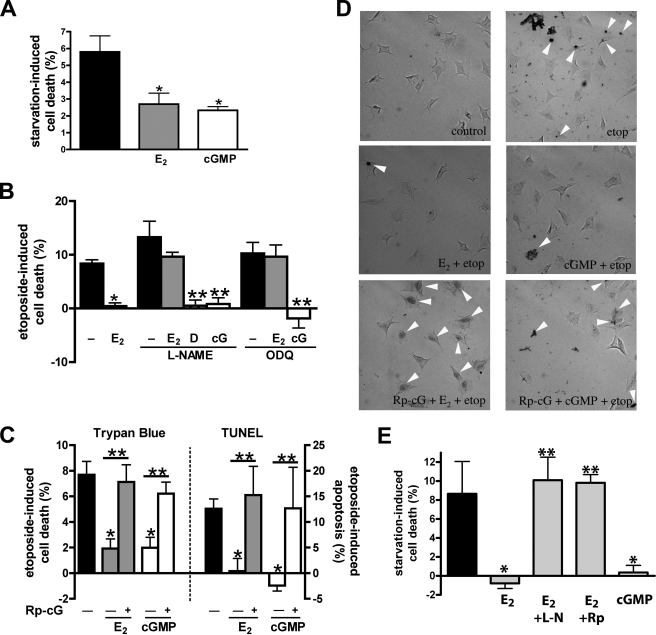
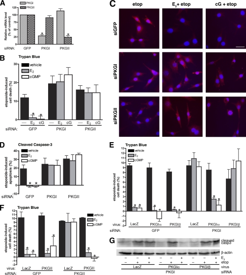
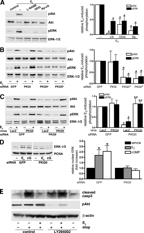
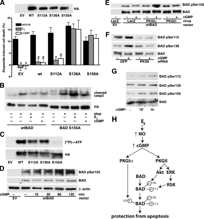
Estrogens promote bone health in part by increasing osteocyte survival, an effect that requires activation of the protein kinases Akt and ERK1/2, but the molecular mechanisms involved are only partly understood. Because estrogens increase nitric oxide (NO) synthesis and NO can have anti-apoptotic effects, we examined the role of NO/cGMP signaling in estrogen regulation of osteocyte survival. Etoposide-induced death of MLO-Y4 osteocyte-like cells, assessed by trypan blue staining, caspase-3 cleavage, and TUNEL assays, was completely prevented when cells were pre-treated with 17β-estradiol. This protective effect was mimicked when cells were pre-treated with a membrane-permeable cGMP analog and blocked by pharmacological inhibitors of NO synthase, soluble guanylate cyclase, or cGMP-dependent protein kinases (PKGs), supporting a requirement for NO/cGMP/PKG signaling downstream of 17β-estradiol. siRNA-mediated knockdown and viral reconstitution of individual PKG isoforms demonstrated that the anti-apoptotic effects of estradiol and cGMP were mediated by PKG Iα and PKG II. Akt and ERK1/2 activation by 17β-estradiol required PKG II, and cGMP mimicked the effects of estradiol on Akt and ERK, including induction of ERK nuclear translocation. cGMP induced BAD phosphorylation on several sites, and experiments with phosphorylation-deficient BAD mutants demonstrated that the anti-apoptotic effects of cGMP and 17β-estradiol required BAD phosphorylation on Ser(136) and Ser(155); these sites were targeted by Akt and PKG I, respectively, and regulate BAD interaction with Bcl-2. In conclusion, 17β-estradiol protects osteocytes against apoptosis by activating the NO/cGMP/PKG cascade; PKG II is required for estradiol-induced activation of ERK and Akt, and PKG Iα contributes to pro-survival signaling by directly phosphorylating BAD.


Figures

FIGURE 1.
FIGURE 1.
Estradiol protects MLO-Y4 osteocytic cells and primary murine osteoblasts from serum starvation- or etoposide-induced apoptosis through NO/cGMP/PKG signaling. A, MLO-Y4 cells were treated with vehicle (black bar), 100 nm estradiol (E2, gray bar), or 100 μm 8-pCPT-cGMP (cGMP, white bar) and serum-starved for 18 h. Starvation-induced cell death was measured by trypan blue uptake as described under “Experimental Procedures.” B, MLO-Y4 cells were treated with 4 mm l-NAME or 10 μm ODQ for 1 h as indicated, and then received vehicle, 100 nm estradiol, 3 μm DETA-NONOate (D), or 100 μm 8-pCPT-cGMP (cG) for 1 h, followed by an 8-h exposure to 50 μm etoposide. Etoposide-induced cell death was measured by trypan blue uptake as in A. C, MLO-Y4 cells were treated with 100 μm Rp-8-CPT-PET-cGMPS (Rp-cG) for 1 h as indicated (+), followed by 100 nm estradiol (gray bars) or 100 μm cGMP (white bars) for 1 h; cells were then exposed to etoposide for 8 h (trypan blue, left graph) or 6 h (TUNEL assay, right graph). D, representative images of TUNEL staining; cells were treated as in C. E, primary murine osteoblasts received vehicle (black bar), estradiol (gray bars), or cGMP (white bar) and were serum-starved for 18 h as in A. Some cells were pre-treated with vehicle, 4 mm l-NAME (l-N) or 100 μm Rp-8-CPT-PET-cGMPS (Rp) for 1 h prior to receiving estradiol as indicated. Cell death was measured by trypan blue uptake. A–C and D: *, p < 0.05 compared with apoptotic stimulus alone; **, p < 0.05 for the comparison between the presence and absence of pharmacological inhibitor.
FIGURE 2.
FIGURE 2.
PKG Iα and PKG II are independently required for estradiol- and cGMP-mediated protection against apoptosis. MLO-Y4 cells were transfected as described under “Experimental Procedures” with siRNA targeting GFP (control), PKG I, or PKG II. A, mRNA levels of PKG I (striped bars) and PKG II (cross-hatched bars) were measured by quantitative RT-PCR 24 h after transfection and normalized to gapdh mRNA levels as described under “Experimental Procedures.” PKG mRNA levels in GFP siRNA-transfected cells were considered 100%; *, p < 0.05 compared with GFP control. B, cells were pretreated with vehicle (black bars), 100 nm estradiol (E2, gray bars) or 100 μm 8-pCPT-cGMP (cGMP, white bars) for 1 h, followed by an 8-h treatment with 50 μm etoposide. Etoposide-induced cell death was measured by trypan blue uptake as described under “Experimental Procedures.” C and D, cells were treated as in B but cells were fixed, permeabilized, and stained for cleaved caspase-3 to mark apoptotic cells. Nuclei were counterstained with Hoechst 33342. D shows the percentage of cells staining positive for cleaved caspase-3. E, 24 h after GFP or PKG I siRNA transfection, cells were infected with adenovirus expressing β-galactosidase (LacZ, control), siRNA-resistant PKG Iα, or PKG Iβ for 24 h. Cells were treated with vehicle (black bars), estradiol (gray bars), or cGMP (white bars) for 1 h prior to exposure to etoposide for 8 h; cell death was quantified as in B. F, cells were treated as in E, except for transfection with PKG II-specific siRNA and subsequent infection with an adenovirus expressing siRNA-resistant PKG II. G, cells were transfected with siRNA targeting PKG I, were infected with the indicated virus, and were treated with vehicle or estradiol prior to etoposide exposure as in E. Cell lysates were analyzed by Western blotting with an antibody specific for cleaved caspase-3; a β-actin-specific antibody was used as a loading control. B–E: *, p < 0.05 compared with control cells treated with vehicle and then exposed to etoposide.
FIGURE 3.
FIGURE 3.
Estradiol-induced Akt and ERK activation in osteocytes is mediated by NO/cGMP and PKG II. A, serum-starved MLO-Y4 cells were treated with 4 mm l-NAME, 10 μm ODQ, or 100 μm Rp-8-CPT-PET-cGMPS (Rp-cG) for 1 h and then stimulated with 100 nm estradiol (E2) for 5 min as indicated. Akt and ERK phosphorylation were assessed by Western blotting using antibodies specific for Akt phosphorylated on Ser473 (pAkt) or ERK-1 phosphorylated on Tyr204 (pERK); loading was assessed using antibodies recognizing Akt or ERK-1/2 irrespective of phosphorylation status. Relative amounts of pAkt (white bars) or pERK (black bars) were quantified by scanning densitometry. In the bar graph on the right, the amounts of pAkt or pERK in cells treated with estradiol alone were assigned a value of 1; *, p < 0.05 compared with cells treated with estradiol alone. B, MLO-Y4 cells were transfected with siRNAs specific for GFP, PKG I, or PKG II (PKG IIa and PKG IIb targeted two different sequences in PKG II). At 24 h post-transfection, cells were serum-starved for 12 h and stimulated with 100 nm estradiol for 5 min. In the bar graph, pAkt and pERK levels in estradiol-treated cells transfected with GFP siRNA were assigned a value of 1; *, p < 0.05 compared with GFP siRNA-transfected cells. C, 24 h after GFP or PKG II siRNA transfection, MLO-Y4 cells were incubated for 10 h with adenoviruses encoding LacZ or siRNA-resistant PKG II as indicated, and then serum-starved for 12 h. Cells were treated with 100 nm estradiol for the last 5 min. In the bar graph, pAkt and pERK levels in estradiol-treated cells transfected with GFP siRNA and infected with LacZ virus were assigned a value of 1; *, p < 0.05 compared with cells transfected with GFP siRNA and infected with LacZ virus; **, p < 0.05 compared with cells transfected with PKG II siRNA and infected with LacZ virus. D, MLO-Y4 cells were transfected with GFP- or PKG II-specific siRNAs, and were treated with vehicle, 100 nm estradiol, or 100 μm 8-pCPT-cGMP for 1 h. Cell homogenates were fractionated by differential centrifugation, and the nuclear fraction was analyzed by Western blotting using antibodies specific for ERK-1/2 and proliferating cell nuclear antigen (PCNA). In the bar graph, the amount of ERK found in vehicle-treated cells was assigned a value of 1; *, p < 0.05 compared with vehicle-treated cells. E, MLO-Y4 cells were treated with vehicle or 10 μm LY294002 for 1 h; cells then received 100 nm estradiol (E2) or additional vehicle for 1 h, followed by exposure to 50 μm etoposide for 8 h as indicated. Lysates were analyzed by Western blotting for cleaved caspase-3 (upper panel), Akt phosphorylated on Ser473 (middle panel), or β-actin (lower panel).
FIGURE 4.
FIGURE 4.
The anti-apoptotic effects of estradiol and cGMP require BAD phosphorylation on Ser136 and Ser155. A, MLO-Y4 cells were transfected with empty vector (EV) and HA epitope-tagged wild-type or mutant BAD constructs as indicated. At 48 h post-transfection, cells were treated with vehicle (black bars), 100 nm estradiol (E2, gray bars), or 100 μm 8-pCPT-cGMP (cGMP, white bar) for 1 h prior to etoposide exposure for 8 h. Etoposide-induced cell death was measured by trypan blue uptake as described under “Experimental Procedures.” The upper panel shows expression of wild-type and mutant BAD proteins in MLO-Y4 cells. *, p < 0.05 compared with vehicle-treated cells. B, MLO-Y4 cells were transfected with wild-type (wt) or mutant BAD (S155A) and treated as in A. Cells were lysed and analyzed by Western blotting with an antibody specific for cleaved caspase-3; BAD expression was shown using an anti-HA antibody. Mutant BAD S155A migrates slightly faster in SDS-PAGE than the wild-type protein. C, cell lysates from 293T cells transfected with empty vector, wild-type, or mutant BAD constructs were incubated with anti-HA antibody coupled to agarose beads, and the immunoprecipitated proteins were subjected to in vitro phosphorylation with purified PKG I in the presence of [γ-32PO4]ATP as described under “Experimental Procedures.” D, MLO-Y4 cells were transfected with empty vector or wild-type BAD as indicated. At 24 h after transfection, cells were infected with PKG Iα adenovirus titered to increase PKG levels by ∼3-fold above endogenous levels to allow for phosphorylation of the transfected BAD. Twenty-four hours later, cells were treated with 100 μm cGMP for the indicated times. BAD phosphorylation on Ser155 was detected using a phospho-specific antibody (upper panel). The middle panel shows the amount of transfected BAD (endogenous BAD was below the limit of detection), and β-actin served as a loading control (lower panel). E, MLO-Y4 cells were transfected with wild-type BAD as in D, but were infected with adenovirus expressing LacZ, PKG Iα, or PKG Iβ, and 24 h later were treated with 100 μm 8-pCPT-cGMP for 1 h. Note that endogenous PKG in LacZ-infected cells is not sufficient for phosphorylation of overexpressed BAD. F, MLO-Y4 cells were transfected with siRNA targeting GFP or PKG II, transfected with wild-type BAD, and infected with PKG Iα virus as described in D; they were treated with 100 μm 8-pCPT-cGMP for the last 1 h. G, primary murine osteoblasts were serum-starved for 16 h and treated with 100 μm 8-pCPT-cGMP for the indicated time. Endogenous BAD phosphorylation was assessed with phospho-specific antibodies. H, model showing estradiol-induced NO/cGMP signaling converging on BAD phosphorylation through activation of PKG Iα and PKG II for protection from etoposide-induced apoptosis. BAD phosphorylation on Ser136 (by Akt, downstream of PKG II) and on Ser155 (by PKG Iα) are sequential, and both phosphorylation events are required for the anti-apoptotic effects of estradiol and cGMP. BAD phosphorylation on Ser112 is mediated by ribosomal S6 kinase downstream of ERK (40), but was not necessary for estradiol- and cGMP-mediated protection from apoptosis.

Similar articles

Cited by

References

    1. Raisz L. G. (2005) Pathogenesis of osteoporosis. Concepts, conflicts, and prospects. J. Clin. Invest. 115, 3318–3325 - PMC - PubMed
    1. Manolagas S. C. (2010) From estrogen-centric to aging and oxidative stress. A revised perspective of the pathogenesis of osteoporosis. Endocr. Rev. 31, 266–300 - PMC - PubMed
    1. Almeida M., Han L., Martin-Millan M., Plotkin L. I., Stewart S. A., Roberson P. K., Kousteni S., O'Brien C. A., Bellido T., Parfitt A. M., Weinstein R. S., Jilka R. L., Manolagas S. C. (2007) Skeletal involution by age-associated oxidative stress and its acceleration by loss of sex steroids. J. Biol. Chem. 282, 27285–27297 - PMC - PubMed
    1. Tomkinson A., Reeve J., Shaw R. W., Noble B. S. (1997) The death of osteocytes via apoptosis accompanies estrogen withdrawal in human bone. J. Clin. Endocrinol. Metab. 82, 3128–3135 - PubMed
    1. Almeida M., Martin-Millan M., Ambrogini E., Bradsher R., 3rd, Han L., Chen X. D., Roberson P. K., Weinstein R. S., O'Brien C. A., Jilka R. L., Manolagas S. C. (2010) Estrogens attenuate oxidative stress and the differentiation and apoptosis of osteoblasts by DNA-binding-independent actions of the ERα. J. Bone Miner. Res. 25, 769–781 - PMC - PubMed

Publication types

MeSH terms