

MicroRNA alterations of pancreatic intraepithelial neoplasias
- PMID: 22114139
- PMCID: PMC3288338
- DOI: 10.1158/1078-0432.CCR-11-2347
MicroRNA alterations of pancreatic intraepithelial neoplasias
Abstract
Purpose: MicroRNA (miRNA) alterations are likely to contribute to the development of pancreatic cancer and may serve as markers for the early detection of pancreatic neoplasia.
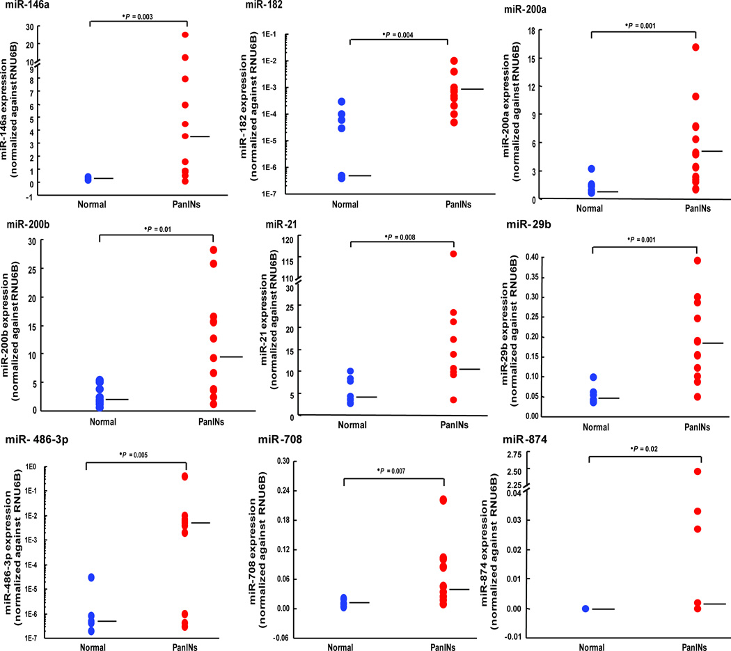
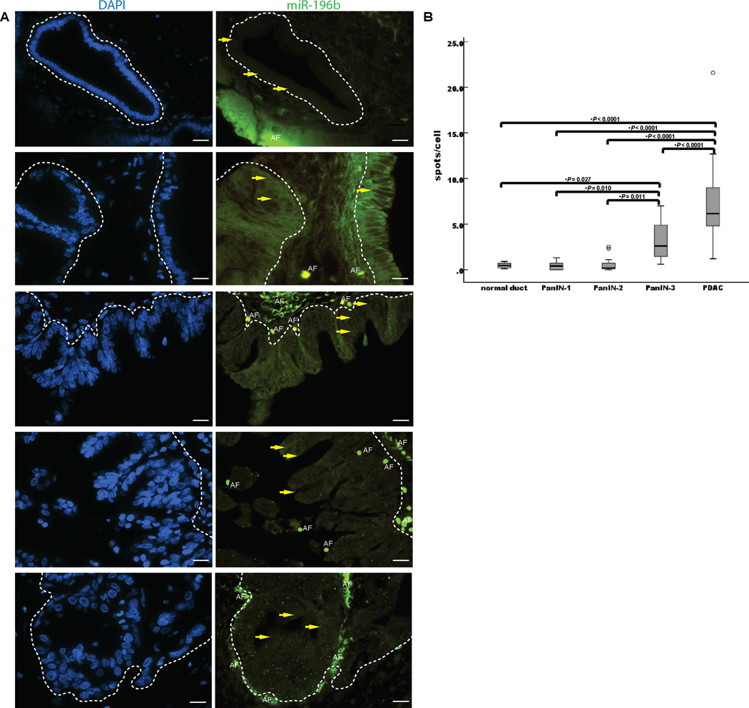
Experimental design: To identify the miRNA alterations that arise during the development of pancreatic cancer, we determined the levels of 735 miRNAs in 34 pancreatic intraepithelial neoplasias (PanIN) and 15 normal pancreatic duct samples isolated by laser capture microdissection using TaqMan miRNA microarrays. Differential expression of selected miRNAs was confirmed by FISH analysis and by quantitative real-time reverse transcription PCR (qRT-PCR) analysis of selected candidate miRNAs in an independent set of PanIN and normal duct samples.
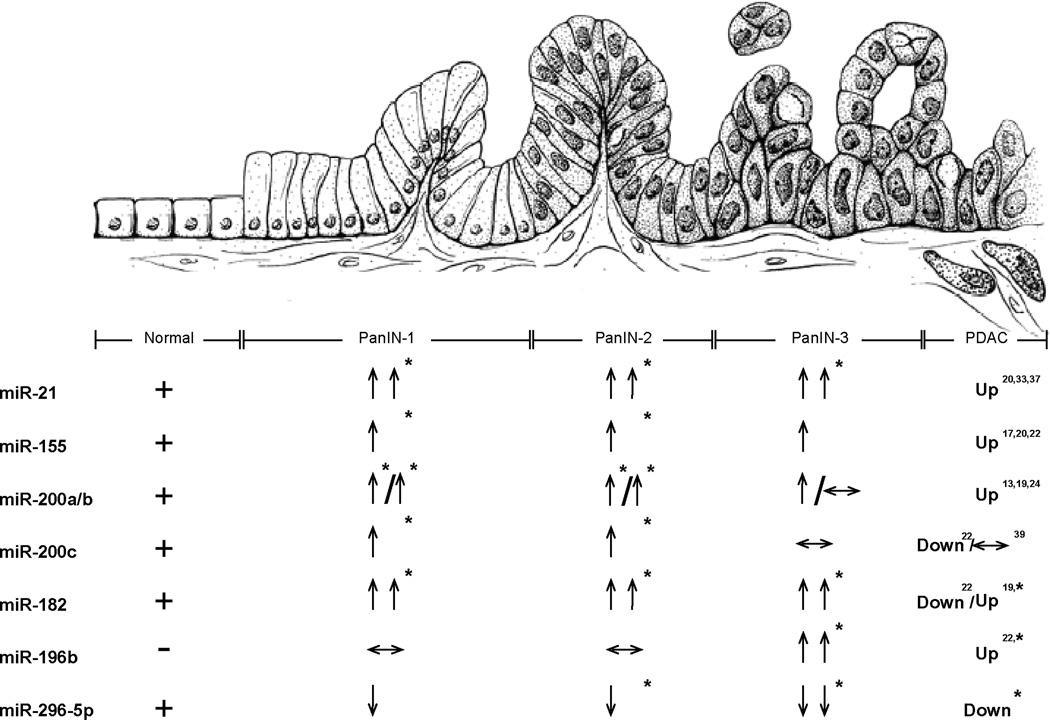
Results: We identified 107 aberrantly expressed miRNAs in different PanIN grades compared with normal pancreatic duct samples and 35 aberrantly expressed miRNAs in PanIN-3 lesions compared with normal pancreatic duct samples. These differentially expressed miRNAs included those that have been previously identified as differentially expressed in pancreatic ductal adenocarcinomas (PDAC; including miR-21, miR-200a/b/c, miR-216a/b, miR-217, miR-146a, miR-155, miR-182, miR-196b, miR-203, miR-222, miR-338-3p, miR-486-3p, etc.) as well as miRNAs not previously described as differentially expressed in these lesions (miR-125b, miR-296-5p, miR-183*, miR-603, miR-625/*, miR-708, etc.). miR-196b was the most selectively differentially expressed miRNA in PanIN-3 lesions.
Conclusions: Many miRNAs undergo aberrant expression in PanIN lesions and are likely to be important in the development of PDAC. The miRNAs, such as miR-196b, whose expression is limited to PanIN-3 lesions or pancreatic cancers could be useful as diagnostic markers.
©2011 AACR.
Conflict of interest statement
Figures

Similar articles
-
MicroRNAs as diagnostic markers for pancreatic ductal adenocarcinoma and its precursor, pancreatic intraepithelial neoplasm.Cancer Genet. 2013 Jun;206(6):217-21. doi: 10.1016/j.cancergen.2013.05.020. Epub 2013 Aug 9. Cancer Genet. 2013. PMID: 23933230
-
Gene expression profiles in pancreatic intraepithelial neoplasia reflect the effects of Hedgehog signaling on pancreatic ductal epithelial cells.Cancer Res. 2005 Mar 1;65(5):1619-26. doi: 10.1158/0008-5472.CAN-04-1413. Cancer Res. 2005. PMID: 15753353
-
Aberrant MicroRNA-155 expression is an early event in the multistep progression of pancreatic adenocarcinoma.Pancreatology. 2010;10(1):66-73. doi: 10.1159/000231984. Epub 2010 Mar 20. Pancreatology. 2010. PMID: 20332664 Free PMC article.
-
Update on the pathology and genetics of exocrine pancreatic tumors with ductal phenotype: precursor lesions and new tumor entities.Dig Dis. 2001;19(1):15-23. doi: 10.1159/000050649. Dig Dis. 2001. PMID: 11385247 Review.
-
Molecular pathways in pancreatic carcinogenesis.J Surg Oncol. 2013 Jan;107(1):8-14. doi: 10.1002/jso.23213. Epub 2012 Jul 17. J Surg Oncol. 2013. PMID: 22806689 Free PMC article. Review.
Cited by
-
Current status of miRNA-targeting therapeutics and preclinical studies against gastroenterological carcinoma.Mol Cell Ther. 2013 Dec 13;1:5. doi: 10.1186/2052-8426-1-5. eCollection 2013. Mol Cell Ther. 2013. PMID: 26056570 Free PMC article. Review.
-
Nickel's Role in Pancreatic Ductal Adenocarcinoma: Potential Involvement of microRNAs.Toxics. 2022 Mar 21;10(3):148. doi: 10.3390/toxics10030148. Toxics. 2022. PMID: 35324773 Free PMC article.
-
miR-128 induces pancreas cancer cell apoptosis by targeting MDM4.Exp Ther Med. 2018 Jun;15(6):5017-5022. doi: 10.3892/etm.2018.6047. Epub 2018 Apr 10. Exp Ther Med. 2018. PMID: 29805525 Free PMC article.
-
Identification of microRNAs as potential biomarker for gastric cancer by system biological analysis.Biomed Res Int. 2014;2014:901428. doi: 10.1155/2014/901428. Epub 2014 May 28. Biomed Res Int. 2014. PMID: 24982912 Free PMC article.
-
MicroRNA-372 acts as a double-edged sword in human cancers.Heliyon. 2023 May 9;9(5):e15991. doi: 10.1016/j.heliyon.2023.e15991. eCollection 2023 May. Heliyon. 2023. PMID: 37251909 Free PMC article. Review.
References
-
- Jemal A, Siegel R, Xu J, Ward E. Cancer statistics, 2010. CA Cancer J Clin. 2010;60:277–300. - PubMed
-
- Hruban RH, Takaori K, Klimstra DS, Adsay NVA-SJ, Biankin AV, Biankin SA, Compton C, Fukushima N, Furukawa T, Goggins M, Kato Y, Kloppel G, Longnecker DS, Luttges J, Maitra A, Offerhaus GJ, Shimizu M, Yonezawa S. An Illustrated Consensus on the Classification of Pancreatic Intraepithelial Neoplasia and Intraductal Papillary Mucinous Neoplasms. Am J Surg Pathol. 2004;28:977–987. - PubMed
-
- Canto MI, Goggins M, Hruban RH, Petersen GM, Giardiello FM, Yeo C, et al. Screening for early pancreatic neoplasia in high-risk individuals: a prospective controlled study. Clin Gastroenterol Hepatol. 2006;4:766–781. quiz 665. - PubMed
-
- Canto MI, Goggins M, Yeo CJ, Griffin C, Axilbund JE, Brune K, et al. Screening for pancreatic neoplasia in high-risk individuals: an EUS-based approach. Clin Gastroenterol Hepatol. 2004;2:606–621. - PubMed
Publication types
MeSH terms
Substances
Grants and funding
LinkOut - more resources
Full Text Sources
Other Literature Sources
Medical
