. 2011 Nov;7(11):e1002362.
doi: 10.1371/journal.ppat.1002362. Epub 2011 Nov 10.

The human herpesvirus-7 (HHV-7) U21 immunoevasin subverts NK-mediated cytoxicity through modulation of MICA and MICB

Affiliations

The human herpesvirus-7 (HHV-7) U21 immunoevasin subverts NK-mediated cytoxicity through modulation of MICA and MICB

Christine L Schneider et al. PLoS Pathog. 2011 Nov.

Abstract

Herpesviruses have evolved numerous immune evasion strategies to facilitate establishment of lifelong persistent infections. Many herpesviruses encode gene products devoted to preventing viral antigen presentation as a means of escaping detection by cytotoxic T lymphocytes. The human herpesvirus-7 (HHV-7) U21 gene product, for example, is an immunoevasin that binds to class I major histocompatibility complex molecules and redirects them to the lysosomal compartment. Virus infection can also induce the upregulation of surface ligands that activate NK cells. Accordingly, the herpesviruses have evolved a diverse array of mechanisms to prevent NK cell engagement of NK-activating ligands on virus-infected cells. Here we demonstrate that the HHV-7 U21 gene product interferes with NK recognition. U21 can bind to the NK activating ligand ULBP1 and reroute it to the lysosomal compartment. In addition, U21 downregulates the surface expression of the NK activating ligands MICA and MICB, resulting in a reduction in NK-mediated cytotoxicity. These results suggest that this single viral protein may interfere both with CTL-mediated recognition through the downregulation of class I MHC molecules as well as NK-mediated recognition through downregulation of NK activating ligands.


Conflict of interest statement

The authors have declared that no competing interests exist.

Figures

Figure 1
Figure 1. Generation of stable cell lines expressing NK ligands and U21.
(A) Schematic representation of class I MHC molecules and NKG2D ligands, depicting α1, α2, and α3 domains of class I MHC molecules, and their overall structural similarity to MICA/B and ULBP molecules. (B) Cell lysates (20 µg) immunoblotted with an antibody directed against U21 or GAPDH, as indicated. Flow cytometric analysis of U373 cells expressing NK ligands or NK ligands and U21. Cells were labeled with antibodies directed against (C) ULBP1 (m295), (D) ULBP2, (E) ULBP3, (F) MICA, and (G) MICB (m360) followed by a PE-conjugated secondary antibody, or with FITC-W6/32 (class I MHC) (H-L). Unlabeled cells (H-L) or cells labeled with the secondary antibody alone (C-G) are indicated by the filled dark gray trace. Control cells labeled with antibodies directed against each NK ligand (C-G) or class I MHC molecules (H-L) are indicated by the filled light gray traces. U21-expressing cells labeled with antibodies directed against each NK ligand (C-G) or class I MHC molecules (H-L) are indicated by the black traces.
Figure 2
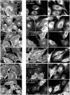
Figure 2. U21 reroutes ULBP1 and MICB to a punctate perinuclear compartment.
Left panels: U373 cells (A), and U373 cells expressing each NK ligand and control vector were immunolabeled with antibodies directed against properly-folded class I MHC molecules (W6/32)(A), or against ULBP1 (m295)(D), ULBP2 (G), ULBP3 (J), MICA (M), or MICB (m360) (P), as noted. Middle and right panels: Cells expressing each NK-ligand and U21, as indicated, were double-labeled with antibodies directed against class I MHC molecules (B), ULBP1 (E), ULBP2 (H), ULBP3 (K), MICA (N), or MICB (Q) and anti-U21 (panels C,F,I,L,O, and R). Asterisks in panels E and F denote cells expressing U21 (F) that also exhibit relocalization of ULBP1. Asterisks in panels N, O, Q, and R indicate cells expressing U21 (O and R) that also exhibit reduced labeling of MICA or MICB. The arrowheads in panels Q and R denote a cell with low apparent levels of U21 (R) and normal surface expression of MICB.
Figure 3
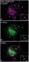
Figure 3. ULBP1 colocalizes with the lysosomal marker lamp2.
(A-C) U373 cells expressing ULBP1 and U21 were double-labeled with antibodies directed against ULBP1 (magenta) and lamp2 (green). The merged image is shown in panel C. The arrowheads denote single coincident puncta, and the boxed regions in the bottom right corner of each panel are enlargements of the smaller boxed region with arrowhead in panel C.
Figure 4
Figure 4. U21 targets ULBP1 for lysosomal degradation.
(A) U373 cells expressing ULBP1 were pulse-labeled for 15 minutes and chased for 0, 2, or 6 hours. ULBP1 was recovered from Triton X-100 lysates with anti-ULBP1 (m295) and treated with either Endo H or PNGase:F. Migration of ULBP1 with and without its N-linked glycan are indicated, as is the approximate position of the 36 kDa molecular weight marker. (B) Cells, as indicated, were pulse-labeled for 15 minutes, chased for 0, 2, and 6 hours and either ULBP1 (m295) or U21 were recovered from Triton X-100 lysates. Where indicated, cells were treated with leupeptin (leu) and folimycin (foli). Migration of U21, class I MHC heavy chain (MHC I), and mature (ULBP1mat) and immature (ULBP1ER) forms of ULBP1 are indicated, as are the approximate positions of molecular weight markers. (C) As a measure of the stability of ULBP1, the percentage of ULBP1 remaining after the 6 hr chase was compared to the percentage of ULBP1 remaining after the 2 hr chase point. The data represent the quantification from three independent experiments (n = 3). Error bars indicate the standard deviation.
Figure 5
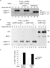
Figure 5. U21 interacts with ULBP1.
(A) U373 and ULBP1-U21 cells were labeled for 2 hrs, and either U21 or ULBP1 (m295) was recovered from digitonin lysates. Migration positions of U21, class I MHC heavy chain, and ULBP1 are indicated. Arrows indicate potential coimmunoprecipitating polypeptides identical in size to ULBP1. (B) Cells, as indicated, were incubated in the presence of folimycin and leupeptin, and U21 was recovered from digitonin lysates. Lysates and immunoprecipitations were immunoblotted with anti-HA and anti-U21 antibodies. Migration positions of mature (HA-ULBP1mat) and immature (HA-ULBP1ER) HA-ULBP1 and U21 are noted.
Figure 6
Figure 6. U21 expression destabilizes MICB.
(A) MICB cells or MICB cells expressing U21 were pulse-labeled for 15 minutes and chased for 0, 2, or 6 hours. MICB (BMO2) and U21 were recovered from Triton X-100 lysates. Migration positions of MICB, U21, and class I MHC heavy chains are indicated, as are approximate molecular weight markers. Note that U21 and MICB migrate at nearly identical positions. (B) Quantification of (A) showing the percent MICB remaining after a 2 hr chase relative to the 0 hr chase point (n = 1). (C) The data represent the quantification of MICB stability in MICB or MICB-U21 cells from three independent experiments (n = 3). Error bars indicate the standard deviation.
Figure 7
Figure 7. U21 expression reduces steady state levels of mature and secreted MICB.
(A) Immunoblot analysis of lysates or supernatants from MICB or MICB-U21 cells. Lysates and concentrated supernatants were prepared from 4×10⁵ MICB or MICB-U21 cells. (B) MICB or MICB-U21 cells were pulse-labeled for 15 min and chased for 0, 0.5, 1, 2, and 4 hrs. MICB was recovered from Triton X-100 lysates (panel i) or cell supernatants (panel ii). Migration positions of MICB, soluble MICB (sMICB), and molecular weight markers are indicated. In U21-expressing cells, the faster-migrating mature (MICB*) and secreted (sMICB*) MICB are noted separately at the right.
Figure 8
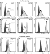
Figure 8. U21 down-regulates and destabilizes MICA and MICB in K562 cells.
(A-F) Flow cytometric analysis of K562 cells expressing ZsGreen or U21-ZsGreen labeled with antibodies to (A) ULBP1 (m295), (B) ULBP2, (C) ULBP3, (D) MICA, and (E) MICB (m360), or (F) ICAM-1 followed by a PE-conjugated secondary antibody. Cells labeled with secondary antibody alone are indicated by the filled dark gray trace. Control ZsGreen-expressing cells are indicated by the filled light gray trace, and ZsGreen-U21 cells are indicated by the black trace. (G-I) Cells were fixed, permeabilized, and labeled with antibodies directed against (G) MICA, (H) MICB (m360), or (I) TfR. Cells labeled with secondary antibody alone are indicated by the filled dark gray trace. Control ZsGreen-expressing cells are indicated by the filled light gray trace, and ZsGreen-U21 cells are indicated by the black trace.
Figure 9
Figure 9. Expression of U21 in K562 cells reduces sensitivity to NKL-mediated cytotoxicity.
(A) NK cytotoxicity assay. Target K562 cells expressing ZsGreen or U21-ZsGreen were incubated in the presence of NKL effector cells at the indicated E:T ratios. The graphs shown are single representative experiment done in triplicate and the error bars indicate the standard deviation between replicate samples (n = 1). (B-C) Target K562 cells expressing ZsGreen (B) or U21-ZsGreen (C) as indicated were incubated with 10 µg/ml IgG1 directed against, MICA, MICB (m360), or a combination of anti-MICA and anti-MICB, or no antibody (control) before addition of NKL effector cells at the indicated E:T ratios. The graphs shown are a single representative experiment performed in triplicate. Error bars indicate the standard deviation between the replicate samples (n = 1). (D) Compilation and statistical analysis of data from 3 independent experiments performed in triplicate (n = 3). The antibody blocking experiment was also performed once in triplicate (n = 1) using an antibody directed against ULBP1 (m295) or an IgG1 isotype control (m479). *pvalue<0.05, **pvalue<0.01, ***pvalue<0.001.

Similar articles

Cited by

References

    1. Tanaka-Taya K, Kondo T, Mukai T, Miyoshi H, Yamamoto Y, et al. Seroepidemiological study of human herpesvirus-6 and -7 in children of different ages and detection of these two viruses in throat swabs by polymerase chain reaction. J Med Virol. 1996;48:88–94. - PubMed
    1. Hansen TH, Bouvier M. MHC class I antigen presentation: learning from viral evasion strategies. Nat Rev Immunol. 2009;9:503–513. - PubMed
    1. Powers C, DeFilippis V, Malouli D, Fruh K. Cytomegalovirus immune evasion. Curr Top Microbiol Immunol. 2008;325:333–359. - PubMed
    1. Yewdell JW, Hill AB. Viral interference with antigen presentation. Nat Immunol. 2002;3:1019–1025. - PubMed
    1. Champsaur M, Lanier LL. Effect of NKG2D ligand expression on host immune responses. Immunol Rev. 2010;235:267–285. - PMC - PubMed

Publication types

MeSH terms