. 2012 Mar;40(5):2294-311.
doi: 10.1093/nar/gkr765. Epub 2011 Nov 15.

Structural analysis of an eIF3 subcomplex reveals conserved interactions required for a stable and proper translation pre-initiation complex assembly

Affiliations

Structural analysis of an eIF3 subcomplex reveals conserved interactions required for a stable and proper translation pre-initiation complex assembly

Anna Herrmannová et al. Nucleic Acids Res. 2012 Mar.

Abstract

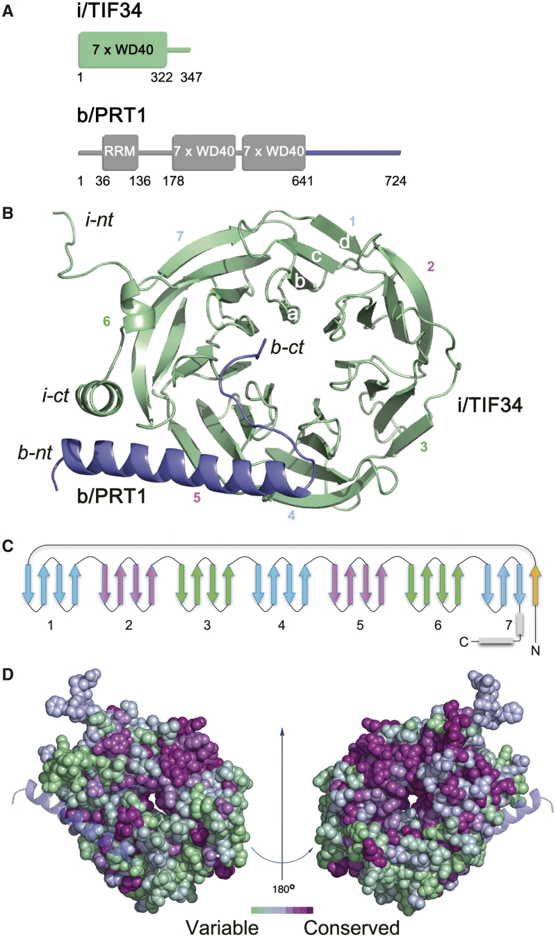
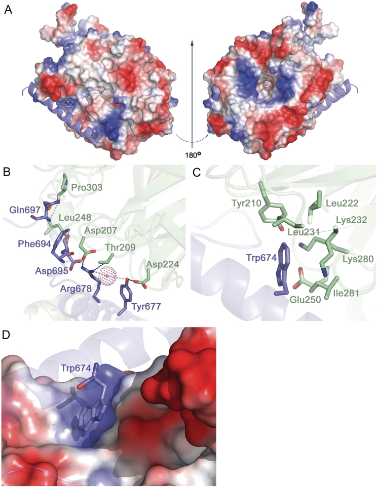
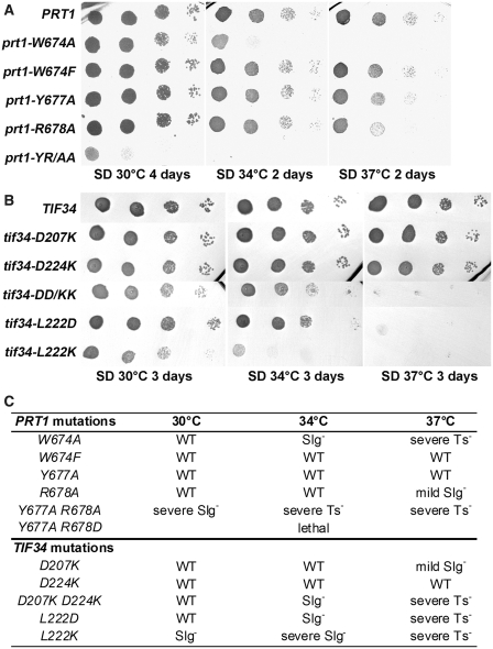
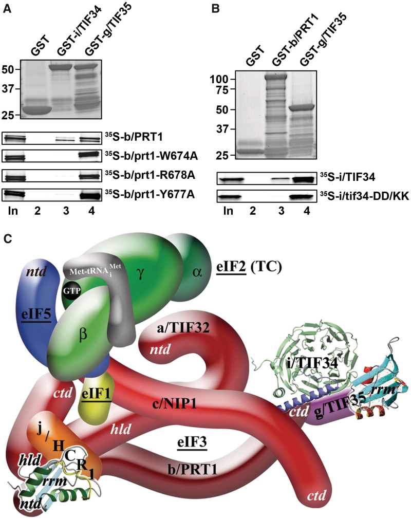
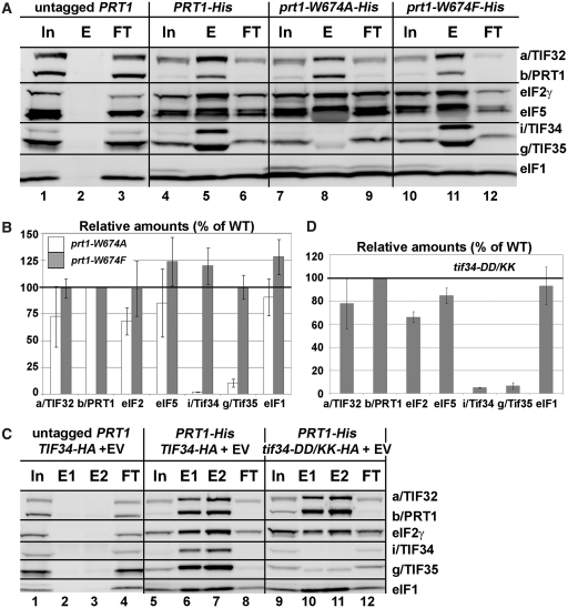
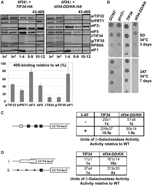
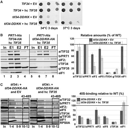
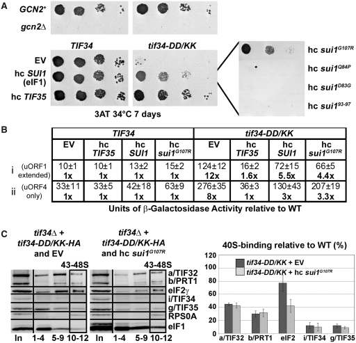
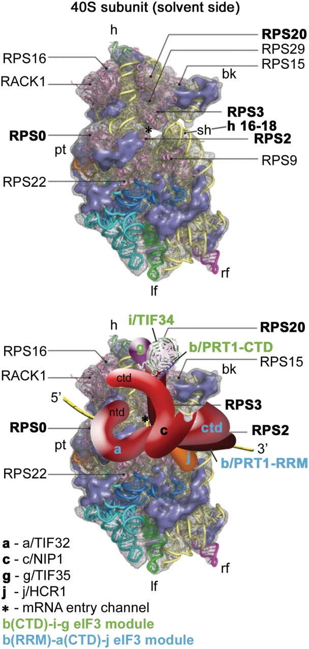
Translation initiation factor eIF3 acts as the key orchestrator of the canonical initiation pathway in eukaryotes, yet its structure is greatly unexplored. We report the 2.2 Å resolution crystal structure of the complex between the yeast seven-bladed β-propeller eIF3i/TIF34 and a C-terminal α-helix of eIF3b/PRT1, which reveals universally conserved interactions. Mutating these interactions displays severe growth defects and eliminates association of eIF3i/TIF34 and strikingly also eIF3g/TIF35 with eIF3 and 40S subunits in vivo. Unexpectedly, 40S-association of the remaining eIF3 subcomplex and eIF5 is likewise destabilized resulting in formation of aberrant pre-initiation complexes (PICs) containing eIF2 and eIF1, which critically compromises scanning arrest on mRNA at its AUG start codon suggesting that the contacts between mRNA and ribosomal decoding site are impaired. Remarkably, overexpression of eIF3g/TIF35 suppresses the leaky scanning and growth defects most probably by preventing these aberrant PICs to form. Leaky scanning is also partially suppressed by eIF1, one of the key regulators of AUG recognition, and its mutant sui1(G107R) but the mechanism differs. We conclude that the C-terminus of eIF3b/PRT1 orchestrates co-operative recruitment of eIF3i/TIF34 and eIF3g/TIF35 to the 40S subunit for a stable and proper assembly of 48S pre-initiation complexes necessary for stringent AUG recognition on mRNAs.


Figures

Figure 1.
Figure 1.
Structure of i/TIF34–b/PRT1 complex. (A) Schematic drawing of the predicted protein domains of yeast i/TIF34 (top) and b/PRT1 subunits of eIF3 (bottom). The folded domains and their boundaries are indicated. Predicted unstructured regions are shown as lines. (B) Overview of the structure: cartoon representation of i/TIF34 (green) and b/PRT1(654–700) (blue). WD-40 blades 1–7, β-strands a–d of blade 1 and N-terminus (nt) and C-terminus (ct) are labeled. The complex is shown from the bottom side of the β-propeller, where loops occur between strands a–b and c–d. (C) Topology diagram of the i/TIF34 fold. (D) Space-filling view of the i/TIF34 in complex with b/PRT1 (bottom view, left and top view, right), colored according to sequence conservation. A gradient of green to purple indicates the degree of phylogenetic conservation, with variable shown as green and most conserved as dark purple. The conservation heat plot of the i/TIF34 surface was generated by ConSurf using multiple alignment of Human, Drosophila, Arabidopsis and Saccharomyces cerevisiae i/TIF34 protein homologues. All structural figures were generated using PyMOL (http://www.pymol.org).
Figure 2.
Figure 2.
Molecular details of i/TIF34 and b/PRT1 interactions. (A) Electrostatic potential (±5kT/e) of the solvent-accessible surface of i/TIF34 in complex with b/PRT1(654–700) rendered on the molecular surface of the complex. A gradient of blue to red shows positive to negative charge, respectively, as calculated using PyMOL built-in APBS tools (55). PQR file for analysis of Poisson–Boltzmann electrostatics calculations was generated using PDB2PQR tool (56) and further used for APBS. b/PRT1(654–700) is shown as a cartoon in blue. View from the b/PRT1 binding site is shown on the left; the ‘reverse’ side of i/TIF34 shown on the right has negative charge clustering at the highly conserved blade 1 (upper part), blades 4 and 5 (the bottom part) and positive charge around the central cavity. (B–C) The i/TIF34-b/PRT1 binding interface involves highly conserved amino acids from both proteins. i/TIF34 is shown in green, b/PRT1 in blue. (B) Y677 and R678 from b/PRT1 form H-bonds (blue) with D207, T209 and D224 from i/TIF34 (only interacting side chains are shown). Other residues making contacts (light blue) via main chain atoms are also shown in sticks. One water molecule (shown in dots) is in close proximity (light pink) to R678 NH1 (2.6 Å) and Y677 OH (3.3 Å). (C) b/PRT1 W674 is surrounded by hydrophobic and charged amino acids (only side chains are shown), which form a shallow pocket. (D) Surface representation of the i/TIF34 hydrophobic pocket, which accommodates b/PRT1 W674. This interaction serves as a ‘lock’ for the i/TIF34 and b/PRT1 interaction interface.
Figure 3.
Figure 3.
Phenotypic and biochemical analysis of i/TIF34 and b/PRT1 mutations that disrupt subunit interactions. (A) The prt1-W674A and -YR/AA but not -W674F mutations produce severe slow growth and temperature sensitive phenotypes. The YAH06 (prt1Δ) strain was transformed with the corresponding plasmids carrying individual mutant alleles and the resident pCR52 (PRT1,URA3) covering plasmid was evicted on 5-FOA. The resulting strains were then spotted in four serial 10-fold dilutions on SD medium and incubated at 30, 34 and 37°C. (B) The tif34-DD/KK, -L222D and L222K mutations produce severe slow growth and temperature sensitive phenotypes. The H450 (tif34Δ) strain was transformed with the corresponding plasmids carrying individual mutant alleles and the resident YEp-i/TIF34-U (TIF34, URA3) covering plasmid was evicted on 5-FOA. The resulting strains were then spotted in four serial 10-fold dilutions on SD medium and incubated at 30, 34 and 37°C. (C) Summary of phenotypes of mutations analyzed in this study.
Figure 4.
Figure 4.
The tif34-DD/KK mutation impairs the direct interaction between i/TIF34 and b/PRT1 in vitro and the revised 3D model of eIF3 in the MFC. (A) The prt1-W674A, -Y677A, and -R678A mutations impair the direct interaction between b/PRT1 and i/TIF34 in vitro. Full-length i/TIF34 (lane 3) and g/TIF35 (lane 4) fused to GST, and GST alone (lane 2), were tested for binding to 35S-labeled wt b/PRT1 and its mutant derivatives; 10% of input amounts added to each reaction is shown in lane 1 (In). (B) Full-length b/PRT1 (lane 3) and g/TIF35 (lane 4) fused to GST, and GST alone (lane 2), were tested for binding to 35S-labeled wt i/TIF34 and the DD/KK mutant derivative. (C) A revised 3D model of eIF3 and its associated eIFs in the MFC (based on the data from (9); ntd, N-terminal domain; ctd, C-terminal domain; hld, HCR1-like domain; rrm, RNA recognition motif; TC, ternary complex). The NMR structure of the interaction between the RRM of human eIF3b (green and light blue) and the N-terminal peptide of human eIF3j (yellow) (12), the NMR structure of the C-terminal RRM of human eIF3g (red and sky-blue) (5), and the X-ray structure of the yeast i/TIF34–b/PRT1 complex (this study), were used to replace the original schematic representations of the corresponding molecules.
Figure 5.
Figure 5.
Disrupting the b/PRT1–i/TIF34 interaction eliminates association of the i/TIF34-g/TIF35 mini-module from the MFC in vivo. (A and B) WCEs prepared from YAH06 (prt1Δ) bearing untagged b/PRT1 (lanes 1–3), 8xHis-tagged b/PRT1 (lanes 4–6), and two of its mutant derivatives (lanes 7–9 and 10–12) were incubated with Ni2+ agarose and the bound proteins were eluted and subjected to western blot analysis with the antibodies indicated in each row. (In) lanes contained 5% of the input WCEs; (E) lanes contained 100% of eluate from the resin; (FT) lanes contained 5% of the flow through. (B) The Western signals for indicated proteins in the E fractions of the wt PRT1-His and its mutants were quantified, normalized for the amounts of the wt b/PRT1 in these fractions and plotted in the histogram as percentages of the corresponding values calculated for the wt b/PRT1. (C and D) WCEs were prepared from YAH12 (prt1Δ tif34Δ) bearing untagged PRT1 and wt TIF34 (lanes 1–4) and from YAH11 (prt1Δ tif34Δ) bearing 8xHis-tagged PRT1 and either wt TIF34 plus empty vector (lanes 5–8) or mutant tif34-DD/KK plus empty vector (lanes 9–12) and analyzed analogously to (A and B).
Figure 6.
Figure 6.
Disruption of the b/PRT1–i/TIF34 interaction prevents 40S-binding of the i/TIF34–g/TIF35 mini-module and dramatically increases leaky scanning over the AUG start site producing a severe Gcn- phenotype. (A) Physical detachment of i/TIF34 and g/TIF35 from the rest of eIF3 selectively affects stability of pre-initiation complexes in vivo. Transformants of H450 (tif34Δ) bearing wt or mutant i/TIF34-HA were grown in SD medium at 34 C to an OD600 of approximately 1.5 and cross-linked with 2% HCHO prior to harvesting. WCEs were prepared, separated on a 7.5–30% sucrose gradient by centrifugation at 41 000 rpm for 5 h and subjected to western blot analysis (note that the anti-RPS0A antibodies were generated in this study; see Supplementary Data). Fractions 1–4, 5–9, and 10–12 (43–48S) were pooled; lanes ‘In0’ and ‘In6’ show samples of the input WCEs (5%) that were processed immediately before (h0) or after (h6) incubation for 6 h on ice, mimicking the duration of the HCHO fractionation experiment to document the stability of the factors of interest in WCEs. Proportions of the 40S-bound proteins relative to the amount of 40S subunits were calculated using NIH ImageJ from three independent experiments. The resulting values obtained with the wt strain were set to 100% and those obtained with mutant strains were expressed as percentages of the wt (SDs are given). This experiment was repeated seven times with similar results. (B) tif34-DD/KK imparts the Gcn- phenotype. H417 (GCN2), H418 (gcn2Δ) and H450 (tif34Δ) bearing wt or mutant i/TIF34-HA were spotted in four serial 10-fold dilutions on SD (upper panel) or SD containing 30 mM 3-AT (lower panel) and then incubated at 34°C for 3 and 7 days, respectively. (C) tif34-DD/KK severely prevents derepression of GCN4-lacZ upon starvation. The H450 strains as in panel A were transformed with the GCN4-lacZ reporter p180 and grown in SD medium at 34°C to an OD600 of approximately 1. The β-galactosidase activities were measured in the WCEs and expressed in units of nmol of O-nitrophenyl-β-d-galactopyranoside hydrolyzed per minute per mg of protein. To induce GCN4-lacZ expression, strains were grown in minimal medium to an OD600 approximately 0.5 and then treated with 10 mM 3-AT for 6 h. The table gives mean values and standard deviations obtained from at least six independent measurements with three independent transformants, and activities with 3-AT-induction relative to those without induction. (D) Detachment of the i/TIF34–g/TIF35 mini-module from eIF3 provokes unusually severe leaky scanning defect. The H450 strains as in (A) were transformed with the GCN4-lacZ reporter plasmids pM226 (i) and plig102-3 (ii) and analyzed as in (C). The table gives activities in mutant relative to wt cells.
Figure 7.
Figure 7.
Increased gene dosage of TIF35 partially suppresses growth defects of tif34-DD/KK mutant by preventing formation of the aberrant PICs. (A) High copy expression of TIF35 partially suppresses growth phenotypes of DD/KK. The H450 strains as in Figure 6A were transformed with empty vector or hc TIF35 and spotted in three serial 10-fold dilutions on SD medium and incubated at 34°C and 37°C for 3 days. (B) High dosage of g/TIF35 destabilizes formation of the MFC in vivo. The YAH12 (prt1Δ tif34Δ) strains as in Figure 5C were transformed with hc TIF35 and subjected to Ni2+-chelation chromatography as described in Figure 5A. The histogram shown on the right combines data from Figure 5C and D and this panel; the data were obtained in parallel experiments carried out at the same time. (C) High dosage of g/TIF35 prevents formation of aberrant TC-containing PICs in vivo. The H450 transformants as in (A) were subjected to formaldehyde cross-linking as described in Figure 6A. Proportions of the 40S-bound proteins relative to the amount of 40S subunits are shown in the histogram on the right. The resulting values obtained with the wt strain were set to 100% and those obtained with the DD/KK strain transformed with empty vector or high copy TIF35 were expressed as percentages of the former (SDs are given).
Figure 8.
Figure 8.
Increased gene dosage of TIF35, SUI1 (eIF1), and its mutant allele sui1G107R suppresses the Gcn phenotype of tif34-DD/KK as well as its severe leaky scanning defect; and high copy sui1G107R disrupts aberrant PICs in vivo. (A) The H450 strains as in Figure 6A were transformed with empty vector, hc SUI1 (eIF1) or its mutant alleles, and with hc TIF35, respectively, spotted in four serial 10-fold dilutions on 3-AT containing SD media and tested for growth at 34°C for 7 days. (B) The strains as in (A) were further transformed with constructs shown in Figure 6D and analyzed as described in there. (C) High dosage of sui1G107R disrupts the aberrant TC-containing PICs in vivo. The H450 transformants as in panel A were subjected to formaldehyde cross-linking as described in Figure 6A. Proportions of the 40S-bound proteins relative to the amount of 40S subunits are shown in the histogram on the right. The resulting values obtained with the wt strain were set to 100% and those obtained with the DD/KK strain transformed with empty vector or hc sui1G107R were expressed as percentages of the former (SDs are given).
Figure 9.
Figure 9.
A model of two eIF3 modules bound to the opposite termini of the scaffold b/PRT1 subunit situated near the mRNA entry channel of the 40S subunit. (Upper panel) The Cryo-EM reconstruction of the 40S subunit is shown from the solvent side with ribosomal RNA represented as tubes. Ribosomal proteins, with known bacterial homologs and placement, are shown as pink cartoons and labeled (adapted from (57)). Positions of RPS0, 2, 3 and 20 and 18S rRNA helices 16–18 are highlighted in bold. The mRNA entry channel is designated by an asterisk. (Lower panel) Hypothetical location of S. cerevisiae eIF3 on the back side of the 40S subunit based on the data presented in this study and elsewhere, including the interactions between RPS0 and a/TIF32-NTD; RPS2 and j/HCR1; RPS2 and 3 and a/TIF32-CTD; helices 16-18 of 18S rRNA and a/TIF32-CTD; and RPS3 and 20 and g/TIF35 (see text for details). The schematic representations of b/PRT1-CTD and i/TIF34 were replaced with the X-ray structure as in Figure 4C. Two eIF3 modules represented by the b/PRT1-CTD–i/TIF34–g/TIF35 and the b/PRT1-RRM–a/TIF32-CTD–j/HCR1 are color-coded in green and blue, respectively. The yellow lines represent mRNA.

Similar articles

Cited by

References

    1. Hinnebusch AG. eIF3: a versatile scaffold for translation initiation complexes. Trends Biochem Sci. 2006;31:553–562. - PubMed
    1. Jackson RJ, Hellen CUT, Pestova TV. The mechanism of eukaryotic translation initiation and principles of its regulation. Nat. Rev. Mol. Cell Biol. 2010;11:113–127. - PMC - PubMed
    1. Phan L, Schoenfeld LW, Valášek L, Nielsen KH, Hinnebusch AG. A subcomplex of three eIF3 subunits binds eIF1 and eIF5 and stimulates ribosome binding of mRNA and tRNAiMet. EMBO J. 2001;20:2954–2965. - PMC - PubMed
    1. Naranda T, Kainuma M, McMillan SE, Hershey JWB. The 39-kilodalton subunit of eukaryotic translation initiation factor 3 is essential for the complex's integrity and for cell viability in Saccharomyces cerevisiae. Mol. Cell. Biol. 1997;17:145–153. - PMC - PubMed
    1. Cuchalová L, Kouba T, Herrmannová A, Danyi I, Chiu W-l, Valášek L. The RNA recognition motif of eukaryotic translation initiation factor 3g (eIF3g) is required for resumption of scanning of posttermination ribosomes for reinitiation on GCN4 and together with eIF3i stimulates linear scanning. Mol. Cell. Biol. 2010;30:4671–4686. - PMC - PubMed

Publication types

MeSH terms

Substances

Associated data