

Cellular therapies supplement: the role of granulocyte macrophage colony-stimulating factor and dendritic cells in regulatory T-cell homeostasis and expansion
- PMID: 22074627
- PMCID: PMC4122334
- DOI: 10.1111/j.1537-2995.2011.03379.x
Cellular therapies supplement: the role of granulocyte macrophage colony-stimulating factor and dendritic cells in regulatory T-cell homeostasis and expansion
Abstract
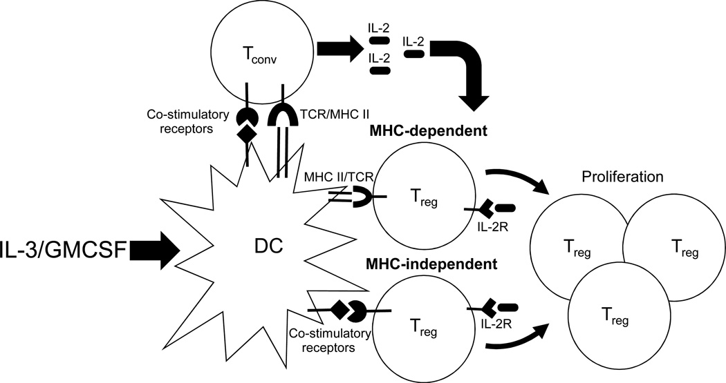
Regulatory T cells are a subset of T cells with inhibitory function that are critical for protection against autoimmunity and immunopathology. A failure to maintain adequate regulatory T-cell numbers in the periphery results in autoimmune manifestations, highlighting the importance of the continuous maintenance of peripheral regulatory T cells. The cellular and molecular requirements for regulatory T-cell homeostasis and expansion are not fully understood but involve a complex interplay among dendritic cells, conventional T cells, and regulatory T cells. In addition, soluble factors such as the cytokine granulocyte macrophage colony-stimulating factor may play a role in enhancing these interactions. In this review, we discuss our National Blood Foundation-funded studies relating to the role of granulocyte macrophage colony-stimulating factor and dendritic cells in controlling regulatory T-cell homeostasis and expansion.
© 2011 American Association of Blood Banks.
Conflict of interest statement
Conflicts of interest: None
Figures


Similar articles
-
Induction of inhibitory central nervous system-derived and stimulatory blood-derived dendritic cells suggests a dual role for granulocyte-macrophage colony-stimulating factor in central nervous system inflammation.Brain. 2010 Jun;133(Pt 6):1637-54. doi: 10.1093/brain/awq081. Epub 2010 Apr 27. Brain. 2010. PMID: 20424288
-
IL-10-producing CD4+CD25+ regulatory T cells play a critical role in granulocyte-macrophage colony-stimulating factor-induced suppression of experimental autoimmune thyroiditis.J Immunol. 2005 Jun 1;174(11):7006-13. doi: 10.4049/jimmunol.174.11.7006. J Immunol. 2005. PMID: 15905543
-
Expansion of immunostimulatory dendritic cells among the myeloid progeny of human CD34+ bone marrow precursors cultured with c-kit ligand, granulocyte-macrophage colony-stimulating factor, and TNF-alpha.J Immunol. 1995 Jun 1;154(11):5851-61. J Immunol. 1995. PMID: 7538534
-
Enhancing the clinical activity of granulocyte-macrophage colony-stimulating factor-secreting tumor cell vaccines.Immunol Rev. 2008 Apr;222:287-98. doi: 10.1111/j.1600-065X.2008.00618.x. Immunol Rev. 2008. PMID: 18364009 Review.
-
Therapeutic potential of FOXP3(+) regulatory T cells and their interactions with dendritic cells.Hum Immunol. 2009 May;70(5):294-9. doi: 10.1016/j.humimm.2009.02.007. Epub 2009 Feb 21. Hum Immunol. 2009. PMID: 19236900 Free PMC article. Review.
Cited by
-
Therapy model for advanced intracerebral B16 mouse melanoma using radiation therapy combined with immunotherapy.Cancer Immunol Immunother. 2013 Jul;62(7):1187-97. doi: 10.1007/s00262-013-1423-9. Epub 2013 Apr 25. Cancer Immunol Immunother. 2013. PMID: 23615842 Free PMC article.
-
Adult Renal Stem/Progenitor Cells Can Modulate T Regulatory Cells and Double Negative T Cells.Int J Mol Sci. 2020 Dec 29;22(1):274. doi: 10.3390/ijms22010274. Int J Mol Sci. 2020. PMID: 33383950 Free PMC article.
-
Skin-derived TSLP systemically expands regulatory T cells.J Autoimmun. 2017 May;79:39-52. doi: 10.1016/j.jaut.2017.01.003. Epub 2017 Jan 23. J Autoimmun. 2017. PMID: 28126203 Free PMC article.
-
The innate immune system stimulating cytokine GM-CSF improves learning/memory and interneuron and astrocyte brain pathology in Dp16 Down syndrome mice and improves learning/memory in wild-type mice.Neurobiol Dis. 2022 Jun 15;168:105694. doi: 10.1016/j.nbd.2022.105694. Epub 2022 Mar 18. Neurobiol Dis. 2022. PMID: 35307513 Free PMC article.
-
Immunotherapy for Parkinson's disease.Neurobiol Dis. 2020 Apr;137:104760. doi: 10.1016/j.nbd.2020.104760. Epub 2020 Jan 21. Neurobiol Dis. 2020. PMID: 31978602 Free PMC article. Review.
References
-
- Kappler JW, Roehm N, Marrack P. T cell tolerance by clonal elimination in the thymus. Cell. 1987;49:273–280. - PubMed
-
- Kisielow P, Bluthmann H, Staerz UD, et al. Tolerance in T-cell-receptor transgenic mice involves deletion of nonmature CD4+8+ thymocytes. Nature. 1988;333:742–746. - PubMed
-
- Liston A, Rudensky AY. Thymic development and peripheral homeostasis of regulatory T cells Curr Opin Immunol. 2007:176–185. - PubMed
-
- Fontenot JD, Gavin MA, Rudensky AY. Foxp3 programs the development and function of CD4+CD25+ regulatory T cells. Nat Immunol. 2003;4:330–336. - PubMed
-
- Sakaguchi S, Sakaguchi N, Asano M, et al. Immunologic self-tolerance maintained by activated T cells expressing IL-2 receptor alpha-chains (CD25) Breakdown of a single mechanism of self-tolerance causes various autoimmune diseases J Immunol. 1995:1151–1164. - PubMed
Publication types
MeSH terms
Substances
Grants and funding
LinkOut - more resources
Full Text Sources
