. 2011 Nov 9;31(45):16241-50.
doi: 10.1523/JNEUROSCI.3667-11.2011.

CX3CR1 deficiency leads to impairment of hippocampal cognitive function and synaptic plasticity

Affiliations

CX3CR1 deficiency leads to impairment of hippocampal cognitive function and synaptic plasticity

Justin T Rogers et al. J Neurosci. .

Abstract

The protective/neurotoxic role of fractalkine (CX3CL1) and its receptor CX3C chemokine receptor 1 (CX3CR1) signaling in neurodegenerative disease is an intricate and highly debated research topic and it is becoming even more complicated as new studies reveal discordant results. It appears that the CX3CL1/CX3CR1 axis plays a direct role in neurodegeneration and/or neuroprotection depending on the CNS insult. However, all the above studies focused on the role of CX3CL1/CX3CR1 signaling in pathological conditions, ignoring the relevance of CX3CL1/CX3CR1 signaling under physiological conditions. No approach to date has been taken to decipher the significance of defects in CX3CL1/CX3CR1 signaling in physiological condition. In the present study we used CX3CR1⁻/⁻, CX3CR1⁺/⁻, and wild-type mice to investigate the physiological role of CX3CR1 receptor in cognition and synaptic plasticity. Our results demonstrate for the first time that mice lacking the CX3CR1 receptor show contextual fear conditioning and Morris water maze deficits. CX3CR1 deficiency also affects motor learning. Importantly, mice lacking the receptor have a significant impairment in long-term potentiation (LTP). Infusion with IL-1β receptor antagonist significantly reversed the deficit in cognitive function and impairment in LTP. Our results reveal that under physiological conditions, disruption in CX3CL1 signaling will lead to impairment in cognitive function and synaptic plasticity via increased action of IL-1β.


Figures

Figure 1.
Figure 1.
a, b, CX3CR1−/− mice show decreased hippocampal neurogenesis in a gene-dose-dependent manner. Unbiased stereology revealed a significant decrease in the number of DCX+ cells in the SGZ of adult male CX3CR1−/− and CX3CR1+/− mice compared to wild-type mice (p < 0.0001). a, CX3CR1−/− mice were also significantly different from CX3CR1+/− mice (p < 0.001). Quantification of the number of cells that were proliferating during the preceding 24 h, as determined by the incorporation of BrdU, showed significantly fewer in the CX3CR1−/− and CX3CR1+/− mice compared to wild-types (p < 0.0001). b, CX3CR1−/− mice were also significantly different from CX3CR1+/− mice (p < 0.05). White bars, WT; gray bars, CX3CR1+/− mice; black bars, CX3CR1−/−. All data are presented as mean ± SEM. ***p < 0.0001; **p < 0.001; *p < 0.05.
Figure 2.
Figure 2.
a–c, CX3CR1−/− and CX3CR1+/− mice show motor learning impairment but normal spontaneous activity. a, On the first day of training (Trials 1–4) no difference between genotypes was observed in the learning ability in the rotarod task. On the second day of training (Trials 5–8) as expected, wild-type mice learned the rotarod task as demonstrated by their ability to remain on the rod for longer periods. Neither the CX3CR1−/− nor the CX3CR1+/− mice showed significant improvement in motor coordination with training when compared to wild-type (repeated-measure ANOVA; p < 0.0001). White circles, WT; gray circles, CX3CR1+/−; black circles, CX3CR1−/−. All data are presented as mean ± SEM. *p < 0.01. b, Comparison of the motor performance of each group of mice on Trial 1 versus Trial 8 shows that wild-type performed significantly better than CX3CR1−/− and CX3CR1+/− mice (linear regression; **p < 0.001). c, CX3CR −/− and CX3CR1+/− mice show normal spontaneous locomotor activity. Wild-type, CX3CR1−/− and CX3CR1+/− mice do not show any difference in total distance traveled in the open field. White bar, WT; gray bar, CX3CR1+/−; black bar, CX3CR1−/−. All data are presented as mean ± SEM.
Figure 3.
Figure 3.
CX3CR1−/− mice show impairment in contextual fear conditioning and Morris water maze memory function. CX3CR1−/− (black circles) and CX3CR1+/− (gray circles) are compared to littermate wild-type mice (white circles). a, A tone (solid bar) was paired with a foot shock (arrowhead) at 2 and 4 min. Freezing behavior is shown on the day of training for CX3CR1−/−, CX3CR1+/−, and wild-type mice and is comparable in all groups. b, CX3CR1−/− and CX3CR1+/− mice showed significant reduced freezing response compared to wild-type when tested 24 h following training. c, CX3CR1−/− and CX3CR1+/− mice showed normal freezing to cue component compared to controls. **p < 0.01. d, e, The hidden-platform version of the Morris water maze task. d, Mean latency to escape from a pool to hidden platform across training days. e, A first probe test was performed on Day 7, and a second probe test was performed on Day 11 to determine the number of pseudo-platform crossings in the target quadrant (TQ) compared to the opposite quadrant (OP), the left quadrant (LQ), and the right quadrant (RQ). White bar, WT; gray bar, CX3CR1+/−; black bar, CX3CR1−/−. All data are presented as mean ± SEM. p < 0.01. f, Average swim speed. No differences were seen in the overall swim distance between WT, CX3CR1−/− and CX3CR1+/− mice during the probe test.
Figure 4.
Figure 4.
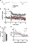
a, b, CX3CR1 deficiency leads to impairments in LTP. LTP was induced with HFS (2 1 s, 100 Hz bursts separated by 20 s; b, arrow) after 20 min of baseline recording, and changes in fEPSP slope are expressed as a percentage of baseline (a). b, Representative fEPSP traces taken from hippocampal slices of (left to right) wild-type, CX3CR1+/− and CX3CR1−/− mice. Both CX3CR1−/− (black) and CX3CR1+/− (red) have deficiencies in LTP of area CA1 compared to wild-type controls (white). c, The last 5 min of fEPSPs slope recordings were averaged for wild-type (n = 12), CX3CR1−/− (n = 10), and CX3CR1+/− (n = 9) mice (**p < 0.05). d, PPF was induced with the use of paired pulses given with an initial delay of 20 ms, and the time to the second pulse was increased 20 ms incrementally until a final delay of 300 ms was reached. There was no significant PPF differences between experimental groups. All data are presented as mean ± SEM. p < 0.01.
Figure 5.
Figure 5.
a, CX3CR1−/− and CX3CR1+/− mice show increased hippocampal protein levels of IL-1β compared to littermate WTs as measured by ELISA. b, Western blot analysis of hippocampi from wild-type, CX3CR1−/−, and CX3CR1+/− mice shows an increase in phospho (p)-p38 protein in CX3CR1−/− and CX3CR1+/− mice compared to that of wild-type (right blot; near-infrared image displayed in gray scale). Relative p-p38 expression normalized to β-actin shows the highest expression in CX3CR1−/− and CX3CR1+/− mice. c, TNFα cerebellar protein levels are significantly increased in CX3CR1−/− and CX3CR1+/− mice compared to wild-type mice as measured by ELISA. d, The area of Iba-1 staining is significant higher in CX3CR1−/− mice compared to wild-type and CX3CR1+/− mice as measured by immunohistochemistry. White bar, WT; gray bar, CX3CR1+/−; black bar, CX3CR1−/−. e, f, Representative photomicrographs of the Iba-1+ cells in the dentate gyrus of wild-type (e) and CX3CR1-deficient mice (f). All data are presented as mean ± SEM. p < 0.01. **p < 0.005; *p < 0.05.
Figure 6.
Figure 6.
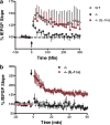
a, b, CX3CR1 deficiency leads to impairments in synaptic plasticity which are rescued through IL-1β antagonism. PTP was induced with HFS (1 s, 100 Hz bursts; arrow) after 1 min of baseline recording. LTP was induced with HFS (2 1 s, 100 Hz bursts separated by 20 s; arrow) after 20 min of baseline recording. Changes in fEPSP slope are expressed as a percentage of baseline. a, The IL-1β antagonist, IL-1ra (100 μg/ml), rescues the PTP deficit (red; n = 7) in hippocampi taken from CX3CR1−/− (black; n = 8) in area CA1 to levels seen in wild-type (white; n = 9). b, The LTP deficit seen in hippocampi taken from CX3CR1−/− (black; n = 10) mice is rescued with the IL-1β antagonist IL-1ra (100 μg/ml) (red; n = 11).
Figure 7.
Figure 7.
IL-1ra reversed the deficit in contextual fear conditioning and Morris water maze memory function induced by the loss of CX3CR1 but not the deficit in motor learning. a, CX3CR1−/− ACSF-treated (blue squares) and CX3CR1−/− IL-1ra-treated mice (red squares) are compared to wild-type ACSF-treated mice (open circles), wild-type mice treated with heat-inactivated IL-1ra (black squares), and wild-type mice treated with IL-1ra (green squares). On the first day of training (Trials 1–4), no difference between groups was observed in the learning ability on the rotarod task. On the second day of training (Trials 5–8), all wild-type mouse groups learned the rotarod task as demonstrated by their ability to remain on the rod for longer periods. Neither the CX3CR1−/− ACSF-treated nor CX3CR1−/− IL-1ra-treated mice showed significant improvement in motor coordination with training when compared to wild-type. b, A tone (solid bar) was paired with a foot shock (arrowhead) at 2 and 4 min. Freezing behavior is shown on the day of training for CX3CR1−/− ACSF-treated mice (blue squares), CX3CR1−/− IL-1ra-treated mice (red squares), wild-type ACSF-treated mice (white circles), wild-type treated with heat-inactivated IL-1ra (black squares), and wild-type IL-1ra-treated mice (green squares) and is comparable in all groups. c, CX3CR1−/− control mice showed significantly reduced freezing compared to all wild-type groups when tested 24 h after training. d, CX3CR1−/− treated with IL-1ra (red squares) showed a freezing response similar to that of wild-type groups. d–f, The hidden-platform version of Morris water maze task. Mean latency to escape from a pool to hidden platform across training days (d). A probe test was performed on Day 10 to determine the number of pseudo-platform crossings in the target quadrant (TQ) compared to the opposite quadrant (OP) (e). White bar, WT ACSF-treated mice; light gray bar, wild-type treated with heat-inactivated IL-1ra; dark gray bar, wild-type treated with IL-1ra; black bar, CX3CR1−/− ACSF-treated mice; black/white bar, CX3CR1−/− IL-1ra-treated mice. f shows the time spent in the target platform zone. CX3CR1−/− ACSF-treated mice (black bar) spent significantly less time in the target zone compared to wild-type controls [white bar, wild-type ACSF-treated mice; light gray bar, wild-type heat-inactivated IL-1ra-treated mice; dark gray bar, wild-type IL-1ra-treated mice]. CX3CR1−/− mice treated with IL-1ra spent the same amount of time in the target quadrant compared to wild-type controls. All data are presented as mean ± SEM. p < 0.01. *p < 0.05 (CX3CR1−/− control vs wild-type controls); ##p < 0.001 (CX3CR1−/− IL-1ra vs CX3CR1−/−).

Similar articles

Cited by

References

    1. Aloisi F. Immune function of microglia. Glia. 2001;36:165–179. - PubMed
    1. Bachstetter AD, Morganti JM, Jernberg J, Schlunk A, Mitchell SH, Brewster KW, Hudson CE, Cole MJ, Harrison JK, Bickford PC, Gemma C. Fractalkine and CX(3)CR1 regulate hippocampal neurogenesis in adult and aged rats. Neurobiol Aging. 2011;32:2030–2044. - PMC - PubMed
    1. Batchelor PE, Liberatore GT, Wong JY, Porritt MJ, Frerichs F, Donnan GA, Howells DW. Activated macrophages and microglia induce dopaminergic sprouting in the injured striatum and express brain-derived neurotrophic factor and glial cell line-derived neurotrophic factor. J Neurosci. 1999;19:1708–1716. - PMC - PubMed
    1. Bazan JF, Bacon KB, Hardiman G, Wang W, Soo K, Rossi D, Greaves DR, Zlotnik A, Schall TJ. A new class of membrane-bound chemokine with a CX3C motif. Nature. 1997;385:640–644. - PubMed
    1. Beffert U, Weeber EJ, Durudas A, Qiu S, Masiulis I, Sweatt JD, Li WP, Adelmann G, Frotscher M, Hammer RE, Herz J. Modulation of synaptic plasticity and memory by Reelin involves differential splicing of the lipoprotein receptor Apoer2. Neuron. 2005;47:567–579. - PubMed

Publication types

MeSH terms