. 2011 Dec;21(12):1723-39.
doi: 10.1038/cr.2011.176. Epub 2011 Nov 8.

S phase-dependent interaction with DNMT1 dictates the role of UHRF1 but not UHRF2 in DNA methylation maintenance

Affiliations

S phase-dependent interaction with DNMT1 dictates the role of UHRF1 but not UHRF2 in DNA methylation maintenance

Jiqin Zhang et al. Cell Res. 2011 Dec.

Abstract

Recent studies demonstrate that UHRF1 is required for DNA methylation maintenance by targeting DNMT1 to DNA replication foci, presumably through its unique hemi-methylated DNA-binding activity and interaction with DNMT1. UHRF2, another member of the UHRF family proteins, is highly similar to UHRF1 in both sequence and structure, raising questions about its role in DNA methylation. In this study, we demonstrate that, like UHRF1, UHRF2 also binds preferentially to methylated histone H3 lysine 9 (H3K9) through its conserved tudor domain and hemi-methylated DNA through the SET and Ring associated domain. Like UHRF1, UHRF2 is enriched in pericentric heterochromatin. The heterochromatin localization depends to large extent on its methylated H3K9-binding activity and to less extent on its methylated DNA-binding activity. Coimmunoprecipitation experiments demonstrate that both UHRF1 and UHRF2 interact with DNMT1, DNMT3a, DNMT3b and G9a. Despite all these conserved functions, we find that UHRF2 is not able to rescue the DNA methylation defect in Uhrf1 null mouse embryonic stem cells. This can be attributed to the inability for UHRF2 to recruit DNMT1 to replication foci during S phase of the cell cycle. Indeed, we find that while UHRF1 interacts with DNMT1 in an S phase-dependent manner in cells, UHRF2 does not. Thus, our study demonstrates that UHRF2 and UHRF1 are not functionally redundant in DNA methylation maintenance and reveals the cell-cycle-dependent interaction between UHRF1 and DNMT1 as a key regulatory mechanism targeting DNMT1 for DNA methylation.


Figures
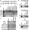
Figure 1
Figure 1
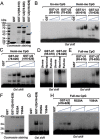
UHRF2 recognizes specifically H3K9 methylation by its tandem tudor domain. (A) A diagram illustrating the structure and sequence similarity between human UHRF1 and UHRF2. UBL, ubiquitin-like domain; TD, tandem tudor domain; PHD, plant homeodomain; SRA, SET and ring associated domain; Ring, really interesting new gene domain. (B) Binding of UHRF2 to a panel of methylated histone H3 and H4 peptides in comparison to other known histone-binding proteins. Pull-downs with HeLa nuclear extracts and various histone peptides were performed and the binding of UHRF1, UHRF2, HP1α, PHF8 and HDAC1 were revealed by western blot analysis. (C) The tandem tudor domain determines the H3K9me2 binding specificity. In vitro synthesized, 35S-met-labeled UHRF2 and deletion or point mutation mutants were subjected to pull-down assays with H3 and H3K9me2 and H3K9me3 peptides. The binding of various UHRF2 proteins were revealed by autoradiography. (D) Recombinant UHRF1 and UHRF2 bound H3K9me2/3 peptides in vitro. Purified GST-UHRF1 (aa 1-436) and GST-UHRF2 (aa 1-408) were subjected to pull down assays and the proteins bound to the peptides were resolved by SDS-PAGE and revealed by Coomassie blue staining.
Figure 2
Figure 2
UHRF2 binds hemi- and fully methylated DNA through its SRA domain. (A) Coomassie blue staining gel showing purified GST-fusions of UHRF1 and UHRF2. The regions of UHRF1 or UHRF2 in number of amino acids fused to GST were as indicated. Arrows indicate the positions of GST-fusion proteins with the expected sizes. Also indicated are protein size markers. (B) Gel mobility shift assay showing binding of GST-UHRF1 (95-610) and GST-UHRF2 (78-626) to hemi-methylated but not un-methylated DNA probes. An increasing amount of recombinant proteins (0.25 μg, 0.5 μg and 1 μg) were used for gel shift. (C) The recombinant UHRF2 containing SRA domain alone, GST-UHRF2 (416-626), is capable of binding hemi-methylated DNA probe. 0.25, 0.5 and 1 μg of proteins were used for gel shift. (D) Comparison of GST-UHRF1 (95-610) and GST-UHRF2 (78-626) in binding of un-methylated, hemi-methylated (one strand methylated) and fully methylated (both strands methylated) DNA probe. (E) Increasing amount of GST-UHRF1 (95-610) and GST-UHRF2 (78-626) were compared for binding of fully methylated DNA probe. Three concentrations used were 0.25, 0.5 and 1 μg, respectively. (F) Coomassie blue staining gel showing purified GST, GST-UHRF2 (416-626), and GST-UHRF2 (416-626) with R520A or Y584A mutation. (G) Gel mobility shift assay showing impaired hemi-methylated DNA binding activity for R520A and Y584A mutants. (H) R520A and Y584A mutants also showed impaired binding activity for fully methylated DNA probe. Three concentrations of proteins used were 0.25, 0.5 and 1 μg, respectively.
Figure 3
Figure 3
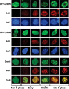
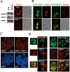
UHRF1 and UHRF2 are enriched at pericentric heterochromatins independent of cell cycle, whereas DNMT1 is enriched at pericentric heterochromatins only in middle and late S phase. NIH3T3 cells were transfected with GFP-UHRF1 or GFP-UHRF2 and blocked at S phase by aphidicolin treatment. The cells were then released from S phase block and labeled with BrdU (red) and also stained with 4′,6-diamidino-2-phenylindole (DAPI). DNMT1 was revealed by immunostaining using a DNMT1 specific antibody. The DAPI staining foci represent pericentric heterochromatins.
Figure 4
Figure 4
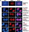
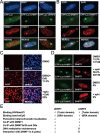
The H3K9me2/3 binding activity is the primary determinant for pericentric heterochromatin association of UHRF2. (A) The pericentric heterochromatin (HC) association for endogenous UHRF2 in NIH3T3 cells. (B) The pericentric heterochromatin localization for wild-type, methylated H3K9-binding deficient (WF143/144AA and FW262/263AA) and hemi-methylated DNA binding deficient (R520A and Y584A) mutants was analyzed in NIH3T3 using anti-Flag antibody cells. After counting large quantity of cells, the percentage of cells with UHRF2 and DAPI co-localization with DAPI foci representing pericentric heterochromatin (HC) was calculated for wild-type UHRF2 and each mutant.
Figure 5
Figure 5
UHRF2 forms neither heterodimers with UHRF1 nor homodimers with itself. (A) UHRF2 did not coimmunoprecipitate with UHRF1. The interaction between UHRF1 and UHRF2 was tested via coimmunoprecipitation between GFP-UHRF2 and Flag-UHRF1 transiently expressed in 293T cells. (B) UHRF2 did not form homodimers or oligomers. The interaction between UHRF2 and UHRF2 was tested via coimmunoprecipitation between GFP-UHRF2 and Flag-UHRF2 transiently expressed in 293T cells. (C) No co-localization between UHRF1 and UHRF2 was observed in DG44-CHO cells. As DG44-CHO cells contain hundreds and thousands of Lac operons integrated in a single genomic site, expression of CFP-Lac-UHRF1 or CFP-Lac-UHRF2 all led to the observation of a bright CFP foci. However, Flag-UHRF2 was not recruited to the bright CFP-Lac-UHRF1 foci and Flag-UHRF1 was not recruited to the bright CFP-Lac-UHRF2 foci.
Figure 6
Figure 6
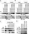
UHRF2 interacts with DNMT1, DNMT3a, DNMT3b and G9a. (A) UHRF1 co-immunoprecipitated with DNMT1, DNMT3a and DNMT3b. GFP-tagged UHRF1 was expressed alone or together with DNMT1, DNMT3a and DNMT3b, respectively in 293T cells and subjected to immunoprecipitation analysis using anti-Flag antibody and western blot analysis using UHRF1 and Flag antibodies. (B) UHRF2 co-immunoprecipitated with DNMT1, DNMT3a and DNMT3b. The co-immunoprecipitation experiments were performed as above except GFP-UHRF2 was used. (C) UHRF1 and UHRF2 in HeLa nuclear extracts co-immunoprecipitated with DNMT1. Note that low levels of UHRF1 were detected in immunoprecipitation of UHRF2 and vice versa, most likely due to slight cross-reaction in western blot between UHRF1 and UHRF2 antibodies. (D) Both UHRF1 and UHRF2 co-immunoprecipitated with G9a. GFP-tagged UHRF2 or UHRF1 were expressed alone or together with Flag-G9a in 293T cells and subjected to IP-western analysis using antibodies as indicated.
Figure 7
Figure 7
Overexpression of UHRF2 in mouse Uhrf1−/− ES cells rescues neither DNA methylation defect nor pericentric heterochromatin localization of DNMT1 in S phase. (A) Confirmation of DNA methylation defect in Uhrf1−/− ES cells by anti-mC immunostaining. Left panel shows western blot analysis of whole cell extracts derived from wild-type E14 and Uhrf1−/− ES cells. Right panel shows representative 5-meC immunostaining data for E14 and Uhrf1−/− ES cells. Also shown are the phase contrast images. (B) Ectopic expression of GFP-UHRF1 but not GFP-UHRF2 rescued DNA methylation defect in Uhrf1−/− ES cells. Note both GFP-UHRF1 and GFP-UHRF2 exhibited a focal staining pattern, in agreement with their expected pericentric heterochromatin localization. (C) Immunostaining for DNMT1 confirmed absence of focal staining pattern (pericentric heterochromatin targeting) for DNMT1 in Uhrf1−/− cells. The cells with DNMT1 focal staining in E14 were circled. Also shown are DAPI staining images. (D) Expression of GFP-UHRF1 but not GFP-UHRF2 restored correct pericentric heterochromatin targeting of DNMT1. The Uhrf1−/− ES cells were transfected with GFP-UHRF1 or GFP-UHRF2 and then subjected to immunostaining for DNMT1. The merged image revealed the same focal localization patterns for DNMT1 and GFP-UHRF1. Left panel shows enlarged images of the cells marked by rectangle. Note that no focal staining pattern for DNMT1 was observed for GFP-UHRF2 expressing cells.
Figure 8
Figure 8
UHRF1 but not UHRF2 interacts with DNMT1 in DG44-CHO cells and this interaction is S phase dependent. (A) The interactions between DNMT1 and UHRF1, and DNMT1 and UHRF2 were analyzed in DG44-CHO cells using CFP-Lac-UHRF1, CFP-lac-UHRF2 and Flag-DNMT1. Expression of control CFP-Lac, CFP-Lac-UHRF1 and CFP-Lac-UHRF2 all resulted in a bright foci in cells. However, recruitment of Flag-DNMT1 was observed only for CFP-Lac-UHRF1 cells in about 10% - 15% cells expressing both CFP-Lac-UHRF1 and Flag-DNMT1. (B) The interaction with DNMT1 was reciprocally analyzed using CFP-Lac-DNMT1 and Flag-UHRF1 and Flag-UHRF2 in DG44-CHO cells. The colocalization with CFP-Lac-DNMT1 was observed only for Flag-UHRF1 but not Flag-UHRF2. Note the colocalization was also observed on other regions enriched with CFP-Lac-UHRF1. This interaction is again observed only in 10% - 15% cells coexpressing both CFP-Lac-DNMT1 and Flag-UHRF1. (C) Aphidicolin treatment enriched DG44-CHO cells in S phase. DG44-CHO cells were treated with or without 1 mg/ml aphidicolin for 20 h. Aphidicolin was removed and cells were incubated in the presence of 10 μM EdU for 2 h and processed for EdU immunostaining (left panel) and merged figures with DAPI staining (right panel). (D) Aphidicolin treatment similarly increased the DG44-CHO cells with UHRF1 and DNMT1 colocalization.

Similar articles

Cited by

References

    1. Law JA, Jacobsen SE. Establishing, maintaining and modifying DNA methylation patterns in plants and animals. Nat Rev Genet. 2010;11:204–220. - PMC - PubMed
    1. Taberlay PC, Jones PA. DNA methylation and cancer. Prog Drug Res. 2011;67:1–23. - PubMed
    1. Chen T, Li E. Structure and function of eukaryotic DNA methyltransferases. Curr Top Dev Bio. 2004;60:55–89. - PubMed
    1. Leonhardt H, Page AW, Weier HU, Bestor TH. A targeting sequence directs DNA methyltransferase to sites of DNA replication in mammalian nuclei. Cell. 1992;71:865–873. - PubMed
    1. Liu Y, Oakeley EJ, Sun L, Jost JP. Multiple domains are involved in the targeting of the mouse DNA methyltransferase to the DNA replication foci. Nucleic Acids Res. 1998;26:1038–1045. - PMC - PubMed

Publication types