Review
. 2012 Feb;121(1):1-20.
doi: 10.1007/s00412-011-0347-4. Epub 2011 Nov 3.

DNA glycosylases: in DNA repair and beyond

Affiliations
Review

DNA glycosylases: in DNA repair and beyond

Angelika L Jacobs et al. Chromosoma. 2012 Feb.

Abstract

The base excision repair machinery protects DNA in cells from the damaging effects of oxidation, alkylation, and deamination; it is specialized to fix single-base damage in the form of small chemical modifications. Base modifications can be mutagenic and/or cytotoxic, depending on how they interfere with the template function of the DNA during replication and transcription. DNA glycosylases play a key role in the elimination of such DNA lesions; they recognize and excise damaged bases, thereby initiating a repair process that restores the regular DNA structure with high accuracy. All glycosylases share a common mode of action for damage recognition; they flip bases out of the DNA helix into a selective active site pocket, the architecture of which permits a sensitive detection of even minor base irregularities. Within the past few years, it has become clear that nature has exploited this ability to read the chemical structure of DNA bases for purposes other than canonical DNA repair. DNA glycosylases have been brought into context with molecular processes relating to innate and adaptive immunity as well as to the control of DNA methylation and epigenetic stability. Here, we summarize the key structural and mechanistic features of DNA glycosylases with a special focus on the mammalian enzymes, and then review the evidence for the newly emerging biological functions beyond the protection of genome integrity.


Figures
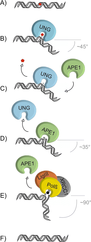
Fig. 1
Fig. 1
The core pathway short-patch BER. The base-excision repair pathway addresses single-base lesions (a). BER is initiated by a DNA glycosylase, e.g., UNG, specifically recognizing and binding a base lesion. Upon encountering a substrate base, e.g., uracil for UNG, the glycosylase flips the base out of the base-stack into its catalytic site pocket where specific contacts examine the substrate base and position it for nucleophilic attack to the N-glycosidic bond (b). Release of the substrate base results in an abasic site (c), which is further processed by the AP-endonuclease, APE1, that cleaves the phosphate backbone 5’ to the abasic site, producing a 3’OH and a 5’deoxyribose-phosphate moiety (5’dRP) (d). Polymerase β (Polβ) hydrolyzes the 5’dRP and fills in the single nucleotide gap, which is subsequently sealed by the DNA ligase III (LigIII), supported by the scaffold protein XRCC1 (e), thus restoring the original base sequence (f). The increase of DNA bending from UNG to Polβ might support the directionality of the handover from one BER factor to the next
Fig. 2
Fig. 2
Mechanism of base removal by monofunctional and bifunctional glycosylases. While all DNA glycosylases share a common principle of action, they differ in details of damage search, recognition, and excision. Among the monofunctional DNA glycosylases, UNG searches for uracil in DNA by oscillating between an open conformation, making unspecific contacts with the DNA, and a closed conformation for base-interrogation (a), involving conserved structures at the mouth of the catalytic site pocket (b). Pre-selected bases are flipped out of the DNA duplex and inserted into the catalytic pocket, where specific hydrogen bonds align the base for nucleophilic attack by an activated water molecule strategically positioned by a conserved aspartic acid residue (red asterisk, c). TDG differs from UNG by its non-specific interaction with DNA through a clamp formed by the N-terminal domain (a), its larger catalytic pocket that accommodates a broader range of substrates, and its ability to involve the opposing base in lesion recognition (b). The catalytic residue in TDG is an asparagine (black asterisk) that positions, but does not activate, a water molecule which can then act as a weak nucleophile (c). TDG stays firmly bound to the abasic site upon base release (d) until SUMOylation induces dissociation by neutralizing the N-terminal DNA binding activity (e). Similar to UNG, the bifunctional OGG1 pre-selects bases for flipping by interactions with conserved amino-acid residues at the mouth of the catalytic cavity (a, b), and similar to TDG, it also takes the opposing base into account for substrate selection (c). Bifunctional glycosylases use a conserved amino-acid residue (blue asterisk) for nucleophilic attack, which results in a covalent intermediate between the glycosylase and the DNA substrate (d). Resolution of this intermediate produces a DNA nick that is further processed by APE1 (e)
Fig. 3
Fig. 3
The base-flipping intermediate captured in a crystal structure of substrate bound human UNG. The UNG double mutant L272R/D145N, stabilizing the glycosylase–substrate complex, was co-crystallized with an oligonucleotide bearing a U•G mismatch. Uracil (red) is flipped into an extrahelical position (a) and inserted deeply into the tight fitting active site pocket of UNG (b), where it is positioned by specific molecular interactions for the nucleophilic attack on the N-glycosidic bond (Slupphaug et al. 1996)
Fig. 4
Fig. 4
Uracil DNA glycosylase in antibody diversification. UNG plays a central role in somatic hypermutation (SHM) and class switch recombination (CSR). UNG2 is targeted to immunoglobulin loci in activated B-cells by AID converting cytosine to uracil in single-stranded DNA during transcription. While replication across unprocessed uracil itself will generate C → T mutations, uracil excision by UNG provides for a wider range of mutations both at the C•G and nearby A•T base pairs; (1) by generating non-instructive AP-sites which, upon DNA replication give rise to transition or transversion mutations, (2) by initiating long-patch BER or, (3) following cleavage by APE1, by providing a DNA strand nick for activation of MMR. Error-prone synthesis associated with long-patch BER and MMR would then produce mutations at A•T base pairs. Similarly, DNA strand breaks occurring through BER of deaminated cytosines in switch regions of immunoglobulin heavy chain loci may induce CSR (blue panel)
Fig. 5
Fig. 5
Possible pathways of active DNA demethylation involving BER. Enzymatic removal of 5-meC has been suggested to be accomplished by different DNA glycosylase mediated mechanisms. Direct excision of 5-meC by mammalian DNA glycosylases has been tested with contradicting results, suggesting the possibility that posttranslational modification might induce a shift in the substrate spectrum, allowing for the removal of 5-meC. (a) Another possibility is the enzymatic conversion of 5-meC to substrates more favorable to DNA glycosylases, such as deamination to thymine (b), hydroxylation to 5-hydroxymethylcytosine (c) and/or further oxidation or deamination of 5-hmC to 5-formylcytosine (5-fC) and 5-carboxylcytosine (5-caC) (d) or 5-hydroxymethyluracil (e), respectively. All these processes could be accomplished by a cooperation of 5-meC deaminases like AID, hydroxylases like the TET proteins, and DNA glycosylases like TDG (G•T, G•5hmU, G•fC, G•caC), MBD4 (G•T, ?) and possibly SMUG1 (G•T, G•5hmU, ?)

Similar articles

Cited by

References

    1. Almeida KH, Sobol RW. A unified view of base excision repair: lesion-dependent protein complexes regulated by post-translational modification. DNA Repair (Amst) 2007;6:695–711. doi: 10.1016/j.dnarep.2007.01.009. - DOI - PMC - PubMed
    1. Al-Tassan N, Chmiel NH, Maynard J, Fleming N, Livingston AL, Williams GT, Hodges AK, Davies DR, David SS, Sampson JR, Cheadle JP. Inherited variants of MYH associated with somatic G:C– > T:A mutations in colorectal tumors. Nat Genet. 2002;30:227–232. doi: 10.1038/ng828. - DOI - PubMed
    1. An Q, Robins P, Lindahl T, Barnes DE. C – > T mutagenesis and gamma-radiation sensitivity due to deficiency in the Smug1 and Ung DNA glycosylases. EMBO J. 2005;24:2205–2213. doi: 10.1038/sj.emboj.7600689. - DOI - PMC - PubMed
    1. An Q, Robins P, Lindahl T, Barnes DE. 5-Fluorouracil incorporated into DNA is excised by the Smug1 DNA glycosylase to reduce drug cytotoxicity. Cancer Res. 2007;67:940–945. doi: 10.1158/0008-5472.CAN-06-2960. - DOI - PubMed
    1. Aravind L, Koonin EV (2000) The alpha/beta fold uracil DNA glycosylases: a common origin with diverse fates. Genome Biol 1, RESEARCH0007 - PMC - PubMed

Publication types

Substances

LinkOut - more resources