

1,25-Dihydroxyvitamin D3 inhibits proliferation but not the suppressive function of regulatory T cells in the absence of antigen-presenting cells
- PMID: 22044285
- PMCID: PMC3230799
- DOI: 10.1111/j.1365-2567.2011.03507.x
1,25-Dihydroxyvitamin D3 inhibits proliferation but not the suppressive function of regulatory T cells in the absence of antigen-presenting cells
Abstract
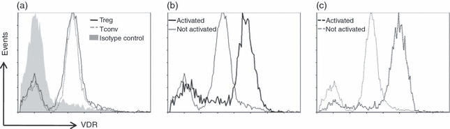
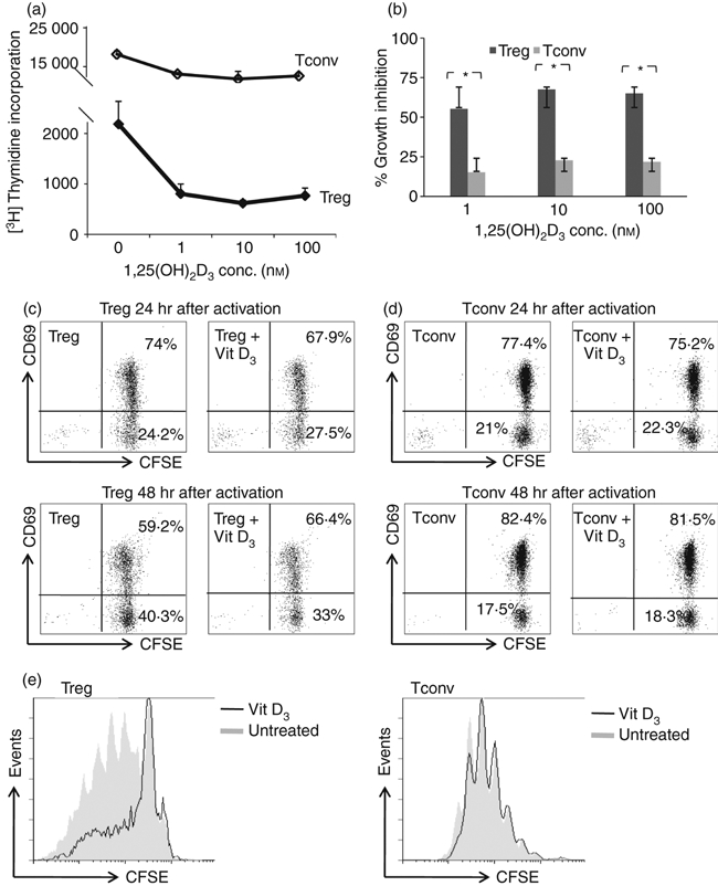
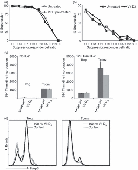
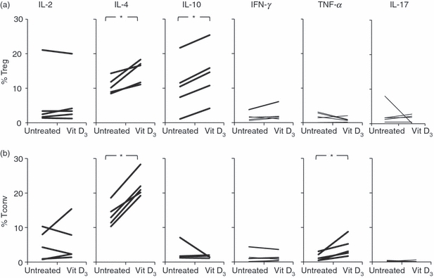
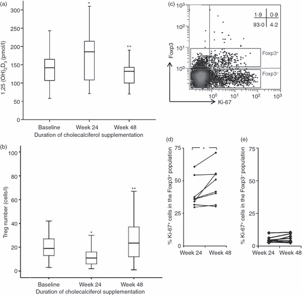
Vitamin D3 is known to induce regulatory T (Treg) cells by rendering antigen-presenting cells tolerogenic, its direct effect on human naturally occurring Treg cells is unclear. Here, we investigated if and how 1,25-dihydroxyvitamin D(3) [1,25(OH)2D3] can directly affect the proliferation and function of human naturally occurring Treg cells in vitro. First, we demonstrated that these Treg cells express vitamin D receptors that were up-regulated following anti-CD3/CD28-bead stimulation. 1,25(OH)2D3 inhibited proliferation of Treg cells even when exogenous interleukin-2 was provided. Treg cells were more susceptible to the inhibitory effect of 1,25(OH)2D3 than conventional T cells(.) 1,25(OH)2D3 neither affected the anergic state nor the suppressive function of Treg cells but induced a subtle increase in interleukin-10-secreting cells. The cell-division-inhibiting effect of 1,25(OH)2D3 on Treg cells was also demonstrated in vivo by supplementing vitamin D-deficient HIV-1-infected patients with 2000 IU cholecalciferol (vitamin D3). Increased serum 1,25(OH)2D3 levels were associated with a drop in the number and percentage of Treg cells, which may be attributed to a decrease in the proliferating Foxp3+ Treg cell population. In conclusion, 1,25(OH)2D3 directly affects Treg cell growth and promotes interleukin-10 production without apparent effects on activation status and suppressive phenotype whereas in vivo, high serum 1,25(OH)2D3 levels are associated with reduced Treg cell proliferation and a reduced number of Treg cells.
© 2011 The Authors. Immunology © 2011 Blackwell Publishing Ltd.
Figures
Similar articles
-
1α,25-dihydroxyvitamin D3 in combination with transforming growth factor-β increases the frequency of Foxp3⁺ regulatory T cells through preferential expansion and usage of interleukin-2.Immunology. 2014 Sep;143(1):52-60. doi: 10.1111/imm.12289. Immunology. 2014. PMID: 24673126 Free PMC article.
-
Vitamin D-binding protein controls T cell responses to vitamin D.BMC Immunol. 2014 Sep 18;15:35. doi: 10.1186/s12865-014-0035-2. BMC Immunol. 2014. PMID: 25230725 Free PMC article.
-
Vitamin D deficiency in pregnant women impairs regulatory T cell function.J Steroid Biochem Mol Biol. 2015 Mar;147:48-55. doi: 10.1016/j.jsbmb.2014.11.020. Epub 2014 Nov 21. J Steroid Biochem Mol Biol. 2015. PMID: 25448751
-
Vitamin D and systemic cancer: is this relevant to malignant melanoma?Br J Dermatol. 2002 Aug;147(2):197-213. doi: 10.1046/j.1365-2133.2002.04960.x. Br J Dermatol. 2002. PMID: 12174089 Review.
-
Vitamin D status, 1,25-dihydroxyvitamin D3, and the immune system.Am J Clin Nutr. 2004 Dec;80(6 Suppl):1717S-20S. doi: 10.1093/ajcn/80.6.1717S. Am J Clin Nutr. 2004. PMID: 15585793 Review.
Cited by
-
Vitamin D status is not associated with inflammatory cytokine levels during experimental human endotoxaemia.Clin Exp Immunol. 2013 Feb;171(2):231-6. doi: 10.1111/cei.12006. Clin Exp Immunol. 2013. PMID: 23286950 Free PMC article.
-
Host and Environmental Factors Influencing Individual Human Cytokine Responses.Cell. 2016 Nov 3;167(4):1111-1124.e13. doi: 10.1016/j.cell.2016.10.018. Cell. 2016. PMID: 27814508 Free PMC article.
-
Vitamin D Supplementation in Central Nervous System Demyelinating Disease-Enough Is Enough.Int J Mol Sci. 2019 Jan 8;20(1):218. doi: 10.3390/ijms20010218. Int J Mol Sci. 2019. PMID: 30626090 Free PMC article. Review.
-
Analysis of the Seasonal Fluctuation of γδ T Cells and Its Potential Relation with Vitamin D3.Cells. 2022 Apr 26;11(9):1460. doi: 10.3390/cells11091460. Cells. 2022. PMID: 35563767 Free PMC article.
-
Pharmacological and pharmacokinetic studies with vitamin D-loaded nanoemulsions in asthma model.Inflammation. 2014 Jun;37(3):723-8. doi: 10.1007/s10753-013-9790-0. Inflammation. 2014. PMID: 24326945
References
-
- Lips P. Vitamin D physiology. Prog Biophys Mol Biol. 2006;92:4–8. - PubMed
-
- Mahon BD, Wittke A, Weaver V, Cantorna MT. The targets of vitamin D depend on the differentiation and activation status of CD4 positive T cells. J Cell Biochem. 2003;89:922–32. - PubMed
-
- Veldman CM, Cantorna MT, Deluca HF. Expression of 1,25-dihydroxyvitamin D3 receptor in the immune system. Arch Biochem Biophys. 2000;374:334–8. - PubMed
-
- Hewison M, Freeman L, Hughes SV, et al. Differential regulation of vitamin D receptor and its ligand in human monocyte-derived dendritic cells. J Immunol. 2003;170:5382–90. - PubMed
-
- Sigmundsdottir H, Pan J, Debes GF, et al. DCs metabolize sunlight-induced vitamin D3 to ‘program’ T cell attraction to the epidermal chemokine CCL27. Nat Immunol. 2007;8:285–93. - PubMed
Publication types
MeSH terms
Substances
LinkOut - more resources
Full Text Sources
Medical
