. 2011 Oct 31;195(3):499-513.
doi: 10.1083/jcb.201104128.

The C terminus of talin links integrins to cell cycle progression

Affiliations

The C terminus of talin links integrins to cell cycle progression

Pengbo Wang et al. J Cell Biol. .

Abstract

Integrins are cell adhesion receptors that sense the extracellular matrix (ECM) environment. One of their functions is to regulate cell fate decisions, although the question of how integrins initiate intracellular signaling is not fully resolved. In this paper, we examine the role of talin, an adapter protein at cell-matrix attachment sites, in outside-in signaling. We used lentiviral small hairpin ribonucleic acid to deplete talin in mammary epithelial cells. These cells still attached to the ECM in an integrin-dependent manner and spread. They had a normal actin cytoskeleton, but vinculin, paxillin, focal adhesion kinase (FAK), and integrin-linked kinase were not recruited to adhesion sites. Talin-deficient cells showed proliferation defects, and reexpressing a tail portion of the talin rod, but not its head domain, restored integrin-mediated FAK phosphorylation, suppressed p21 expression, and rescued cell cycle. Thus, talin recruits and activates focal adhesion proteins required for proliferation via the C terminus of its rod domain. Our study reveals a new function for talin, which is to link integrin adhesions with cell cycle progression.


Figures
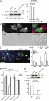
Figure 1.
Figure 1.
Talin1 is required for MEC proliferation. (A, left) MECs were infected with either shTln1 or GFP-only lentivirus or mock infected (3 h), cultured (48 h), FACS sorted to isolate GFP+ cells, and immunoblotted with antibodies to talin1, β1 integrin, and actin. (right) Relative talin levels were quantified by an infrared imaging system (Odyssey; LI-COR Biosciences; actin is the loading control). Note that in this and subsequent figures, n = 3 or more independent experiments. (B) Mock, GFP-only, or shTln1-infected cells were cultured for 48 h, replated, cultured a further 72 h, and then examined by fluorescence (top) and phase-contrast microscopy (bottom). The talin-depleted cells spread but did not become confluent. Talin localized to adhesions of uninfected MECs (red) but was absent in shTln1-infected cells (green). (C) GFP-only or shTln1-infected cells were replated, cultured overnight, and labeled with BrdU (2 h) or stained for phospho-Histone3. (left) Representative field after BrdU labeling. The arrow points to GFP-positive cells in S phase, whereas the arrowhead shows that the GFP-positive cells in talin knockdown cells did not incorporate BrdU into their DNA. Quantification of proliferation in infected cells was GFP only = 60 ± 5.9% and shTln1/GFP = 21 ± 3.6%. (right) Quantification of proliferation by phospho-Histone3 (pH3+) in infected cells. (D) MECs were cotransfected with either shTln1 or empty pLVTHM plasmid (pLV) together with shRNA-resistant human full-length talin1 (tlnFL), cultured (48 h), replated (overnight), and labeled with EdU (2 h). EdU+ cells were quantified in both the infected (GFP) and uninfected (unin) control cells on the same dishes. (E) GFP-only or shTln1-infected 4T1 cells were cultured (48 h), FACS sorted to isolate GFP+ cells, and either immunoblotted with antibodies to talin1 and actin or cultured overnight and labeled with BrdU (2 h). Error bars indicate SEM. Bars: (B, top) 20 µm; (B, bottom) 100 µm; (C) 50 µM.
Figure 2.
Figure 2.
Depletion of talin1 does not affect β1 integrin activation or the MEC cytoskeleton. (A, left and middle) Cells were cotransfected with shRNAmir for β1 integrin (sh-β1mir), shRNA for talin1 (shTln1), and the empty vectors (pLB2 and pLVTHM [pLV]). After 48 h, GFP-positive cells sorted by FACS were used in an adhesion assay on FN. (left) Blot showing that both β1 integrin and talin1 proteins were effectively deleted by using shRNA. (middle) Depletion of β1 integrin almost completely blocked cell attachment, but in contrast, talin knockdown only slightly reduced adhesion. (right) GFP-only or shTln1-infected MECs were FACS sorted to isolate GFP+ cells and used for adhesion assays on FN, laminin (LM), vitronectin (VN), or collagen I (CO). (B, top) FACS-sorted GFP-only or shTln1-infected cells were cultured overnight and stained for β1 integrin and phalloidin. (bottom) Costaining of talin with F-actin in shTln1-infected cells. (bottom left) Representative images. (bottom right) The intensity of total actin fluorescence per micrometer squared in control GFP-negative cells and in cells with talin knockdown (GFP positive). The GFP-positive cells depleted of talin showed normal microfilament distribution (arrows). The yellow lines demarcate the cell periphery. (C) FACS-sorted GFP-only or shTln1-infected cells were cultured overnight, briefly detergent extracted before fixation, and stained for either total (top) or active (bottom) β1 integrin (9EG7). Arrows point to β1 integrin–containing adhesion complexes in talin-depleted cells. Error bars indicate SEM. Bars, 15 µm.
Figure 3.
Figure 3.
Cell cycle is defective in talin-depleted MECs regardless of their spread size. (A) GFP-only or shTln1-infected MECs were replated, cultured overnight, and labeled with BrdU. Infected cells were visualized by GFP fluorescence, and the area of cell spreading was quantified and divided into seven groups ranging from <50 to >300 µm2. (left) Size distribution. (right) Quantification of BrdU staining. White bars: GFP-only infected cells. Black bars: GFP/shTln1-infected cells. (B) GFP-only or shTln1-infected MECs were replated on FN-coated micropatterned coverslips and then cultured overnight so that the cells spread to the same extent. They were then labeled with EdU. (left) Representative images. Note that the cells expressing shTln1 were defective in proliferation. (right) Quantification of proliferation data. Error bars indicate SEM. Bar, 15 µm.
Figure 4.
Figure 4.
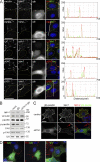
Talin1 is required for recruitment of adhesion complex proteins. (A) GFP-positive talin1-deficient cells were replated (overnight) and coimmunostained for vinculin (vin), paxillin (pax), ILK, pFAK, or pY20 together with antitalin–Alexa Fluor 568. GFP-expressing cells lack talin (encircled by yellow lines), whereas the adjacent uninfected cells contain talin adhesions. Fluorescence intensity profiles, to the right of the fluorescence images, depict the area of the line drawn in image overlays. (B) FACS-sorted mock-infected, GFP-only, or shTln1-infected cells were immunoblotted for adhesion proteins. (C) GFP-paxillin was expressed in MECs together with either shTln1 or control pSilencer. GFP-paxillin localizes to talin-containing adhesions in control cells (white arrows). In talin-depleted cells, GFP-paxillin does not localize to adhesions and is cytosolic. Red arrows point to talin in an adjacent untransfected cell. (D) MECs transduced with shTln1 were replated onto FN-coated coverslips, and after 48 h, the cells were immunostained for vinculin, paxillin, or talin. Note the absence of vinculin and paxillin in talin (Tln)-depleted cells. Bars: (A) 15 µm; (C and D) 10 µm.
Figure 5.
Figure 5.
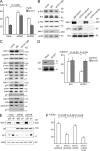
FAK links talin with the cell cycle. (A) MECs were cotransfected with shTln1 or empty vector (pSilencer [pSi]) together with vectors encoding wt-myrFAK-V5 (wtFAK) or an autophosphorylation site mutant, mu-myrFAK (muFAK). Replated cells were then labeled with EdU to evaluate proliferation. (B, left) FACS-sorted mock-infected, GFP-only, or shTln1-infected cells cultured in the continuous presence of serum were immunoblotted for signaling proteins Erk and Akt. Actin is the loading control. (right) Alternatively, cells were serum starved, then treated with or without serum for 6 h, and immunoblotted for talin, pErk, and total Erk. (C) Cells infected as in B (left) were blotted for positive and negative Cdk regulators, the cyclins, and the Cdk inhibitors. The lines separate independent blots, each showing talin knockdown and loading controls for either actin or Erk. (D, left) To deplete MECs of p21, they were transfected with either shRNA for p21 (sh-p21) or empty pLVTHM (pLV; mock) and cultured (48 h). After sorting for GFP-positive cells, whole-cell lysates were immunoblotted for p21. Erk was using as a loading control. The p21 band was confirmed by virtue of its strong up-regulation in aphidicolin-treated cells (not depicted). (right) MECs were cotransfected with either sh-p21 or mock (pLVTHM) together with either shTln1 or pLVTHM (control for shTln1), cultured (48 h), and labeled with EdU. Note that shTln1 blocks proliferation, but this is rescued by cotransfection with sh-p21. (E) MECs transfected as in A were extracted and immunoblotted for p21. Note that shTln1 induces expression of p21, which is reversed by cotransfection with wt-myrFAK but not mu-myrFAK. (F) MECs were transfected with controls (pLV + pcDNA6) or with vectors for either just talin knockdown (shTln1 + pcDNA6), talin knockdown and wt-myrFAK rescue (shTln1 + wtFAK), or talin knockdown and FAK rescue together with excess p21 (shTln1 + wtFAK + p21). Note that talin depletion blocked proliferation, which was rescued by wt-myrFAK, but the rescue was prevented by overexpressed p21. Error bars indicate SEM.
Figure 6.
Figure 6.
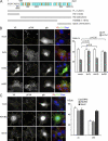
The talin rod domain is required for efficient cell cycle in MECs. (A) Map of human talin1 domains, with amino acid numbers. The gray boxes denote constructs used for rescue experiments. The N-terminal head contains four FERM domains F0–F3. The C-terminal rod contains 62 amphipathic α helices, shown as ovals. The vinculin binding sites (VBS) are in orange. The positions of actin-binding sites (ABS), integrin binding sites (IBS), and the dimerization domain (DS) are shown. The figure was adapted from Critchley (2009). FL, full length. (B, left) MECs were cotransfected with shTln1 and vectors encoding human talin1-V5 tag fusion proteins for full-length talin (tlnFL), the talin head (tlnHD), and rod domain (tlnRD). After 48 h, cells were replated (overnight) and costained for pFAK and V5. (right) Cells expressing human talin1 constructs with either shTln1 or empty plasmid (pSilencer [pSi]) were labeled with EdU to evaluate proliferation. (C, left) MECs were cotransfected with shTln1 and vectors encoding human talin1-V5 tag fusion proteins for the N-terminal half of talin (HDVBS) or the C-terminal portion (talinC). After 48 h, cells were replated (overnight) and costained for pFAK and V5. (right) Cells expressing human talinC, HDVBS, or control pcDNA6 together with either shTln1 or pSilencer were labeled with EdU to evaluate proliferation. The yellow lines demarcate the cell periphery. Error bars indicate SEM. Bars, 15 µm.
Figure 7.
Figure 7.
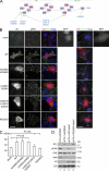
The C-terminal talin dimerization motif is necessary for cell cycle. (A) Map of talin1 C-terminal α-helical bundles, with specific helix numbers (shown in pink if they run antiparallel), and locations of the IBS2 double helical bundle and the C-terminal ABS helical bundle coupled to the DS domain. Mutations within the IBS and VBS sites and the dimerization domain are as shown. Each mutant was constructed as a V5 tag fusion in pcDNA6. (B) MECs were cotransfected with shTln1 and vectors encoding wild-type (WT) talinC or talinC mutations in ΔIBS (K2085D/K2089D and L2094A/I2095A), ΔVBS (V2087G/L2091S/I2352G), and ΔDS (R2526G). shTln1 together with empty pcDNA6 was used as a mock. Cells were costained for V5 (to identify location of transfected talinC constructs) and either pFAK (left) or vinculin (right). Note that wild-type talinC (second row) and the ΔIBS mutants (third and fourth rows) localized to adhesion complexes and recruited pFAK and vinculin, whereas ΔVBS and ΔDS (fifth and sixth rows) did not. The yellow lines demarcate the cell periphery. (C) Similar cultures to B were stained for EdU, and the percentage of EdU staining in the transfected cells was determined. Note that there was no significant difference in proliferation between MECs with talin1-only knockdown (i.e., mock) and those expressing shTln1 together with talinC ΔVBS or ΔDS. P-values are compared with mock. (D) MECs were transfected similarly to those in B, but instead of immunostaining, GFP+ cells were FACS sorted, and whole-cell lysates were blotted for p21, pFAK, total FAK, talin, the V5 epitope, and Erk as a loading control. Note that the cells expressing shTln1 with control pcDNA6 vector (lane 2) contained no pFAK and expressed p21 (these cells are equivalent to mock in C, in which cell cycle was suppressed). FAK was phosphorylated, and p21 was largely abolished by rescue with wild-type talinC (lane 3) but not with talinC-ΔDS (lane 4; these cells are equivalent to wild type and R2526G in C). Error bars indicate SEM. Bars, 15 µm.
Figure 8.
Figure 8.
Talin is a bidirectional transducer of integrin signals. Our study reveals that talin is a bidirectional transducer of integrin signals, both activating adhesion and regulating cell behavior. Talin’s head activates integrins, providing outward conformational changes that lead to high affinity interactions of the extracellular portion of integrins with ECM proteins. This has been well established in the literature. Our new results show that talin’s C-terminal tail is involved in recruiting vinculin and FAK to adhesion complexes and that this domain of talin is involved with cell cycle progression via p21 suppression. P, phosphorylation.

Similar articles

Cited by

References

    1. Anthis N.J., Wegener K.L., Ye F., Kim C., Goult B.T., Lowe E.D., Vakonakis I., Bate N., Critchley D.R., Ginsberg M.H., Campbell I.D. 2009. The structure of an integrin/talin complex reveals the basis of inside-out signal transduction. EMBO J. 28:3623–3632 10.1038/emboj.2009.287 - DOI - PMC - PubMed
    1. Barsukov I.L., Prescot A., Bate N., Patel B., Floyd D.N., Bhanji N., Bagshaw C.R., Letinic K., Di Paolo G., De Camilli P., et al. 2003. Phosphatidylinositol phosphate kinase type 1gamma and beta1-integrin cytoplasmic domain bind to the same region in the talin FERM domain. J. Biol. Chem. 278:31202–31209 10.1074/jbc.M303850200 - DOI - PubMed
    1. Bryant P., Zheng Q., Pumiglia K. 2006. Focal adhesion kinase controls cellular levels of p27/Kip1 and p21/Cip1 through Skp2-dependent and -independent mechanisms. Mol. Cell. Biol. 26:4201–4213 10.1128/MCB.01612-05 - DOI - PMC - PubMed
    1. Bustelo X.R., Sauzeau V., Berenjeno I.M. 2007. GTP-binding proteins of the Rho/Rac family: regulation, effectors and functions in vivo. Bioessays. 29:356–370 10.1002/bies.20558 - DOI - PMC - PubMed
    1. Cabodi S., Tinnirello A., Di Stefano P., Bisarò B., Ambrosino E., Castellano I., Sapino A., Arisio R., Cavallo F., Forni G., et al. 2006. p130Cas as a new regulator of mammary epithelial cell proliferation, survival, and HER2-neu oncogene-dependent breast tumorigenesis. Cancer Res. 66:4672–4680 10.1158/0008-5472.CAN-05-2909 - DOI - PubMed

Publication types