Review
. 2012 Jan;64(1):16-64.
doi: 10.1124/pr.110.002790. Epub 2011 Oct 28.

The blood-testis barrier and its implications for male contraception

Affiliations
Review

The blood-testis barrier and its implications for male contraception

C Yan Cheng et al. Pharmacol Rev. 2012 Jan.

Abstract

The blood-testis barrier (BTB) is one of the tightest blood-tissue barriers in the mammalian body. It divides the seminiferous epithelium into the basal and the apical (adluminal) compartments. Meiosis I and II, spermiogenesis, and spermiation all take place in a specialized microenvironment behind the BTB in the apical compartment, but spermatogonial renewal and differentiation and cell cycle progression up to the preleptotene spermatocyte stage take place outside of the BTB in the basal compartment of the epithelium. However, the BTB is not a static ultrastructure. Instead, it undergoes extensive restructuring during the seminiferous epithelial cycle of spermatogenesis at stage VIII to allow the transit of preleptotene spermatocytes at the BTB. Yet the immunological barrier conferred by the BTB cannot be compromised, even transiently, during the epithelial cycle to avoid the production of antibodies against meiotic and postmeiotic germ cells. Studies have demonstrated that some unlikely partners, namely adhesion protein complexes (e.g., occludin-ZO-1, N-cadherin-β-catenin, claudin-5-ZO-1), steroids (e.g., testosterone, estradiol-17β), nonreceptor protein kinases (e.g., focal adhesion kinase, c-Src, c-Yes), polarity proteins (e.g., PAR6, Cdc42, 14-3-3), endocytic vesicle proteins (e.g., clathrin, caveolin, dynamin 2), and actin regulatory proteins (e.g., Eps8, Arp2/3 complex), are working together, apparently under the overall influence of cytokines (e.g., transforming growth factor-β3, tumor necrosis factor-α, interleukin-1α). In short, a "new" BTB is created behind spermatocytes in transit while the "old" BTB above transiting cells undergoes timely degeneration, so that the immunological barrier can be maintained while spermatocytes are traversing the BTB. We also discuss recent findings regarding the molecular mechanisms by which environmental toxicants (e.g., cadmium, bisphenol A) induce testicular injury via their initial actions at the BTB to elicit subsequent damage to germ-cell adhesion, thereby leading to germ-cell loss, reduced sperm count, and male infertility or subfertility. Moreover, we also critically evaluate findings in the field regarding studies on drug transporters in the testis and discuss how these influx and efflux pumps regulate the entry of potential nonhormonal male contraceptives to the apical compartment to exert their effects. Collectively, these findings illustrate multiple potential targets are present at the BTB for innovative contraceptive development and for better delivery of drugs to alleviate toxicant-induced reproductive dysfunction in men.


Figures
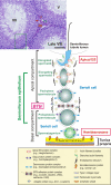
Fig. 1.
Fig. 1.
The location of the BTB in the seminiferous epithelium of adult mammalian testes and its physiological relationship with developing germ cells during spermatogenesis. The micrograph is the cross-section of an adult rat testis showing three seminiferous tubules at stages V, VII, and late VIII of the seminiferous epithelial cycle (see Fig. 2 for detailed description of the epithelial cycle). The green boxed area is a section of the seminiferous epithelium that is magnified and represented by the schematic drawing shown below it, illustrating the intimate relationship between Sertoli and germ cells and the relative location of the BTB. Leydig cells (red arrowheads) that produce testosterone (via steroidogenesis) and a microvessel (e.g., erythrocytes inside the microvessel are denoted by two green arrowheads) found in the interstitial space (In) are noted. The BTB is located near the basement membrane in the tunica propria, which also segregates the epithelium into the basal and the apical (or adluminal) compartment. The BTB is constituted by coexisting TJ, basal ES, desmosome, and gap junction, and the ultrastructural features of the BTB as typified by the actin filament bundles sandwiched between the cisternae of endoplasmic reticulum and the plasma membranes of two apposing Sertoli cells (i.e., the basal ES) shown herein can be seen in the electron micrographs shown in Fig. 3. The apical ES also shares similar ultrastructural features with the basal ES, except that the actin filament bundles that are sandwiched between the cisternae of endoplasmic reticulum and the plasma membranes of the apposing Sertoli cell and elongating spermatids (step 8–19) are restricted only to the Sertoli cell (see also micrographs in Figs. 2 and 3). As discussed in the text, there is cross-talk between the apical ES, the BTB, and the hemidesmosome. These three ultrastructures also form a functional axis that regulates and coordinates junction-restructuring events during spermatogenesis. Scale bar, 60 μm.
Fig. 2.
Fig. 2.
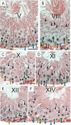
Cross-sections of testes from adult rats illustrating some selected stages of the seminiferous epithelial cycle of spermatogenesis. The stages of the seminiferous epithelial cycle shown herein are tubules at stage V (A), VIII (B), X (C), XI (D), XII (E), and XIV (F). The sections herein are paraffin sections stained with hematoxylin and eosin. During spermatogenesis, the seminiferous epithelium (composed of Sertoli and germ cells) undergoes cyclic changes as the result of germ cell development, which is manifested by notable changes on the morphology and relative location of the spermatids (purple arrowheads, such as acrosome formation, elongation of the tail), round spermatids (white arrowheads) and spermatocytes (red and green arrowheads), and can be divided into 6, 12, and 14 stages in human, mouse, and rat, respectively (Leblond and Clermont, 1952; Parvinen, 1982; de Kretser and Kerr, 1988; Hess, 1990; Hess and de Franca, 2008). The duration of a complete seminiferous epithelial cycle takes ∼8.6 days (Oakberg, 1956) and ∼12.8 days (Clermont et al., 1959) to complete in mouse and rat, respectively, when a specific section of a tubule is monitored by stereomicroscopy to define changes from stages I to XII and I to XIV in these species. But because certain phases of the spermatogenesis, such as type A spermatogonial renewal and their subsequent differentiation, take longer time to complete, spermatogenesis in mouse, rat, and human was estimated to take 34 to 35, 48 to 53, and 64 days, respectively, to complete in studies using [3H]thymidine or 32P-thymidine incorporation (de Kretser and Kerr, 1988) (Table 2). In stage V tubule (A), the heads of elongating spermatids almost make contact with the Sertoli cell nucleus (yellow arrowheads) located near the basement membrane of the tunica propria (green asterisk). Yellow arrowhead, Sertoli cell; blue arrowhead, type B spermatogonium; gray arrowhead, A1 spermatogonium; green arrowhead, pachytene spermatocyte; white arrowhead, step 5 spermatid; purple arrowhead, step 17 spermatid. In stage VIII tubule (B), elongated spermatids line up at the luminal edge of the epithelium to prepare for sperm release at spermiation. Yellow arrowhead, Sertoli cell; blue arrowhead, A1, spermatogonium; red arrowhead, preleptotene spermatocyte; green arrowhead, pachytene spermatocyte; white arrowhead; step 8 spermatid; purple arrowhead, step 19 spermatid; orange arrowhead, Leydig cell in the interstitium. In stage X tubule (C), yellow arrowhead, Sertoli cell; blue arrowhead, A2 spermatogonium; red arrowhead, leptotene spermatocyte; green arrowhead, pachytene spermatocyte; purple arrowhead, step 10 spermatid; orange arrowhead, Leydig cell. In stage XI tubule (D), yellow arrowhead, Sertoli cell; blue arrowhead, A2 spermatogonium; red arrowhead, leptotene spermatocyte; green arrowhead, pachytene spermatocyte; purple arrowhead, step 11 spermatid. In stage XII tubule (E), yellow arrowhead, Sertoli cell; blue arrowhead, type A2 spermatogonium; red arrowhead, zygotene spermatocyte; green arrowhead, pachytene spermatocyte; purple arrowhead, step 12 spermatid. At stage XIV (F), meiosis I and II occur, and the two secondary spermatocytes that arise from meiosis I can be seen (red asterisks); also two step 1 spermatids that arise from telophase of meiosis II are also seen (blue asterisks). Yellow arrowhead, Sertoli cell; blue arrowhead, A3 spermatogonium; green arrowhead, early pachytene spermatocyte; purple arrowhead, step 14 spermatid; orange arrowhead, Leydig cell. Scale bar, 25 μm (A–F).
Fig. 3.
Fig. 3.
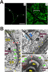
Cellular, functional and ultrastructural features of the blood-testis barrier (BTB) in mammalian testes during the seminiferous epithelial cycle of spermatogenesis. A, an intact and functional BTB is found in each of the three adjacent seminiferous tubules as shown in a, in which FITC (Mr 389.39) administered to an adult rat (∼300 g b.wt.) via the jugular vein was unable to pass through the BTB located near the basement membrane of the tunica propria (see broken white-line) to enter the apical compartment in each of these tubules (see the white bracket), even though FITC traversed the TJ barrier in the microvessels in the interstitial space (see green fluorescence in the interstitium annotated by the white arrowheads) (a). In the right panel, this rat was treated with CdCl2 (3 mg/kg b.wt. i.p.) for 3 days (b), which is known to disrupt the BTB integrity, before administration of FITC at the jugular vein. The BTB was found to be disrupted in all three tubules in this cross-section of testis because FITC entered the apical compartment “freely,” reaching the lumen of the seminiferous tubule (see the white bracket). Scale bars, 50 μm (a and b). B, the anatomy of the BTB. In a, this is an electron micrograph shown the typical ultrastructural features of the BTB created by two adjacent Sertoli cells lying on the basement membrane (yellow asterisks). Shown here are the basal ES, typified by the actin filament bundles (green arrowheads) sandwiched between cisternae of the endoplasmic reticulum (ER) and apposing plasma membranes of two Sertoli cells (apposing red arrowheads), TJ (yellow arrowheads) coexisting with the basal ES. In b, the desmosome (denoted by the blue arrowheads), which is also part of the components of the BTB, typified by the presence of electron dense substances along the two adjacent Sertoli cell and the absence of the distinctive actin filament bundles since desmosome is an intermediate-based cell-cell anchoring junction type. In c, the electron micrograph illustrates the apical ES surrounding the head of this elongating spermatid, in which the actin filament bundles (green arrowheads) are sandwiched between cisternae of the ER and the apposing plasma membranes of the Sertoli cell and the elongating spermatid (apposing red arrowheads), similar to the basal ES at the BTB shown in a, except that this typical features of the ES restricted only to the Sertoli cell. Scale bars, 0.5 μm (a), 1 μm (b), and 0.1 μm (c). Ac, acrosome; Nu, nucleus.
Fig. 4.
Fig. 4.
A schematic illustration of spermatogenesis in rodents and humans involving cell division (mitosis, meiosis) and cell differentiation shows the significance of the BTB that segregates germ cells in the basal or the apical (adluminal) compartment of the seminiferous epithelium. The bracketed number below each germ cell type represents the number of daughter cells derived from an earlier progenitor cell via either mitosis (black arrow), meiosis [green bar, which includes meiosis I (blue arrow) and meiosis II (green arrow)], or transformation/differentiation without involving cell division (red arrow). In theory, 4096 elongated spermatids (ES) and thus spermatozoa are formed from a single Asingle spermatogonium in rodents, but >75% of germ cells (e.g., spermatogonia, spermatocytes, and spermatids) undergo apoptosis and degeneration, so that the number of spermatozoa derived from a single spermatogonium is considerably less. A, type A spermatogonium; In, intermediate spermatogonium; B, type B spermatogonium; Spc, spermatocyte; sS, secondary spermatocyte; rS, round spermatid, which undergoes spermiogenesis involving step 1-to-19 and 1-to-6 spermatids in rats and humans, respectively, to form elongated spermatids; ES, elongated spermatid, which transforms and differentiates to spermatozoan before spermiation; As, Asingle spermatogonium; Apr, Apaired spermatogonium; Aal, Aaligned spermatogonium; A1-A4 are differentiated spermatogonia. The BTB physically divides the seminiferous epithelium into the basal and apical (adluminal) compartment. All the events of spermatogonial mitotic division and differentiation take place in the basal compartment, when type B spermatogonia differentiate into preleptotene spermatocytes, which are the germ cells in transit at the BTB so that meiosis I/II and spermiogenesis take place behind the BTB in the apical compartment until spermiation (i.e., the release of sperm into the tubule lumen).
Fig. 5.
Fig. 5.
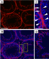
Distribution of the actin-based cytoskeleton in the seminiferous tubules of adult rat testes. A, cross-section of an adult rat testis in which F-actin was visualized by staining with rhodamine-conjugated phalloidin (Invitrogen, Carlsbad, CA) (red fluorescence), and cell nuclei were stained with 4′,6-diamidino-2-phenylindole and shown in the merged image (B). The two boxed areas in B are magnified and shown in C and D illustrating the relative location of BTB in C as denoted by white arrowheads, and F-actin is also predominant in the tunica propria (see white arrows) associated with peritubular myoid cells. Extensive F-actin network is also detected at the apical ES, surrounding the heads of the elongating spermatids, as shown in D. Scale bars, 80 μm (A and B) and 20 μm (C and D).
Fig. 6.
Fig. 6.
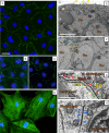
Morphology and ultrastructural features of the Sertoli cell BTB in vitro. Sertoli cells were cultured at ∼0.02 × 10⁶ cells/cm2 (A–C) or at 0.0125 × 10⁶ cells/cm2 (D) on Matrigel-coated glass coverslips for 4 days. Thereafter, cells were fixed in 4% paraformaldehyde (w/v) in phosphate-buffered saline (10 mM sodium phosphate and 0.15 M NaCl, pH 7.4 at 22°C), permeabilized in 0.1% Triton X-100 (v/v) in phosphate-buffered saline, and stained with a monospecific antibody against ZO-1 (A, an TJ adaptor protein), occludin (B, a TJ-integral membrane protein), N-cadherin (C, a basal ES integral membrane protein), or stained for F-actin using FITC-conjugated phalloidin (Sigma-Aldrich, St. Louis, MO) (D). Cells were mounted in ProLong Gold antifade reagent with 4,6-diamidino-2-phenylindole (Invitrogen) to visualize cell nuclei. Both TJ and basal ES were established in these Sertoli cells cultured in vitro, confirming earlier findings that a functional TJ permeability barrier is established in Sertoli cells cultured in vitro, by measuring transepithelial electrical resistance across the Sertoli cell epithelium (Grima et al., 1998; Lui et al., 2001). The distinctive actin filament network is also found in these Sertoli cells as shown in D. E to H, electron micrographs of Sertoli cells (0.5 × 10⁶ cells/cm2) cultured in vitro on Matrigel-coated dishes for 4 days, and the ultrastructural features of the BTB detected in vivo are also found in this Sertoli cell epithelium (F–H), illustrating that a functional BTB has been established. E, two adjacent Sertoli cells with the distinctive cell nuclei (Nu) are noted, and microvilli are also found (yellow arrowheads), which are the typical features of Sertoli cells cultured in vitro. F, three adjacent Sertoli cells are noted (Nu) and the functional BTB encircled by the blue and yellow brackets are magnified and shown in G and H, respectively. G, basal ES is typified by the presence of actin filament bundles (yellow arrowheads), which are sandwiched between cisternae of the endoplasmic reticulum (ER) and the apposing Sertoli cell plasma membranes (see apposing green arrowheads), and TJ (red arrowheads) is found to coexist with basal ES. H, functional desmosome (blue arrowheads), which is typified by the presence of electron-dense substances on both sides of the two adjacent Sertoli cells, is also noted. Desmosome shown in H and the basal ES and TJ shown in G are the magnified images from the corresponding areas in F, illustrating the ultrastructures of the Sertoli cell BTB in vitro. In short, these observations illustrate that a functional BTB was established in these Sertoli cells cultured in vitro, mimicking the BTB in vivo by having the ultrastructures of the BTB found in vivo. Scale bars, 20 μm (A–D), 5 μm (E and F), and 1 μm (G and H).
Fig. 7.
Fig. 7.
A schematic drawing illustrating the molecular architecture of the constituent proteins at the BTB. The BTB is typified by the presence of actin filament bundles sandwiched between cisternae of the endoplasmic reticulum and the apposing plasma membranes of two adjacent Sertoli cells. Cell adhesion at the BTB is conferred by the presence of several integral membrane proteins and their adaptors, such as TJ protein complexes (occludin-ZO-1, claudin-ZO-1, JAM-ZO-1), basal ES protein complexes (cadherin-catenin, nectin-afadin), gap junction protein complex (connexin-43-plakophilin-2), and desmosome protein complex (desmoglein-2/desmocollin-2-plakophilin/plakoglobin). Adaptors in these protein complexes (e.g., ZO-1, β-catenin, afadin, plakophilin, plakoglobin) also recruit additional adaptors [e.g., zyxin, axin, Wiskott-Aldrich syndrome protein (WASP), ponsin] and regulatory proteins to the site, including nonreceptor protein kinases [such as Src family kinases (e.g., c-Src, c-Yes), Fer kinase, FAK], polarity proteins [e.g., PAR6, protein associated with Lin seven-1 (PALS1), PALS1-associated tight junction protein (PATJ), atypical PKC (aPKC)], GTPases (e.g., Rab8B, Cdc42), and MAPKs [e.g., p38 MAPK and JNK/stress-activated protein kinase (SAPK)]. The actin network is also maintained by the actin capping and bundling protein Esp8 and the actin nucleation Arp2/3 protein complex. Also present is the intermediate filament near the desmoglein-desmocollin complex, and the tubulin network with the motor proteins (e.g., myosin VIIA, dynein, kinesin) near the actin filament to facilitate preleptotene spermatocyte transit at the site.
Fig. 8.
Fig. 8.
A schematic drawing illustrating functional domains in nonreceptor protein kinases FAK and Src. Both FAK and c-Src (the transforming, sarcoma-inducing gene of Rous sarcoma virus) are mediators of integrin-based signaling, most notably at the focal adhesion complex (also known as focal contact, which is an actin-based cell-matrix anchoring junction. Focal contact is not found in the testis. Instead, FAK and c-Src are components of the TJ and basal ES at the BTB, as well as the apical ES in the apical compartment). There are at least nine members in the Src kinase family: Src, Yes, Hck, Fyn, Fgr, Lyn, Lck, Blk, and Yrk. c-Src and c-Yes are found at the BTB and are structurally associated with the occludin-ZO-1 and the N-cadherin-β-catenin adhesion complexes. FAK consists of an N-terminal domain that binds β1-integrin, followed by a FERM (band 4.1, ezrin, radixin, moesin homology) domain, a catalytic kinase domain, and a FAT (focal adhesion targeting) domain near its C terminus. Also present are the three Pro-rich regions PRI, PRII, and PRIII, which also serve as the sites for the attachment of a number of adaptors and/or regulatory proteins (such as c-Src, PI-3K, PTEN, p130Cas) after activation of FAK via one or several of its putative phosphorylation sites (e.g., Tyr-397, -407, -576, -577, -861, and -925). Members of the Src kinase family, such as c-Src and c-Yes, consist of four Src homology (SH) domains, SH1 to SH4 (Xu et al., 1997; Chong et al., 2005). The SH4 domain near the N terminus of Src kinase contains the myristoylation and membrane-localization site and a unique domain of 50 to 70 amino acid residues that has no similarity among members of the Src kinase family, thus making each Src kinase member a unique protein. SH2 and SH3 domains are involved in the interaction with phosphorylated Tyr residues of other proteins and Pro-rich regions, respectively. For instance, FAK interacts with c-Src/c-Yes at its SH2 domain. The SH1 domain is the catalytic kinase site. c-Src interacts with several BTB proteins: occludin, N-cadherin, CAR, desmoglein-2, connexin-43, plakophilin-2, β-catenin (Lee and Cheng, 2005; Wang et al., 2007; Li et al., 2009b; Lie et al., 2010b), and myotubularin-related protein 2 (Zhang et al., 2005), conferring proper phosphorylation status in many of the integral membrane proteins at the BTB to regulate cell adhesion. Src has two important phosphorylation sites at Tyr-530 and Tyr-419 near its C terminus. Upon phosphorylation of Tyr-530, Src assumes an inactive locked conformation via interaction between the SH3 and the SH1 (kinase) domain; however, dephosphorylation of Tyr-530 and autophosphorylation of Tyr-419 within the catalytic kinase domain induce Src to assume an active open conformation, making its catalytic domain active to induce Tyr phosphorylation of its substrates. Src and FAK form a functional dual kinase complex to affect multiple cellular functions (Brunton and Frame, 2008; Aleshin and Finn, 2010; Bolós et al., 2010; Cabodi et al., 2010a,b), including the testis (Yan and Cheng, 2006).
Fig. 9.
Fig. 9.
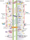
A current model illustrating the maintenance of the immunological barrier integrity during the transit of preleptotene spermatocytes at the BTB during spermatogenesis. This model was prepared based on recent findings in the field as discussed in section IV. Left, schematic drawing of a tubule at stage VII of the epithelial cycle with an intact BTB above a preleptotene spermatocyte differentiated from a type B spermatogonium, showing several adhesion protein complexes of TJ, basal ES, desmosome, and gap junction. The typical actin filament bundles sandwiched in between cisternae of endoplasmic reticulum and the apposing Sertoli cell plasma membranes (or apposing Sertoli cell-elongating spermatid) are also shown at the BTB or at the apical ES. At late stage VII to early VIII of the epithelial cycle (center), cytokines (e.g., TGF-β3, TNFα) and testosterone induce endocytosis of integral membrane proteins (and/or their adaptors) (possibly also mediated by changes in their phosphorylation status induced by FAK and/or Src), so that these proteins are internalized, destabilizing the old BTB site to open up the TJ barrier for the transit of preleptotene spermatocytes. This endocytic vesicle-mediated protein trafficking event is facilitated by the concomitant action of polarity proteins (e.g., PAR6, 14-3-3, PAR3) and the combined action of Arp2/3 protein complex and Eps8. Some of the endocytosed proteins will be targeted for degradation, but others will be transcytosed and recycled to the new BTB site behind the transiting preleptotene spermatocyte. This establishment of a new BTB is also facilitated by de novo synthesis of BTB proteins mediated by testosterone. Also, the transit of spermatocytes across the BTB is facilitated because some of the integral membrane proteins at the BTB are also found in these germ cells (e.g., CAR), so that these proteins can form homotypic interactions between the transiting spermatocytes and Sertoli cells at the BTB to disallow an opened BTB (center). Thus, as shown on the right, these BTB restructuring events that occur at stage VIII of the epithelial cycle concomitant with spermiation will not compromise the integrity of the immunological barrier conferred by the BTB. This model also demonstrates the presence of multiple targets for male contraceptive development. For instance, a disruption of the polarity proteins that are involved in the endocytic vesicle-mediated protein trafficking events would disable the transit of preleptotene spermatocytes at the BTB, halting spermatogenesis. Such action is likely to generate minimal side effects because the site of action is localized at the BTB microenvironment and not systemic.
Fig. 10.
Fig. 10.
Schematic illustration on the molecular architecture of connexin, connexon, hemichannel, and gap junction communication channel in gap junction. A, a typical connexin (e.g., Cx43, Cx26, Cx33) is composed of four transmembrane domains, two extracellular loops, one intracellular loop, and the intracellular N- and C-terminal tails. The C-terminal region confers most of the distinctiveness among connexins, which also contains phosphorylation sites for activation and inactivation and for interactions with binding partners (e.g., ZO-1, c-Src). B, an uncoupled functional connexon (also known as hemichannel) is composed of six connexins, which can be of the same (homomeric) or different (heteromeric) types. C, two coupled and compatible connexons create a gap junction communication channel between two adjacent Sertoli or Sertoli-germ cells, which can be homotypic or heterotypic.
Fig. 11.
Fig. 11.
A schematic drawing illustrating the molecular mechanism underlying BTB disruption induced by a toxicant (e.g., cadmium, BPA). Left, seminiferous epithelium of a normal tubule with an intact BTB. However, toxicants enter the Sertoli cells not at the BTB but instead at the plasma membrane via junction-associated “pores” and/or drug transporters. These toxicants can induce oxidative stress, which, in turn, mediates their effects on kinases (e.g., FAK, c-Src) causing unwanted protein endocytosis, which can destabilize the BTB. Alternatively, toxicants can also activate MAPK (e.g., p38 MAPK, ERK), which can also induce unwanted protein endocytosis. These effects can also be mediated via changes in the homeostasis of the Eps8 and Arp2/3 protein complex, compromising the optimal endocytic vesicle-mediated protein trafficking events, destabilizing cell adhesion at the BTB. The net result leads to a disruption of the BTB, which in turn affects germ cell adhesion in the basal and apical compartment of the epithelium, causing exfoliation of germ cells. These findings also illustrate potential targets that can be tackled to therapeutically manage toxicant-induced BTB disruption and the subsequent male reproductive dysfunction (e.g., reduced sperm count). For instance, the unwanted acceleration of protein endocytosis induced by cadmium or BPA can be prevented by modifying the action of polarity proteins (e.g., 14-3-3, PAR3, PAR6) and/or Arp3/3 complex to stabilize the BTB integrity. Functional studies can now be designed based on this hypothetical model to block or to therapeutically manage toxicant-induced BTB disruption.

Similar articles

Cited by

References

    1. Ahmed S, Goh WI, Bu W. (2010) I-BAR domains, IRSp53 and filopodium formation. Semin Cell Dev Biol 21:350–356 - PubMed
    1. Agency for Toxic Substances and Disease Registry (2008) Cadmium toxicity: ATSDR Case Studies in Environmental Medicine. Agency for Toxic Substances and Disease Registry, Atlanta, GA: Available at: http://www.atsdr.cdc.gov/csem/cadmium/docs/cadmium.pdf - PubMed
    1. Aitken RJ, Findlay JK, Hutt KJ, Kerr JB. (2011) Apoptosis in the germ line. Reproduction 141:139–150 - PubMed
    1. Alberts B, Johnson A, Lewis J, Raff M, Roberts K, Walter P. (2002) Molecular Biology of the Cell, 4th ed, Garland Science, New York
    1. Aleshin A, Finn RS. (2010) SRC: A century of science brought to the clinic. Neoplasia 12:599–607 - PMC - PubMed

Publication types

MeSH terms

Substances