. 2011 Oct 30;7(12):925-34.
doi: 10.1038/nchembio.694.

Identifying polyglutamine protein species in situ that best predict neurodegeneration

Affiliations

Identifying polyglutamine protein species in situ that best predict neurodegeneration

Jason Miller et al. Nat Chem Biol. .

Erratum in

  • Nat Chem Biol. 2012 Mar;8(3):318

Abstract

Polyglutamine (polyQ) stretches exceeding a threshold length confer a toxic function to proteins that contain them and cause at least nine neurological disorders. The basis for this toxicity threshold is unclear. Although polyQ expansions render proteins prone to aggregate into inclusion bodies, this may be a neuronal coping response to more toxic forms of polyQ. The exact structure of these more toxic forms is unknown. Here we show that the monoclonal antibody 3B5H10 recognizes a species of polyQ protein in situ that strongly predicts neuronal death. The epitope selectively appears among some of the many low-molecular-weight conformational states assumed by expanded polyQ and disappears in higher-molecular-weight aggregated forms, such as inclusion bodies. These results suggest that protein monomers and possibly small oligomers containing expanded polyQ stretches can adopt a conformation that is recognized by 3B5H10 and is toxic or closely related to a toxic species.


Figures
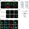
Figure 1
Figure 1
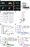
mAb 3B5H10 binds low molecular weight disease-associated polyQ expansions. (a) 3B5H10 preferentially labeled striatal neurons transiently expressing disease-associated polyQ expansions in an exon1 fragment or full-length htt. Striatal neurons transfected with Httex1-eGFP (Q17, Q72) or GFP-Full-Length-Htt (Q17, Q138) (top row; green) were labeled with mAb 3B5H10 (bottom row; red) and imaged by confocal microscopy. Scale bar=10 µm. (b, c) 3B5H10 recognizes disease-associated polyQ expansions in other neurodegeneration-causing proteins. (b) HEK293 extracts containing HA epitope–tagged androgen receptor (AR) (wt=Q25, mutant=Q65) or GST-tagged atrophin fragments (wt=Q19, mutant=Q81) were blotted with 3B5H10 and α-HA or α-GST antibodies, respectively. 3B5H10 preferentially recognized versions with disease-associated polyQ expansions. (c) Striatal neurons transfected with Myc-Ataxin-3 (wt=Q27, mutant=Q78) were labeled with α-Myc polyclonal and 3B5H10 mAb and imaged by confocal microscopy. α-Myc (green) recognizes both wt and mutant ataxin-3, whereas 3B5H10 (red) preferentially labeled mutant ataxin-3. Scale bar=5 µm. (d–e) 3B5H10 recognizes visibly non-aggregated, diffuse forms of mHtt. (d) Protein extracts from HEK293 cells expressing FLAG epitope-tagged mHtt (171-Q68-FLAG) were blotted with α-FLAG or 3B5H10. Aggregated forms of mHtt that are retained in the stacking portion of the gel (see α-FLAG lane) selectively lose 3B5H10 immunoreactivity. (e) Striatal neurons transfected with Httex1-(Q46, Q72, or Q97)-eGFP were labeled with Alexa 647-conjugated 3B5H10 and MW8, MW7, or EM48 α-htt antibodies. Fluorescence from GFP (green), Alexa 647 (blue), and Cy3-conjugated secondary antibodies (red) to detect MW8, MW7, or EM48 was collected with confocal microscopy. Scale bar=10 µm.
Figure 2
Figure 2
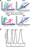
Quantitative binding of α-htt antibodies 3B5H10, EM48, MW1, or MW7 to httex1 is distinguishable and predictable. (a) 3B5H10, EM48, MW1, and MW7 differ significantly in their quantitative binding to diffuse htt in situ, suggesting each recognizes a unique htt species. Quantitative binding for each of the antibodies to httex1 can be estimated by regression analysis when the fluorescence of the eGFP tag fused to httex1 and httex1’s polyQ length are known. Striatal neurons transfected with Httex1-(Q17,Q46,Q72, or Q97)-eGFP were fixed at 24 hours and subjected to immunocytochemistry with one of the four α-htt antibodies. Fluorescence was measured by confocal microscopy (17–48 neurons per condition). For this analysis, only neurons without IBs were measured. (b) The significant data scatter around the linear regression lines in Figure 2a suggested that predicting the amount of antibody binding to a given mHtt-transfected neuron carries significant estimation error. To account for this error, we reanalyzed the data in Figure 2a with Bayesian statistics. The output of Bayesian regression analysis is a probability plot demonstrating how likely the actual regression coefficient (α) is a particular value. Bayesian regression plots for 3B5H10 are presented in Figure 2b, while α for the other antibodies are presented in Supplementary Figure 5 online.
Figure 3
Figure 3
Novel methodology distinguishes which of several simultaneously existing in situ epitopes of diffuse httex1 best predicts neurotoxicity. (a) The survival of individual neurons and the levels of diffuse Httex1-(Q17,Q46,Q72, or Q97)-eGFP they contained were determined by automated microscopy and recorded in a spreadsheet. Next, we used the regression coefficients from Figure 2 to relate diffuse Httex1-Qn-eGFP levels to antibody binding values (bottom; copied from Figure 2a). To account for the inaccuracy inherent in estimating antibody binding values from the graphs at bottom, we technically calculated regression coefficients using Bayesian methods (Fig. 2b, Supplementary Fig. 5 online). Scale bar=25 µm. (b) Using the regression coefficients from the plots in Figure 2, we can estimate how much antibody staining would have occurred in each neuron. These antibody staining values are then noted in the spreadsheet. (c) Finally, using each neuron’s estimated amount of antibody staining and its survival time, we compared the epitopes with each other using Bayesian hierarchical survival (Cox) analysis, which determines the antibody that best predicts degeneration. Hierarchical Bayesian methods ensure that “estimation errors” from each step of the analysis propagate through to the final readout. A more detailed schematic of our approach is illustrated in Supplementary Figure 6 online and detailed in Supplementary Methods online.
Figure 4
Figure 4
A species of htt recognized by 3B5H10 best predicts striatal neurodegeneration. The significance to neurodegeneration of htt species formed in situ and distinguished by 3B5H10, EM48, MW1, or MW7 was assessed by Cox analysis with a hierarchical Bayesian statistical approach (Fig. 3). Each graph plots the Cox coefficient (β) value for a particular antibody on the x axis and the probability of that coefficient value on the y axis. A positive coefficient signifies that antibody staining is associated with decreased survival. A negative coefficient signifies improved survival. (a) Cox coefficient (β) distribution for 3B5H10. (b) Cox coefficient (β) distribution for MW1. (c) Cox coefficient (β) distribution for MW7. (d) Cox coefficient (β) distribution for EM48.
Figure 5
Figure 5
3B5H10 does not recognize large oligomers of mHtt. (a) N-FRET (mean±s.d.) from cortical neurons was high in IBs formed from Httex1-Q97-CFP and Httex1-Q97-YFP (Q97 IBs) but low in regions of neurons with diffuse mHtt (Q97 diffuse) and not significantly different than in neurons with Httex1-Q25-CFP and Httex1-Q25-YFP (Q25) or CFP and YFP (FP only). Scale bar=10 µm. (b) HEK293 extracts containing Htt-171-(Q17, Q40, Q68, Q89, or Q142)-FLAG were loaded on a 0.20-µm membrane slot-blot and probed with α-FLAG or 3B5H10. α-FLAG revealed submicroscopic aggregates of Htt-171-(Qn)-FLAG that were unrecognizable by 3B5H10. (c) Agarose gel electrophoresis of extracts from PC12 cells stably expressing truncated (no polyproline) Httex1-Q103-GFP were blotted with α-GFP or 3B5H10. While oligomeric Httex1-Q103-GFP was present (arrow), 3B5H10 only stained the dye front, which represents monomer and possibly small oligomers. (d) 3B5H10 prevents mHtt aggregation detected by AFM. Httex1-Q53 aggregation was triggered with or without 3B5H10. (e) AFM-detectable species observed with 3B5H10 alone or with 3B5H10 or MW8 added to monomeric Httex1-Q53. 3B5H10 addition to htt stabilizes a 2–3 nm globular species, consistent with the size of a monomeric htt:antibody complex. (f) AFM-detectable species observed with 3B5H10, MW8, buffer, or nothing added to pre-aggregated oligomers of Httex1-Q53. 3B5H10 had similar effects when added to pre-formed fibrils (Supplementary Fig. 10 online). (g) The final size of AFM-detectable species when 3B5H10 is added to monomeric Httex1-Q53 (Fig. 5e) or pre-aggregated oligomers (Fig. 5f) is statistically indistinguishable (Spearman’s rank correlation coefficient). This size is most consistent with a monomeric htt:antibody complex.
Figure 6
Figure 6
Epitope recognized by 3B5H10 is preferentially present in low molecular weight htt species. (a) A solution of Httex1-Q53 was chemically cross-linked and probed with MW7 or 3B5H10. MW7 staining confirms the presence of monomers and small oligomers. The epitope 3B5H10 recognizes is preferentially present in monomers over small oligomers (Full blot: Supplementary Figure 11 online). (b) Monovalent 3B5H10 Fab retains specificity for mHtt over wt-htt, similar to intact bivalent 3B5H10. 3B5H10 Fab was cleaved from the intact IgG by papain proteolysis and purified by ion-exchange and size-exclusion chromatography. HEK293 extracts containing Htt-171-(Q17 or Q68)-FLAG were combined and blotted with i) vehicle, ii) chromatographic fractions corresponding to purified Fab or intact 3B5H10 IgG, or iii) undigested 3B5H10. This was followed by secondary antibody incubation with α-FLAG, α-Fc (which recognizes only intact antibodies), or α-Fab (which recognizes Fabs or intact antibodies). (c) Purified 3B5H10 Fab and Thio-Httex1-Q39-His6 were combined at different molar ratios (Fab:htt = 0.25, 0.5, 1, 1.5, 2) and analyzed by size-exclusion chromatography. A single peak consistent with a 1:1 Fab:htt complex was observed: peaks of pure 3B5H10 Fab or Thio-Httex1-Q39-His6 appeared if either was in molar excess of the other. (d) Purified 3B5H10 Fab and Thio-Httex1-Q39-His6 were combined and analyzed by equilibrium sedimentation analytical ultracentrifugation. The data best fit a model in which 3B5H10 Fab binds httex1 in a 1:1 ratio. At high concentrations, the complex dimerizes. Predictions for this model are overlaid on the raw data curves. The even distribution of residuals suggests no bias in the fit.
Figure 7
Figure 7
The “linear lattice” versus “emergent conformation” hypotheses for expanded polyQ conformation. The linear lattice model posits that a relatively unstructured epitope of polyQ in wt-htt repeats itself as the polyQ stretch expands. The emergent conformation model posits that expansion of the polyQ stretch induces a conformational change, such that wild-type and mutant polyQ are in distinct conformations. As shown in the middle panel, the linear lattice and emergent conformation hypotheses are not mutually exclusive; both models may be simultaneously true for mHtt. Alternatively, some mHtt molecules may exhibit linear lattice epitope repeats, while others may display an emergent conformation, possibly in the same neuron. Assessment of prognostic value for both epitopes by Cox analysis reveals that the emergent conformation, recognized by 3B5H10, is more toxic or more closely related to a toxic species than the linear lattice epitope, recognized by MW1.

Comment in

Similar articles

Cited by

References

    1. THDCR G. A novel gene containing a trinucleotide repeat that is expanded and unstable on Huntington’s disease chromosomes. Cell. 1993;72:971–983. - PubMed
    1. Orr HT, Zoghbi HY. Trinucleotide repeat disorders. Annu. Rev. Neurosci. 2007;30:575–621. - PubMed
    1. Kayed R, et al. Common structure of soluble amyloid oligomers implies common mechanism of pathogenesis. Science. 2003;300:486–489. - PubMed
    1. Ko J, Ou S, Patterson PH. New anti-huntingtin monoclonal antibodies: Implications for huntingtin conformation and its binding proteins. Brain Res. Bull. 2001;56:319–329. - PubMed
    1. Rakhit R, et al. An immunological epitope selective for pathological monomer-misfolded SOD1 in ALS. Nat. Med. 2007;13:754–759. - PubMed

Publication types

MeSH terms