. 2011 Dec;7(12):1415-23.
doi: 10.4161/auto.7.12.17877.

Physical exercise stimulates autophagy in normal skeletal muscles but is detrimental for collagen VI-deficient muscles

Affiliations

Physical exercise stimulates autophagy in normal skeletal muscles but is detrimental for collagen VI-deficient muscles

Paolo Grumati et al. Autophagy. 2011 Dec.

Abstract

Autophagy is a catabolic process that provides the degradation of altered/damaged organelles through the fusion between autophagosomes and lysosomes. Proper regulation of the autophagic flux is fundamental for the homeostasis of skeletal muscles in physiological conditions and in response to stress. Defective as well as excessive autophagy is detrimental for muscle health and has a pathogenic role in several forms of muscle diseases. Recently, we found that defective activation of the autophagic machinery plays a key role in the pathogenesis of muscular dystrophies linked to collagen VI. Impairment of the autophagic flux in collagen VI null (Col6a1–/–) mice causes accumulation of dysfunctional mitochondria and altered sarcoplasmic reticulum, leading to apoptosis and degeneration of muscle fibers. Here we show that physical exercise activates autophagy in skeletal muscles. Notably, physical training exacerbated the dystrophic phenotype of Col6a1–/– mice, where autophagy flux is compromised. Autophagy was not induced in Col6a1–/– muscles after either acute or prolonged exercise, and this led to a marked increase of muscle wasting and apoptosis. These findings indicate that proper activation of autophagy is important for muscle homeostasis during physical activity.


Figures
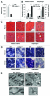
Figure 1.
Figure 1.
Long-term physical exercise is detrimental for Col6a1–/– muscles. (A) Histogram showing the mean distance covered daily by wild-type and Col6a1–/– mice under voluntary running wheel exercise (*p < 0.001, n = 6 each genotype). Error bars indicate s.e.m. (B) Incidence of apoptosis in TA (left) and diaphragm (right) muscles of 5-mo-old wild-type and Col6a1–/– mice housed either in standard conditions (no run) or after 3-mo running (3-m run) wheel exercise (*p < 0.001, n = 6 each group). Error bars indicate s.e.m. (C) Representative H & E staining of cross-sections of TA and diaphragm muscles isolated from wild-type and Col6a1–/– mice in standard conditions (no run) or after 3-mo run wheel exercise. Under standard conditions, Col6a1–/– muscles display various alterations, with centrally nucleated fibers (black arrowheads) and atrophic myofibers (black arrows) in both diaphragm and TA, and signs of inflammation (white asterisk) in diaphragm. After three months of exercise, only a few degenerating myofibers (white arrows) are detected in wild-type muscles, whereas in Col6a1–/– TA centrally nucleated, atrophic and degenerated myofibers are markedly increased. Three months of exercise leads to extensive myofiber degeneration in Col6a1–/– diaphragm. (D) Representative cross-sections of TA and diaphragm muscles stained for SDH. Histochemical reaction for SDH produces a purple-blue staining with a speckled pattern, which is proportional to the number of mitochondria and their oxidative activity. In resting conditions (no run), most myofibers of TA show scattered staining of variable intensities, due to the predominantly glycolytic metabolic activity of this muscle, while the diaphragm shows intense blue staining in all myofibers, reflecting its oxidative metabolism. After exercise (3-m run), Col6a1–/– mice display several abnormally and poorly stained fibers in TA (red asterisks) and a diffuse pale blue staining of diaphragm. Scale bar, 100 μm. (E) Electron micrographs of diaphragm from wild-type and Col6a1–/– mice after 3 mo of running wheel exercise. Col6a1–/– diaphragm displays several ultrastructural defects, while wild-type diaphragm maintains a normal structure. Col6a1–/– diaphragm presents a marked accumulation of dilated sarcoplasmic reticulum (arrowheads), abnormal mitochondria and apoptotic nuclei (upper panels). Many Col6a1–/– mitochondria appear fragmented and some of them with altered cristae, while wild-type mitochondria maintain elongated shape and normal cristae morphology (lower panels). Scale bars, 1 μm (upper panels) or 200 nm (lower panels). mit, mitochondria; nu, nuclei; WT, wild-type.
Figure 2.
Figure 2.
Autophagy is impaired in Col6a1–/– muscles after long-term physical exercise. (A) Western blot analysis for Akt phosphorylation and LC3 lipidation in TA (left) and diaphragm (right) of 5-mo-old wild-type and Col6a1–/– mice housed either in standard conditions (no run) or after 3-mo run wheel exercise. (B) Densitometric quantifications of P-Akt levels and of LC3-II vs LC3-I ratio, as determined by western blots of TA (left) and diaphragm (right) (*p < 0.05, n = 3).
Figure 3.
Figure 3.
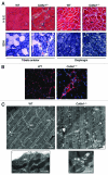
Treadmill exercise causes muscle wasting in Col6a1–/– mice. (A) Representative cross-sections of TA and diaphragm muscles stained with H & E or with histochemical reaction for SDH and isolated from 5-mo-old wild-type and Col6a1–/– mice after 1 h treadmill exercise. H & E staining shows normal morphology in both muscles of wild-type animals after treadmill. Instead, TA from Col6a1–/– mice after treadmill displays severe muscle degeneration characterized by inflammation (white asterisk), degenerating myofibers (white arrows), centrally nucleated fibers (black arrowheads), and atrophic myofibers (black arrows). The extent of muscle degeneration in the diaphragm of Col6a1–/– mice after treadmill is similar to that observed in standard conditions. SDH staining of TA and diaphragm is normal in wild-type mice after treadmill. In Col6a1–/– animals subjected to treadmill several abnormal poorly stained fibers (red asterisks) are present in TA, while the diaphragm displays an apparently normal SDH staining. Scale bar, 100 μm. (B) Representative IgG immunolabeling images of TA cross-sections from wild-type and Col6a1–/– mice after 1 h of treadmill exercise, confirming extensive inflammation in Col6a1–/– TA but not in the corresponding wild-type samples. Scale bar, 100 μm. (C) Electron micrographs of TA from wild-type and Col6a1–/– mice after treadmill exercise. Col6a1–/– TA displays a large number of severe ultrastructural alterations, while wild-type TA presents fibers with dilated sarcoplasmic reticulum and some altered mitochondria. Col6a1–/– TA displays a marked accumulation of dilated sarcoplasmic reticulum (arrowheads) and abnormal mitochondria. Almost all Col6a1–/– mitochondria appear fragmented and many of them with completely swollen cristae, while the majority of wild-type mitochondria maintain elongated shape and normal cristae morphology (lower panels). Scale bars, 1 μm. mit, mitochondria; WT, wild-type.
Figure 4.
Figure 4.
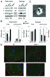
Treadmill exercise induces autophagy in wild-type muscle but not in Col6a1–/– mice. (A) Western blot analysis for Akt phosphorylation and LC3 lipidation in TA (left) and diaphragm (right) muscles from 5-mo-old wild-type and Col6a1–/– mice housed in standard conditions (no run) or after 1 h treadmill exercise (TM). (B) Densitometric quantifications of P-Akt levels and LC3-II vs LC3-I ratio, as determined by western blots of TA (left) and diaphragm (right) (*p < 0.05, n = 3). (C) Electron micrograph of double-membrane (arrowheads) autophagosome in TA of wild-type mice after treadmill exercise. Scale bar, 200 nm. (D) Fluorescence microscope analysis of cryosections of TA from wild-type;GFP-LC3 and Col6a1–/–;GFP-LC3 mice after treadmill exercise. GFP-LC3 puncta (green fluorescence) are abundant in wild-type muscle fibers, but they are scarce and poorly detectable in Col6a1–/– myofibers. Immunohistochemistry for laminin (red fluorescence) was performed to reveal myofiber outline, while Hoechst staining (blue) was used to label nuclei. The lower panels are higher magnifications of the squared areas, clearly showing GFP-LC3 puncta (arrowheads) in wild-type but not in Col6a1–/– myofibers. Scale bar, 25 μm. TM, treadmill; WT, wild-type.

Comment in

Similar articles

Cited by

References

    1. Cecconi F, Levine B. The role of autophagy in mammalian development: cell makeover rather than cell death. Dev Cell. 2008;15:344–57. doi: 10.1016/j.devcel.2008.08.012. - DOI - PMC - PubMed
    1. Levine B, Kroemer G. Autophagy in the pathogenesis of disease. Cell. 2008;132:27–42. doi: 10.1016/j.cell.2007.12.018. - DOI - PMC - PubMed
    1. Maiuri MC, Zalckvar E, Kimchi A, Kroemer G. Self-eating and self-killing: crosstalk between autophagy and apoptosis. Nat Rev Mol Cell Biol. 2007;8:741–52. doi: 10.1038/nrm2239. - DOI - PubMed
    1. Mizushima N, Levine B, Cuervo AM, Klionsky DJ. Autophagy fights disease through cellular self-digestion. Nature. 2008;451:1069–75. doi: 10.1038/nature06639. - DOI - PMC - PubMed
    1. Masiero E, Agatea L, Mammucari C, Blaauw B, Loro E, Komatsu M, et al. Autophagy is required to maintain muscle mass. Cell Metab. 2009;10:507–15. doi: 10.1016/j.cmet.2009.10.008. - DOI - PubMed

Publication types