

doi: 10.1128/JVI.05988-11.
Epub 2011 Oct 19.
Reconstitution of the Kaposi's sarcoma-associated herpesvirus nuclear egress complex and formation of nuclear membrane vesicles by coexpression of ORF67 and ORF69 gene products
Affiliations
- PMID: 22013050
- PMCID: PMC3255883
- DOI: 10.1128/JVI.05988-11
Item in Clipboard
Reconstitution of the Kaposi's sarcoma-associated herpesvirus nuclear egress complex and formation of nuclear membrane vesicles by coexpression of ORF67 and ORF69 gene products
J Virol.
2012 Jan.
Abstract
The Kaposi's sarcoma-associated herpesvirus nuclear egress complex is composed of two proteins, ORF67 and ORF69. In this study, we have recapitulated the KSHV complex by coexpression of these two proteins in insect cells using expression from recombinant baculoviruses. The proteins form a complex at the nuclear membrane as judged by live-cell analysis of protein fusions tagged with green fluorescent protein (GFP) and mCherry. Ultrastructural analysis of infected cells showed that ORF67 expression results in reduplication of the nuclear membrane. When the two proteins are expressed together, numerous virion-size nuclear membrane-derived vesicles were evident at the nuclear margins.
Figures

Cloning, expression, and localization of the KSHV nuclear egress complex proteins. (A) ORF67 (271 amino acids) and ORF69 (302 amino acids) were amplified and cloned into the baculovirus transfer vectors that encode C-terminal, GFP, mCherry, and V5 tags. (B) Amplified baculoviruses were used to infect Sf21 cells to confirm expression of the tagged polypeptides. The proteins were resolved on 4 to 12% Bis-Tris NuPage gels (Invitrogen), and proteins were detected using anti-V5 mouse (Invitrogen), anti-GFP rabbit (Molecular Probes), and anti-DsRed rabbit (BD Biosciences). Protein standards are shown in the left lane, and size is indicated in kilodaltons. (C) Similar infections and methods were used to examine coexpression of ORF67 and ORF69 using V5 and HA tags and Western blot procedures. Cells were infected with the individual viruses or coinfected with viruses expressing V5 and HA fusion proteins as well as a dual-expression virus vector. (D) Sf21 cells were infected with baculoviruses expressing GFP- and mCherry-tagged ORF67 and ORF69. The cells were visualized by confocal microscopy 48 h postinfection. The objective lens was 63×.
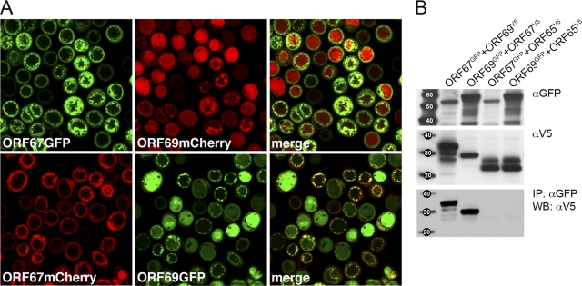
ORF67 and ORF69 interactions observed visually by relocalization of ORF69 to the nuclear boundary and biochemically by coimmunoprecipitation methods. (A) Sf21 cells were coinfected with viruses expressing GFP/mCherry tagged proteins and analyzed by confocal microscopy 48 h postinfection. The objective lens was 63×. The merge of the GFP and mCherry fluorescence shows relocalization of ORF69 to the nuclear margins. (B) ORF67V5 and ORF69V5 are coprecipitated by ORF69GFP and ORF67GFP, respectively (immunoprecipitation [IP] with αGFP followed by Western blot [WB] analysis with αV5). The proteins were processed for Western blots as described in the legend to Fig. 1. The proteins present in the input lysates are shown by the αGFP and αV5 blots (top and middle). Protein standards are shown in the left lane, and size is indicated in kilodaltons.
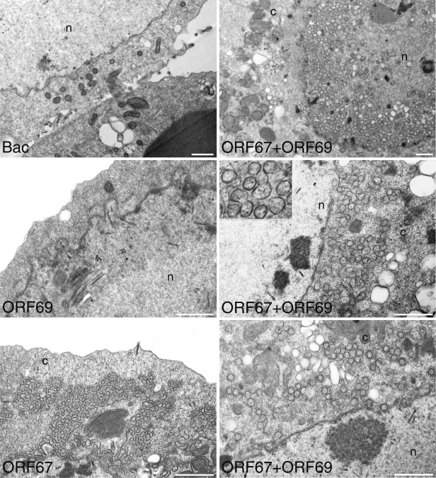
Nuclear membrane remodeling by KSHV ORF67 and vesicle formation by the coexpression of ORF67 and ORF69. Sf21 cells were infected with the baculoviruses expressing ORF67 and ORF69 or infected with a control baculovirus. Infected cells were processed for TEM 66 h postinfection. Extensive nuclear membrane duplication was seen in BacORF67-infected cells; in cells coinfected with BacORF67 and BacORF69, numerous vesicles around the nuclear boundary were observed. These nuclear remodeling phenotypes were not seen in the control virus infections or in BacORF69-infected cells. The nucleus (n) and cytoplasm (c) compartments are indicated. Bars, 1,000 nm.
Similar articles
-
Functional Identification and Characterization of the Nuclear Egress Complex of a Gammaherpesvirus.J Virol. 2019 Nov 26;93(24):e01422-19. doi: 10.1128/JVI.01422-19. Print 2019 Dec 15. J Virol. 2019. PMID: 31554685 Free PMC article.
-
Interactions of the Kaposi's Sarcoma-associated herpesvirus nuclear egress complex: ORF69 is a potent factor for remodeling cellular membranes.J Virol. 2013 Apr;87(7):3915-29. doi: 10.1128/JVI.03418-12. Epub 2013 Jan 30. J Virol. 2013. PMID: 23365436 Free PMC article.
-
Identification and characterization of the product encoded by ORF69 of Kaposi's sarcoma-associated herpesvirus.J Virol. 2008 May;82(9):4562-72. doi: 10.1128/JVI.02400-07. Epub 2008 Feb 27. J Virol. 2008. PMID: 18305046 Free PMC article.
-
The way out: what we know and do not know about herpesvirus nuclear egress.Cell Microbiol. 2013 Feb;15(2):170-8. doi: 10.1111/cmi.12044. Epub 2012 Nov 7. Cell Microbiol. 2013. PMID: 23057731 Review.
-
Pathological Features of Kaposi's Sarcoma-Associated Herpesvirus Infection.Adv Exp Med Biol. 2018;1045:357-376. doi: 10.1007/978-981-10-7230-7_16. Adv Exp Med Biol. 2018. PMID: 29896675 Review.
Cited by
-
Vesicular Nucleo-Cytoplasmic Transport-Herpesviruses as Pioneers in Cell Biology.Viruses. 2016 Sep 27;8(10):266. doi: 10.3390/v8100266. Viruses. 2016. PMID: 27690080 Free PMC article.
-
Mapping of sequences in Pseudorabies virus pUL34 that are required for formation and function of the nuclear egress complex.J Virol. 2013 Apr;87(8):4475-85. doi: 10.1128/JVI.00021-13. Epub 2013 Feb 6. J Virol. 2013. PMID: 23388710 Free PMC article.
-
Comprehensive Analysis of the Tegument Proteins Involved in Capsid Transport and Virion Morphogenesis of Alpha, Beta and Gamma Herpesviruses.Viruses. 2023 Oct 6;15(10):2058. doi: 10.3390/v15102058. Viruses. 2023. PMID: 37896835 Free PMC article. Review.
-
The Human Cytomegalovirus Transmembrane Protein pUL50 Induces Loss of VCP/p97 and Is Regulated by a Small Isoform of pUL50.J Virol. 2020 Jun 16;94(13):e00110-20. doi: 10.1128/JVI.00110-20. Print 2020 Jun 16. J Virol. 2020. PMID: 32321808 Free PMC article.
-
KSHV: pathways to tumorigenesis and persistent infection.Adv Virus Res. 2014;88:111-59. doi: 10.1016/B978-0-12-800098-4.00002-7. Adv Virus Res. 2014. PMID: 24373311 Free PMC article. Review.
References
Publication types
MeSH terms
Substances
Grants and funding
LinkOut - more resources
Full Text Sources
