

Cyclophosphamide perturbs cytosine methylation in Jurkat-T cells through LSD1-mediated stabilization of DNMT1 protein
- PMID: 22007908
- PMCID: PMC3221796
- DOI: 10.1021/tx2003849
Cyclophosphamide perturbs cytosine methylation in Jurkat-T cells through LSD1-mediated stabilization of DNMT1 protein
Abstract
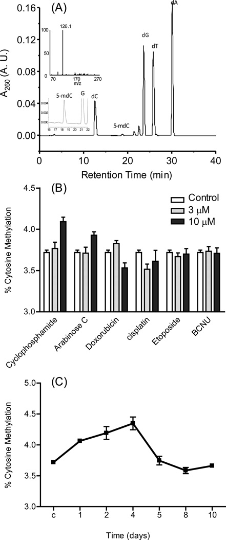
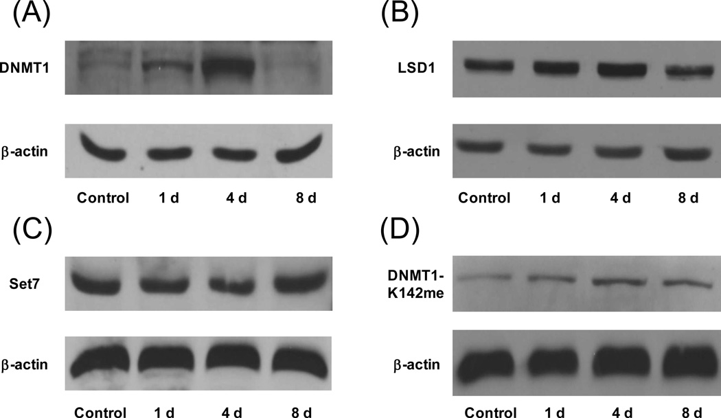
Aberrant cytosine methylation is known to be associated with cancer development. Here, we assessed how common cancer chemotherapeutic agents perturb cytosine methylation in Jurkat-T acute lymphoblastic leukemia cells. We tested six antitumor agents and found that cyclophosphamide induced the most pronounced increase in global DNA cytosine methylation after a 24-h treatment. Long-term treatment with cyclophosphamide led to a time-dependent increase in cytosine methylation level with up to 4 days of treatment, and the extent of cytosine methylation returned to normal level after 8 days. The trend of change in DNA methylation level paralleled that of the expression level of DNMT1 protein, whereas no significant increase in DNMT1 mRNA level was observed. Previous studies showed that the stability of endogenous DNMT1 protein is regulated by lysine methylation through histone lysine methyltransferase Set7 and lysine-specific demethylase 1 (LSD1), with the methylated DNMT1 being the target for proteasomal degradation. We observed that the elevated expression of DNMT1 protein at 4 days of treatment was correlated with the increased expression of LSD1 protein and with the decreased frequency of K142 methylation in DNMT1. Taken together, our results showed that cyclophosphamide perturbed temporarily global cytosine methylation in Jurkat-T cells via regulation of the lysine methylation level in DNMT1.
Figures
Similar articles
-
Loss of LSD1 (lysine-specific demethylase 1) suppresses growth and alters gene expression of human colon cancer cells in a p53- and DNMT1(DNA methyltransferase 1)-independent manner.Biochem J. 2013 Jan 15;449(2):459-68. doi: 10.1042/BJ20121360. Biochem J. 2013. PMID: 23072722 Free PMC article.
-
The lysine demethylase LSD1 (KDM1) is required for maintenance of global DNA methylation.Nat Genet. 2009 Jan;41(1):125-9. doi: 10.1038/ng.268. Epub 2008 Dec 21. Nat Genet. 2009. PMID: 19098913
-
6-Thioguanine reactivates epigenetically silenced genes in acute lymphoblastic leukemia cells by facilitating proteasome-mediated degradation of DNMT1.Cancer Res. 2011 Mar 1;71(5):1904-11. doi: 10.1158/0008-5472.CAN-10-3430. Epub 2011 Jan 14. Cancer Res. 2011. PMID: 21239472 Free PMC article.
-
SET7/9 mediated methylation of non-histone proteins in mammalian cells.Epigenetics. 2009 Aug 16;4(6):383-7. doi: 10.4161/epi.4.6.9450. Epub 2009 Aug 6. Epigenetics. 2009. PMID: 19684477 Free PMC article. Review.
-
Mammalian cytosine DNA methyltransferase Dnmt1: enzymatic mechanism, novel mechanism-based inhibitors, and RNA-directed DNA methylation.Curr Med Chem. 2008;15(1):92-106. doi: 10.2174/092986708783330700. Curr Med Chem. 2008. PMID: 18220765 Review.
Cited by
-
Quantitative assessment of Tet-induced oxidation products of 5-methylcytosine in cellular and tissue DNA.Nucleic Acids Res. 2013 Jul;41(13):6421-9. doi: 10.1093/nar/gkt360. Epub 2013 May 8. Nucleic Acids Res. 2013. PMID: 23658232 Free PMC article.
-
Epigenetic Modifications in T Cells: The Role of DNA Methylation in Salt-Sensitive Hypertension.Hypertension. 2020 Feb;75(2):372-382. doi: 10.1161/HYPERTENSIONAHA.119.13716. Epub 2019 Dec 16. Hypertension. 2020. PMID: 31838911 Free PMC article.
-
Lysine demethylase KDM2A inhibits TET2 to promote DNA methylation and silencing of tumor suppressor genes in breast cancer.Oncogenesis. 2017 Aug 7;6(8):e369. doi: 10.1038/oncsis.2017.71. Oncogenesis. 2017. PMID: 28785073 Free PMC article.
-
Epigenetics in SLE.Curr Rheumatol Rep. 2017 Sep;19(9):58. doi: 10.1007/s11926-017-0685-1. Curr Rheumatol Rep. 2017. PMID: 28752494 Free PMC article. Review.
-
Arginine methylation-dependent LSD1 stability promotes invasion and metastasis of breast cancer.EMBO Rep. 2020 Feb 5;21(2):e48597. doi: 10.15252/embr.201948597. Epub 2019 Dec 12. EMBO Rep. 2020. PMID: 31833203 Free PMC article.
References
-
- Goll MG, Bestor TH. Eukaryotic cytosine methyltransferases. Annu. Rev. Biochem. 2005;74:481–514. - PubMed
-
- Okano M, Bell DW, Haber DA, Li E. DNA methyltransferases Dnmt3a and Dnmt3b are essential for de novo methylation and mammalian development. Cell. 1999;99:247–257. - PubMed
-
- Li E, Bestor TH, Jaenisch R. Targeted mutation of the DNA methyltransferase gene results in embryonic lethality. Cell. 1992;69:915–926. - PubMed
-
- Herman JG, Baylin SB. Gene silencing in cancer in association with promoter hypermethylation. N. Eng. J. Med. 2003;349:2042–2054. - PubMed
Publication types
MeSH terms
Substances
Grants and funding
LinkOut - more resources
Full Text Sources
