. 2011 Nov 8;108(45):E1061-9.
doi: 10.1073/pnas.1108323108. Epub 2011 Oct 17.

Essential PcsB putative peptidoglycan hydrolase interacts with the essential FtsXSpn cell division protein in Streptococcus pneumoniae D39

Affiliations

Essential PcsB putative peptidoglycan hydrolase interacts with the essential FtsXSpn cell division protein in Streptococcus pneumoniae D39

Lok-To Sham et al. Proc Natl Acad Sci U S A. .

Abstract

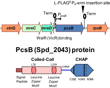
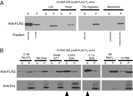
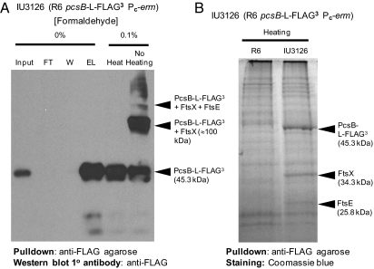
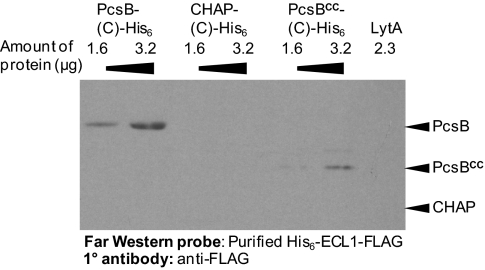
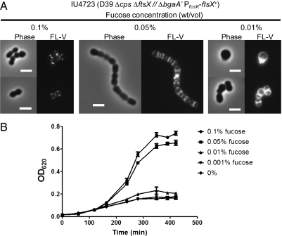
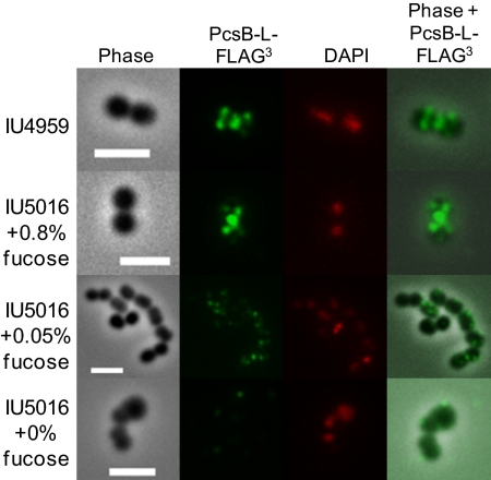
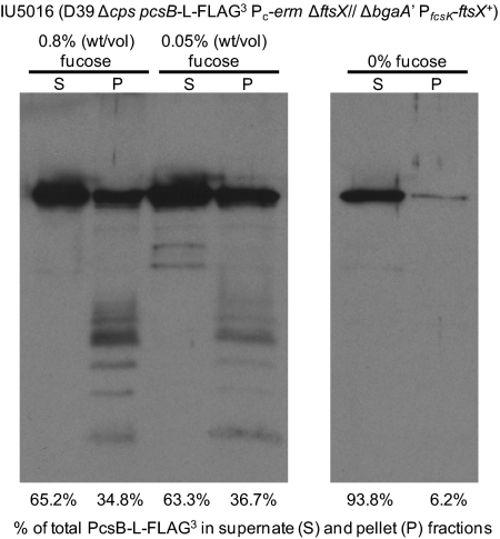
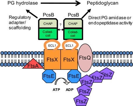
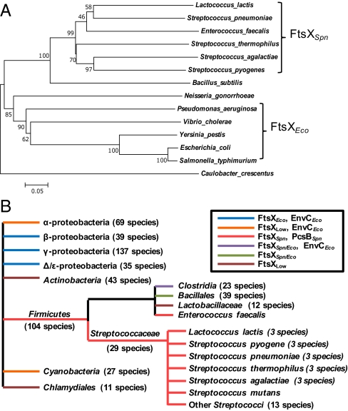
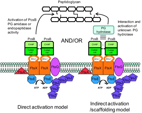
The connection between peptidoglycan remodeling and cell division is poorly understood in ellipsoid-shaped ovococcus bacteria, such as the human respiratory pathogen Streptococcus pneumoniae. In S. pneumoniae, peptidoglycan homeostasis and stress are regulated by the WalRK (VicRK) two-component regulatory system, which positively regulates expression of the essential PcsB cysteine- and histidine-dependent aminohydrolases/peptidases (CHAP)-domain protein. CHAP-domain proteins usually act as peptidoglycan hydrolases, but purified PcsB lacks detectable enzymatic activity. To explore the functions of PcsB, its subcellular localization was determined. Fractionation experiments showed that cell-bound PcsB was located through hydrophobic interactions on the external membrane surface of pneumococcal cells. Immunofluorescent microscopy localized PcsB mainly to the septa and equators of dividing cells. Chemical cross-linking combined with immunoprecipitation showed that PcsB interacts with the cell division complex formed by membrane-bound FtsX(Spn) and cytoplasmic FtsE(Spn) ATPase, which structurally resemble an ABC transporter. Far Western blotting showed that this interaction was likely through the large extracellular loop of FtsX(Spn) and the amino terminal coiled-coil domain of PcsB. Unlike in Bacillus subtilis and Escherichia coli, we show that FtsX(Spn) and FtsE(Spn) are essential in S. pneumoniae. Consistent with an interaction between PcsB and FtsX(Spn), cells depleted of PcsB or FtsX(Spn) had strikingly similar defects in cell division, and depletion of FtsX(Spn) caused mislocalization of PcsB but not the FtsZ(Spn) early-division protein. A model is presented in which the interaction of the FtsEX(Spn) complex with PcsB activates its peptidoglycan hydrolysis activity and couples peptidoglycan remodeling to pneumococcal cell division.


Conflict of interest statement

The authors declare no conflict of interest.

Figures

Fig. 1.
Fig. 1.
Schematic diagrams of the pneumococcal pcsB operon and the PcsB protein. pcsB is a single gene operon between mreCD and rpsB that is regulated positively by the WalRSpn response regulator, which binds upstream of the PpcsB promoter (8). Putative transcription terminators (lollipops) and the insertion point that creates the PcsB-l-FLAG3 fusion protein and adds the selectable Pc-erm marker are indicated. The PcsB polypeptide contains the following segments: a signal peptide that is cleaved off during export (16), a coiled-coil domain containing two putative leucine zipper motifs, an alanine-rich linker, and a CHAP domain containing a characteristic catalytic triad of 3 aa (Cys292-His343-N366). Simultaneous L78S and L219P amino acid changes in the putative leucine zipper motifs of the coiled-coil domain cause a temperature-sensitive phenotype. Additional details in the text.
Fig. 2.
Fig. 2.
PcsB binds to the outer surface of the cell membrane by hydrophobic interactions. (A) Fractionation of S. pneumoniae cells after PcsB-FLAG by Western blotting using anti-FLAG antibody. Supernates (S) or pellets (P) are marked for centrifugation steps, and cell fractions are indicated below the blots. Strain IU1845 was grown exponentially and fractionated as described in Materials and Methods. Secreted PcsB-FLAG was recovered from the culture supernatant by TCA precipitation. Bound PcsB-FLAG was from cell pellets, which were washed briefly with 5 M LiCl or digestion buffer. Washed cells were digested with mutanolysin and lysozyme (PG digestion lanes), and protoplasts were lysed by adding hypotonic buffer (membrane lanes). (B) Release of PcsB-FLAG from the cell surface of IU1845 was performed by washing with the chemicals indicated. Exponentially grown strain IU1845 was washed, and cell-bound (P) or released (S) PcsB-FLAG was detected by Western blotting as described in Materials and Methods. Released PcsB-FLAG was recovered from supernates by trichloroacetic acid (TCA) precipitation. Disruption of cell membrane was monitored by release of intracellular Era protein with anti-Era antibody (38). Arrows indicate large releases of PcsB-FLAG from cell surfaces by low concentrations of SDS that did not disrupt cell membranes. The experiment was performed three times with similar results.
Fig. 3.
Fig. 3.
PcsB forms a complex with division proteins FtsEXSpn in cell membranes. (A) Pull down of PcsB-l-FLAG3 complexes in membranes of exponentially growing cells that were treated with formaldehyde are shown, which was described in Materials and Methods. Bands on Western blots were detected with anti-FLAG antibody. Left lanes, samples that were not cross-linked. FT, flow-through fractions; W, washed fractions; EL, eluted fractions. Right lanes, cross-linked samples that were heated to break cross-links or not heated to maintain complexes. The composition of the bands was determined by MALDI MS as described in Materials and Methods. The experiment was done two times with similar results. (B) Pull down of cross-linked PcsB-l-FLAG3 complexes in membranes detected by Coomassie blue staining. Samples were prepared, heated to break cross-links, and resolved by SDS/PAGE as described in the text and Materials and Methods. Left lane, extract from R6 control strain that does not express a FLAG-tagged protein. Right lane, extract from strain IU3126 expressing PcsB-l-FLAG3. The unique pull-down bands are indicated, and their identities were confirmed by MALDI MS. The experiment was performed three times with similar results.
Fig. 4.
Fig. 4.
Purified PcsB and the coiled-coil (CC) domain of PcsB, but not the CHAP domain of PcsB, interacts with purified ECL1 of FtsXSpn. Proteins were purified, and Far Western blots were performed as described in Materials and Methods. The blot contains the amounts of purified proteins indicated at the top. The blot was probed with purified ECL1-FLAG protein and developed with anti-FLAG antibody. The expected position on the blot of PcsB-His6, PcsBCC-His6, and the CHAP domain are indicated. LytA should run between PcsBCC-His6 and PcsB-His6. No signal was detected on control blots when purified ECL1 or primary anti-FLAG antibody was omitted. The experiment was performed three times with similar results.
Fig. 5.
Fig. 5.
Defective cell morphology and FL-V staining and impaired growth caused by depletion of FtsXSpn. (A) Morphology of cells depleted of FtsXSpn. Depletion of FtsXSpn was brought about in ftsX merodiploid strain IU4723 (D39 Δcps ΔftsX// PfcsK-ftsX+) by decreasing the concentration of fucose in the culture medium as described in Materials and Methods. Samples were taken for phase-contrast microscopy or staining with FL-V and observation by epifluorescent microscopy at OD620 ∼ 0.2 for cultures containing 0.05% or 0.1% (wt/vol) fucose or 300 min after fucose was removed completely from the culture medium. (B) Growth curve (linear scale) of strain IU4723 depleted for FtsXSpn. The experiment was performed three times with similar results. (Scale bar, 2 μm.)
Fig. 6.
Fig. 6.
Localization of PcsB to equators and septa of dividing pneumococcal cells depends on FtsXSpn. Strains IU4959 (D39 Δcps pcsB-l-FLAG3) and IU5016 (D39 Δcps pcsB-l-FLAG3 ΔftsX::P-aad9// PfcsK-ftsX+) were grown exponentially, FtsXSpn was depleted in strain IU5016, and IFM with anti-FLAG antibody and staining with DAPI were performed as described in Materials and Methods. Representative images are shown from three independent experiments. Column 1, phase-contrast images showing cell outlines; column 2, location of PcsB-l-FLAG3 (pseudocolored green); column 3, DAPI staining of DNA nucleoids (pseudocolored red); column 4, overlaid images of columns 1 (phase) and 2 (PcsB-l-FLAG3). (Scale bar, 2 μm.) Additional details in the text.
Fig. 7.
Fig. 7.
PcsB-l-FLAG3 is released to the culture medium when FtsXSpn is severely depleted. Strain IU5016 was grown in BHI broth supplemented with 0.05% (wt/vol) fucose at 37 °C and diluted into fresh BHI broth containing 0.8% (wt/vol) fucose, 0.05% (wt/vol) fucose, or no fucose as described in Materials and Methods. When cultures reached early exponential phase (OD620 ∼ 0.2) or after ∼300 min, whichever came first, cells were collected by centrifugation, and PcsB-l-FLAG3 released to culture supernates (S) or in cell pellets (P) was detected by Western blotting using anti-PcsB antibody as described in the Materials and Methods. Percentages of total PcsB-l-FLAG3 in each sample in the supernatant (S) or pellet (P) are indicated. Phase-contrast microscopic examination of cells at the time samples taken for Western blotting did not reveal obvious lysis. Comparable results were obtained in three independent experiments.
Fig. 8.
Fig. 8.
Model for PcsB and FtsEXSpn interactions and functions in S. pneumoniae. In this model, a direct PG hydrolytic activity of PcsB is regulated by interactions that include binding of the CC domain of PcsB and the ECL1 of FtsXSpn. Membrane-bound FtsXSpn interacts with the cytoplasmic FtsESpn ATPase subunit. This model postulates that PG remodeling activity by PcsB is coordinated with cell division through its interaction with the FtsEXSpn complex, which interacts with FtsZSpn and other division proteins. Interaction of PcsB with the FtsEXSpn complex could also be required for PcsB to activate other PG hydrolases involved in cell division. Additional details in the text.
Fig. 9.
Fig. 9.
Phylogenetic and cooccurrence analysis of FtsXSpn and FtsXEco. (A) Neighbor-joining phylogenetic trees of FtsX in different bacteria were generated as described in Materials and Methods. The percentages of replicate trees in bootstrap tests are shown next to branches in the tree. Results presented here show an interaction between PcsBSpn and FtsXSpn, and parallel findings show an interaction between FtsXEco and EnvCEco (52). Consistent with these different interactions, FtsXSpn and FtsXEco are in distinctly different branches of the FtsX phylogenetic tree. (B) Distribution and cooccurrence of FtsXSpn, FtsXEco, EnvCEco, and PcsBSpn were determined by string analysis as described in Materials and Methods and are diagrammatically represented, where branches indicate groups only and not evolutionary relatedness. FtsXSpn, FtsXEco, EnvCEco, and PcsBSpn indicate homologs in organisms with high (>100 bits) similarity to the corresponding pneumococcal or E. coli proteins. FtsXLow indicates FtsX homologs in organisms with low similarity scores (<100 bits) to either pneumococcal or E. coli FtsX. FtsXSpn/Eco indicates homologs in organisms with high similarity scores (>100 bits) to both FtsXSpn and FtsXEco. Cooccurrence of strong homologs to FtsXEco and EnvCEco occur throughout β-, γ-, and Δ/ε-proteobacteria, whereas cooccurrence of strong homologs of FtsXSpn and PcsBSpn are confined to the Streptococcaceae and Enterococcus faecalis. The Actinobacteria, Clamydiales, Bacillales, and Lactobacillaceae lack strong homologs of EnvCEco and PcsBSpn and presumably have other classes of proteins that interact with their FtsX homologs. Additional details are in the text.
Fig. P1.
Fig. P1.
Models for PcsB and FtsEXSpn interactions and functions in S. pneumoniae. In the direct interaction model (left side), a peptidoglycan hydrolytic activity of PcsB is activated by interactions that include binding of the coiled-coil domain of PcsB and the ECL1 of FtsXSpn, shown herein. Membrane-bound FtsXSpn interacts with the cytoplasmic FtsESpn ATPase subunit. This model postulates that peptidoglycan remodeling by a PcsB hydrolytic activity is coordinated with cell division through its interaction with the FtsEXSpn complex, which interacts with FtsZSpn and other division proteins (4). In the indirect model, peptidoglycan remodeling is catalyzed by activation of an unknown peptidoglycan hydrolase that interacts with PcsB bound to the FtsEXSpn complex. In the indirect model, PcsB acts as a scaffolding protein between this hypothetical peptidoglycan hydrolase and the FtsEXSpn complex. The two models are not mutually exclusive.

Similar articles

Cited by

References

    1. Henriques-Normark B, Normark S. Commensal pathogens, with a focus on Streptococcus pneumoniae, and interactions with the human host. Exp Cell Res. 2010;316:1408–1414. - PubMed
    1. van der Poll T, Opal SM. Pathogenesis, treatment, and prevention of pneumococcal pneumonia. Lancet. 2009;374:1543–1556. - PubMed
    1. Weiser JN. The pneumococcus: Why a commensal misbehaves. J Mol Med (Berl) 2010;88:97–102. - PMC - PubMed
    1. Bancroft EA. Antimicrobial resistance: It's not just for hospitals. JAMA. 2007;298:1803–1804. - PubMed
    1. Black RE, et al. Global, regional, and national causes of child mortality in 2008: A systematic analysis. Lancet. 2010;375:1969–1987. - PubMed

Publication types

MeSH terms

LinkOut - more resources