Review
. 2011 Oct 14;11(11):723-37.
doi: 10.1038/nri3073.

Protective and pathogenic functions of macrophage subsets

Affiliations
Review

Protective and pathogenic functions of macrophage subsets

Peter J Murray et al. Nat Rev Immunol. .

Abstract

Macrophages are strategically located throughout the body tissues, where they ingest and process foreign materials, dead cells and debris and recruit additional macrophages in response to inflammatory signals. They are highly heterogeneous cells that can rapidly change their function in response to local microenvironmental signals. In this Review, we discuss the four stages of orderly inflammation mediated by macrophages: recruitment to tissues; differentiation and activation in situ; conversion to suppressive cells; and restoration of tissue homeostasis. We also discuss the protective and pathogenic functions of the various macrophage subsets in antimicrobial defence, antitumour immune responses, metabolism and obesity, allergy and asthma, tumorigenesis, autoimmunity, atherosclerosis, fibrosis and wound healing. Finally, we briefly discuss the characterization of macrophage heterogeneity in humans.


Figures

Figure 1
Figure 1. Tissue macrophages perform important homeostatic functions
Mononuclear phagocytes are generated from committed haematopoietic stem cells located in the bone marrow. Macrophage precursors are released into the circulation as monocytes and quickly migrate into nearly all tissues of the body, where they differentiate into mature macrophages. Various populations of mature tissue macrophages are strategically located throughout the body and perform important immune surveillance activities, including phagocytosis, antigen presentation and immune suppression.
Figure 2
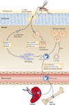
Figure 2. Inter-organ communication is required for macrophage recruitment
During infection and tissue stress, monocyte recruitment has a key role in providing the damaged tissues with adequate numbers of macrophages. The figure depicts an exemplar of the monocyte-to-macrophage recruitment and deposition process. Here, Leishmania major parasites that have infiltrated the skin after a sandfly bite elicit a weak local macrophage response that is insufficient to generate a protective response. The body compensates by depositing platelets on the parasite surface that release platelet-derived growth factor (PDGF). The local PDGF then increases the levels of CC-chemokine ligand 2 (CCL2), possibly by the stimulation of fibroblasts and other PDGF-responsive interstitial cells. CCL2 is a key monocyte attractant that causes monocyte efflux from the bone marrow and presumably the splenic monocyte reservoir. Extravasation of the monocytes is followed by differentiation into macrophages that phagocytose the parasites and present their antigens to T cells. Interferon-γ (IFNγ) production from T cells drives an M1 response that contains parasite growth. TH1, T helper 1.
Figure 3
Figure 3. In situ macrophage proliferation
A recent discovery has shown that, contrary to previous thinking, macrophages can enter the cell cycle and proliferate locally. Thus far, in situ proliferation has been shown to be specific for T helper 2 (TH2)-type responses to worms. In the example shown, a nematode is recognized through unknown mechanisms that may involve basophils, nuocytes and other sentinel lymphocytes and granulocytes. a | Local secretion of interleukin-4 (IL-4) initiates macrophage proliferation in situ, followed by amplification of the IL-4 response, which is mediated by antigen-specific TH2 cells. b | The increase in macrophage numbers has been proposed to play an important part in both killing the worms and driving a resolving phase of the infection. The underlying mechanism of IL-4-induced proliferation may involve multiple signals from the IL-4 receptor (IL-4R), including activation of signal transducer and activator of transcription 6 (STAT6). Individually or collectively, these signals may repress macrophage-activating factor (MAF) and MAFB, causing entry into the cell cycle. c | IL-4 can also cause M1-polarized macrophages to enter the cell cycle. In this case, an M1-polarized macrophage receives dual polarizing signals that drive gene expression characteristic of both M1 and M2 macrophages. ARG1, arginase 1; MAPK, mitogen-activated protein kinase; NO, nitric oxide; PI3K, phosphoinositide 3-kinase; RELMα, resistin-like molecule-α; TNF, tumour necrosis factor.
Figure 4
Figure 4. Distinct macrophage subsets regulate inflammation and wound healing
When tissues are damaged, inflammatory mediators are released, triggering an antifibrinolytic-coagulation cascade that activates clotting and the development of a provisional extracellular matrix (ECM). Platelet activation and degranulation also promotes blood vessel dilation and increased permeability, allowing efficient recruitment of inflammatory monocytes to the site of tissue injury, where they differentiate into macrophages and become activated by various cytokines, such as interferon-γ (IFNγ), that are released from neighbouring inflammatory cells, including neutrophils, natural killer (NK) cells, resident tissue macrophages and T cells. Pattern recognition receptor engagement can also contribute to the activation of resident dendritic cells (DCs) and recruited monocytes. During this initial leukocyte migration phase, inflammatory macrophages often display an M1-like phenotype, producing nitric oxide (NO), reactive oxygen species (ROS), interleukin-1 (IL-1) and tumour necrosis factor (TNF), which are important components of the antimicrobial arsenal. Secretion of matrix metalloproteinases (MMPs) such as MMP2 and MMP9 by inflammatory M1 macrophages also helps to degrade the ECM, facilitating the recruitment of inflammatory cells to the site of tissue injury. If the tissue-damaging irritant persists, activated M1 cells can further exacerbate the inflammatory response by recruiting large numbers of T helper 17 (TH17) cells and neutrophils, leading to substantial tissue damage. The damaged epithelial cells also release alarmins, including IL-25, IL-33 and thymic stromal lymphopoietin (TSLP), which induce IL-4 and IL-13 secretion by a variety of innate and adaptive immune cells, including nuocytes, mast cells, basophils and TH2 cells. When the inflammatory stimulus or pathogen is eliminated, M1 cell activation diminishes, and the alarmins and TH2-type cytokines drive the conversion of the immune response into a wound healing response, which is characterized by the accumulation of M2 macrophages that promote wound healing and fibrosis through the production of MMPs (including MMP12, tissue inhibitor of metalloproteinases 1 (TIMP1), growth factors (including platelet-derived growth factor (PDGF)) and cytokines (such as transforming growth factor-β1 (TGFβ1)). In the final stages of a wounding response, macrophages take on a regulatory/suppressive phenotype, which is characterized by the expression of arginase 1 (ARG1), resistin-like molecule-α (RELMα), programmed death ligand 2 (PDL2) and IL-10, which have all been shown to facilitate the resolution of wound healing and restore homeostasis while limiting the development of fibrosis, in part by suppressing T cell proliferation and collagen synthesis by activated myofibroblasts. M2 macrophages also promote the resolution of wound healing by antagonizing inflammatory M1 responses.

Similar articles

Cited by

References

    1. Geissmann F, et al. Development of monocytes, macrophages, and dendritic cells. Science. 2010;327:656–661. - PMC - PubMed
    1. Mantovani B, Rabinovitch M, Nussenzweig V. Phagocytosis of immune complexes by macrophages. Different roles of the macrophage receptor sites for complement (C3) and for immunoglobulin (IgG) J. Exp. Med. 1972;135:780–792. - PMC - PubMed
    1. Erwig LP, Henson PM. Clearance of apoptotic cells by phagocytes. Cell Death Differ. 2008;15:243–250. - PubMed
    1. Hajishengallis G, Lambris JD. Microbial manipulation of receptor crosstalk in innate immunity. Nature Rev. Immunol. 2011;11:187–200. - PMC - PubMed
    1. Gordon S, Taylor PR. Monocyte and macrophage heterogeneity. Nature Rev. Immunol. 2005;5:953–964. - PubMed

Publication types