. 2012 Feb;93(Pt 2):267-274.
doi: 10.1099/vir.0.036780-0. Epub 2011 Oct 12.

Analysis of subcellular G3BP redistribution during rubella virus infection

Affiliations

Analysis of subcellular G3BP redistribution during rubella virus infection

Jason D Matthews et al. J Gen Virol. 2012 Feb.

Abstract

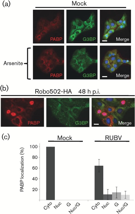
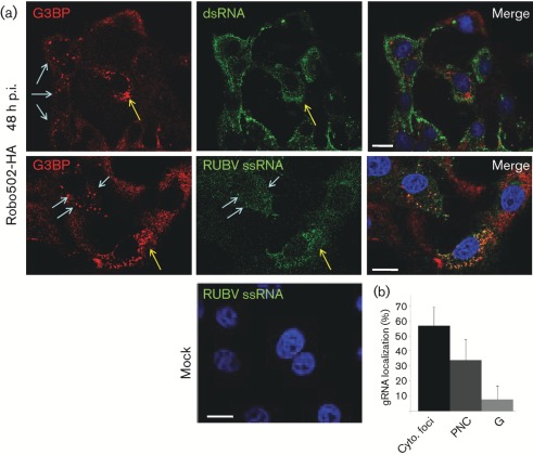
Rubella virus (RUBV) replicates slowly and to low titre in vertebrate cultured cells, with minimal cytopathology. To determine whether a cellular stress response is induced during such an infection, the formation of Ras-GAP-SH3 domain-binding protein (G3BP)-containing stress granules (SGs) in RUBV-infected cells was examined. Late in infection, accumulation of G3BP granules was detected, albeit in fewer than half of infected cells. Active virus RNA replication was required for induction of these granules, but they were found to differ from SGs induced by arsenite treatment both in composition (they did not uniformly contain other SG proteins, such as PABP and TIA-1) and in resistance to cycloheximide treatment. Thus, bona fide SGs do not appear to be induced during RUBV infection. The distribution of G3BP, either on its own or in granules, did not overlap with that of dsRNA-containing replication complexes, indicating that it played no role in virus RNA synthesis. However, G3BP did co-localize with viral ssRNAs in perinuclear clusters, suggesting an interaction that could possibly be important in a post-replicative role in virus replication, such as encapsidation.


Figures

Fig. 1.
Fig. 1.
Analysis of G3BP during RUBV infection. (a) Mock- or Robo502/P150-HA-infected cells (m.o.i. = 3) were analysed at 3, 24 and 48 h p.i. by Western blotting probed against HA (to detect HA-tagged P150), G3BP and calnexin (as an internal control). This experiment was repeated twice with similar results and thus a representative blot is shown. (b) The subcellular distributions of P150 (ii; green) and G3BP (iii; red) in Robo502/P150-GFP-infected cells (m.o.i. = 0.3) at 48 h p.i. are shown, with the merge in (iv). G3BP was detected by rabbit anti-G3BP and donkey anti-rabbit–Alexa Fluor 595. Blue arrows point to G3BP granules and yellow arrows point to perinuclear clusters. Similarly stained mock-infected cells are shown to the left in (i). Nuclei were stained with Hoechst 33342. Bars, 10 µm. (c) G3BP localization was scored in at least 100 mock- or 100 Robo502/P150-HA-infected cells (m.o.i. = 3) in at least 15 randomly chosen fields of view (infected cells were identified by dsRNA staining). G3BP distribution was categorized as solely cytoplasmic (Cyto), present in granules resembling stress granules (G) or present in perinuclear clusters (PNC). Error bars represent sd. These experiments were conducted at least twice.
Fig. 2.
Fig. 2.
Analysis of PABP during late RUBV infection. (a) Untreated (top panels) or arsenite-treated (bottom panels) cells were co-stained for PABP (red) and G3BP (green). PABP was detected by mouse anti-PABP and anti-mouse–Alexa Fluor 595, while G3BP was detected by rabbit anti-G3BP and chicken anti-rabbit–Alexa Fluor 488. Nuclei were stained with Hoechst 33342. Bars, 10 µm. (b) As (a), except that cells were infected with Robo502/P150-HA (m.o.i. = 3) and analysed at 48 h p.i. (c) At 48 h p.i., PABP localization was scored in at least 100 mock- or 100 Robo502/P150-HA-infected cells (m.o.i. = 3) (identified by dsRNA staining) in at least 15 randomly chosen fields of view (from at least two different experiments). PABP distribution was categorized as solely cytoplasmic (Cyto), nuclear (Nuc), granules resembling stress granules (G) or both nuclear and granules (Nuc/G). Error bars represent sd.
Fig. 3.
Fig. 3.
Analysis of G3BP granule composition and dynamics. (a) The localization of PABP in cells containing G3BP granules was tabulated in arsenite-treated (Ars+) Vero cells or Robo502/P150-HA-infected cells (m.o.i. = 3, 48 h p.i.) (at least 100 cells total for each sample). Three distinct localization patterns of PABP were observed in RUBV-infected cells positive for G3BP granules: nuclear (Nuc), granules (G) or both (Nuc/G). (b) The presence of TIA-1 in G3BP granules was determined by co-staining [TIA-1 (rabbit) and G3BP (chicken) antibody] of arsenite-treated Vero cells or Robo502/P150-HA-infected Vero cells (m.o.i. = 3) at 48 h p.i. At least 100 different cells that contained granules were counted from 15 different fields of view from each sample and the percentage of cells that contained G3BP granules with co-localizing TIA-1 was determined. (c) Vero cells treated for 35 min with 0.5 mM sodium arsenite were then exposed to 10 µg cycloheximide ml−1 (arsenite remained on cells during cycloheximide treatment) for an additional 35 min (+), while Robo502/P150-HA-infected cells (m.o.i. = 3) were exposed to 10 µg cycloheximide ml−1 for the entire 70 min (+). Vero cells treated with arsenite for 70 min, but not exposed to cycloheximide, and untreated Robo502/P150-HA-infected cells served as controls (−). The cells were then stained with anti-G3BP antibody, and 100 cells from at least 15 different fields of view were scored for the presence of G3BP granules (infected cells in the Robo502/P150HA-infected culture were identified by dsRNA staining). Error bars represent sd from at least two independent experiments.
Fig. 4.
Fig. 4.
G3BP granules form concomitantly with viral RNA replication and NSP accumulation. Vero (replication non-permissive) or C-Vero (replication-permissive) cells were transfected with RUBrep/P150-GFP in vitro RNA transcripts and analysed for the percentage of GFP-positive (i.e. successfully transfected) cells that contained G3BP granules by immunofluorescence using antibodies against G3BP. (a) Western blotting of Vero and C-Vero cell lysates for FLAG-tagged CP (approx. 30 kDa) is shown at top, whilst below are micrographs showing Vero or C-Vero cells at 48 h post-transfection with RUBrep/P150-GFP transcripts and stained for dsRNA (red) (GFP-tagged P150 is green in these micrographs and nuclei were counterstained with Hoechst 33342). (b) Quantification by ImageQuant of P150 levels on Western blots from 48 h post-transfection lysates (Vero or C-Vero) using GFP antibodies (mean of two independent experiments). (c) Quantification of GFP-positive cells containing G3BP granules. At least 100 cells from 15 different fields of view were analysed from at least two different experiments. Error bars represent sd.
Fig. 5.
Fig. 5.
Analysis of G3BP subcellular localization with viral ss- and dsRNA. (a) In the top panels, Robo502/P150-HA-infected cells (m.o.i. = 3, at 48 h p.i.) were stained red for G3BP with rabbit anti-G3BP/goat anti-rabbit–TRITC conjugate and green for dsRNA using mouse anti-dsRNA/goat anti-mouse–FITC conjugate, with the merged image shown on the right. In the bottom panels, similarly infected cells were probed with nick-translated, dUTP–Alexa Fluor 594-labelled DNA from the Robo502 plasmid to detect viral ssRNA (the FISH probe is pseudo-coloured green) and G3BP (red) as above. Mock-infected cells stained with RUBV-specific probes are shown (bottom). Blue arrows point to SGs and yellow arrows point to perinuclear clusters. Bars, 10 µm. (b) The primary localization of ssRNA [cytoplasmic foci, perinuclear clusters (PNC) or granules (G)] was counted in at least 100 cells from 15 different fields of view (from at least two different experiments). Bars represent sd.

Similar articles

Cited by

References

    1. Afonina E., Stauber R., Pavlakis G. N. (1998). The human poly(A)-binding protein 1 shuttles between the nucleus and the cytoplasm. J Biol Chem 273, 13015–13021 10.1074/jbc.273.21.13015 - DOI - PubMed
    1. Anderson P., Kedersha N. (2002). Visibly stressed: the role of eIF2, TIA-1, and stress granules in protein translation. Cell Stress Chaperones 7, 213–221 10.1379/1466-1268(2002)007<0213:VSTROE>2.0.CO;2 - DOI - PMC - PubMed
    1. Beatch M. D., Hobman T. C. (2000). Rubella virus capsid associates with host cell protein p32 and localizes to mitochondria. J Virol 74, 5569–5576 10.1128/JVI.74.12.5569-5576.2000 - DOI - PMC - PubMed
    1. Beckham C. J., Parker R. (2008). P bodies, stress granules, and viral life cycles. Cell Host Microbe 3, 206–212 10.1016/j.chom.2008.03.004 - DOI - PMC - PubMed
    1. Brune C., Munchel S. E., Fischer N., Podtelejnikov A. V., Weis K. (2005). Yeast poly(A)-binding protein Pab1 shuttles between the nucleus and the cytoplasm and functions in mRNA export. RNA 11, 517–531 10.1261/rna.7291205 - DOI - PMC - PubMed

Publication types

LinkOut - more resources